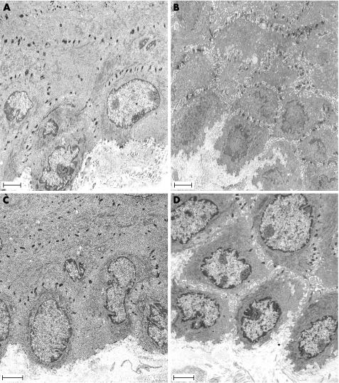
Figure 6 Effect of acute stress on the intercellular spaces of the oesophageal epithelium. Representative photomicrographs of the oesophageal epithelium in (A) control rat, (B) stressed rat, (C) control rat exposed to acid‐pepsin, and (D) stressed rat exposed to acid‐pepsin. An increase in the intercellular spaces was observed in stressed rats (B, D). Scale bars = 2.5 μm.

An official website of the United States government
Here's how you know
Official websites use .gov
A
.gov website belongs to an official
government organization in the United States.
Secure .gov websites use HTTPS
A lock (
) or https:// means you've safely
connected to the .gov website. Share sensitive
information only on official, secure websites.
