Atypical fibroxanthoma (AFX) is a pleomorphic tumour, first described by Helwig in 1961.1 AFX was thought to be a superficial form of malignant fibrous histiocytoma, but some describe AFX as a reactive lesion that can be confused with pleomorphic carcinomas. Clinically, AFX presents as a 1–2 cm rapidly growing, dome‐shaped, polypoid nodule or plaque which may crust or ulcerate.2 AFX is a low‐grade sarcoma and is usually cured by excision; however, recurrence and rare metastases have been reported.3
Histological examination shows a dome‐shaped nodule covered by a thin epidermis, which may be surrounded by a collarette of hyperplastic epithelium.4 Tumour cells are spindle—or epithelioid—shaped, and may have foamy cytoplasm. Cells may be dispersed randomly or arranged in fascicles. Nuclei are large, pleomorphic, and may be hyperchromatic and multiple in number.
Immunohistochemistry is essential in the diagnosis of AFX, because many poorly differentiated cutaneous spindle‐cell neoplasms have similar morphologies but stain differently. AFX is usually negative for S‐100 protein, HMB45, desmin, CD34 and cytokeratin (CK).5,6,7 Periodic acid Schiff stain fails to demonstrate glycogen in cells with foamy cytoplasm.7 AFX is CD68 variably positive and procollagen positive. Rare staining for actin and CD1A has been reported.6
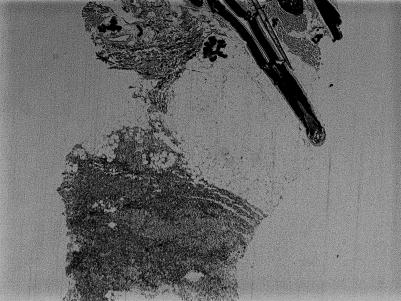
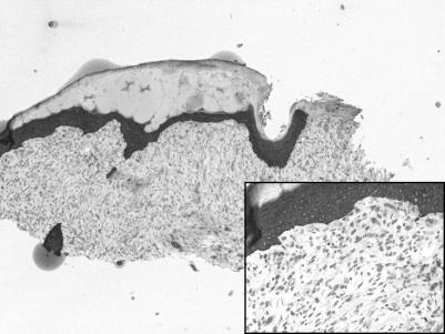
We report two cases of AFX that showed weak aberrant CK positivity. Two Caucasian men, each in his late 70s, presented with scaly, crusted nodules on the scalp and cheek, respectively. Excisional biopsies of both cases showed de‐differentiated fibrohistiocytic neoplasms. Cells from both cases stained positive for procollagen‐1 and CD68, negative for HMB45 and S‐100 protein, and weakly expressed CK (figs 1 and 2). Based on these clinical and histological findings, a diagnosis of AFX was made in both cases.
Figure 1 Atypical fibroxanthoma: case 1, cytokeratin. Note the diffuse, weak cytokeratin positivity in the deep dermis.
Figure 2 Atypical fibroxanthoma: case 2, cytokeratin stain. Note the faint dermal cytokeratin positivity.
Several theories have attempted to explain aberrant staining in cutaneous neoplasms. Tumour cells may inherently have epithelial antigens such as CK, or may take up epithelial antigens in their course of differentiation. Phagocytosis of CKs from other cells and adnexa is also a possibility. Conversely, these neoplasms may actually be de‐differentiated squamous‐cell carcinomas that have lost their epithelial antigens.
It is important to realise the inherent uncertainty in the diagnosis of poorly differentiated cutaneous malignant spindle‐cell neoplasms. The most reliable way to distinguish these neoplasms is immunohistochemistry; however, dermatopathologists must keep in mind the capabilities and limitations of immunohistochemistry. Aberrant CK expression can occur in AFX. It is important not to overinterpret this aberrant staining, which might lead to an erroneous diagnosis.
References
- 1.Helwig E B. Atypical fibroxanthoma. Proceedings of the 18th Annual Tumor Seminar of San Antonio Society of Pathologists, 1961. Texas State J Med 196359664–667. [PubMed] [Google Scholar]
- 2.Fretzin D F, Helwig E B. Atypical fibroxanthoma of the skin. A clinicopathologic study of 140 cases. Cancer 1973311541–1552. [DOI] [PubMed] [Google Scholar]
- 3.Helwig E B, May D. Atypical fibroxanthoma of the skin with metastasis. Cancer 196215717–725. [DOI] [PubMed] [Google Scholar]
- 4.Diaz‐Cascajo C, Weyers W, Borghi S. Pigmented atypical fibroxanthoma. A tumor that may be easily mistaken for malignant melanoma. Am J Dermatopathol 2003251–5. [DOI] [PubMed] [Google Scholar]
- 5.Winkelmann R K, Peters M S. Atypical fibroxanthoma. A study with antibody to S‐100 protein. Arch Dermatol 1985121753–755. [DOI] [PubMed] [Google Scholar]
- 6.Longacre T A, Smoller B R, Rouse R V. Atypical fibroxanthoma. Multiple immunohistochemical profiles. Am J Surg Pathol 1993171199–1209. [DOI] [PubMed] [Google Scholar]
- 7.Requena L, Sangueza O P, Sanchez Yus E.et al Clear‐cell atypical fibroxanthoma: an uncommon histopathologic variant of atypical fibroxanthoma. J Cutan Pathol 199724176–182. [DOI] [PubMed] [Google Scholar]


