Abstract
Objective
To compare real‐time three‐dimensional echocardiography (RT3DE) with two‐dimensional dobutamine stress echocardiography (2DE) for the detection of myocardial ischaemia, with angiographic validation of the results.
Methods
56 patients (mean (SD) age 64.5 (6.2) years, 38 males), referred for coronary angiography, were examined by 2DE and RT3DE during the same dobutamine stress protocol.
Results
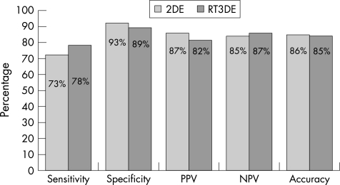
All 56 patients completed the stress protocol uneventfully. The mean (SD) acquisition time for the necessary views to evaluate all segments was 26.3 (2.5) s for RT3DE and 58.8 (3.7) s for 2DE (p<0.001). At peak stress, RT3DE had a higher wall‐motion score index (1.25 (0.24) by 2DE, 1.30 (0.27) by RT3DE; p = 0.014). The regional wall‐motion score for the four apical segments at peak stress was compared; it was 1.35 (0.55) by 2DE and 1.52 (0.69) by RT3DE (p = 0.003). The diagnostic parameters of 2DE versus RT3DE were: sensitivity 73% vs 78%, specificity 93% vs 89% and overall accuracy 86% vs 85%, respectively. In the left anterior descending artery territory, in particular, where RT3DE had higher regional wall‐motion scores, it showed a tendency towards higher sensitivity (85% vs 78%), although this difference did not achieve statistical significance.
Conclusion
RT3DE identifies wall‐motion abnormalities more readily in the apical region than 2DE, which may explain the tendency towards higher sensitivity in the left anterior descending artery territory. RT3DE results were validated using angiography as reference and findings indicate diagnostic equivalence to 2DE, with the advantage of considerable shorter acquisition times.
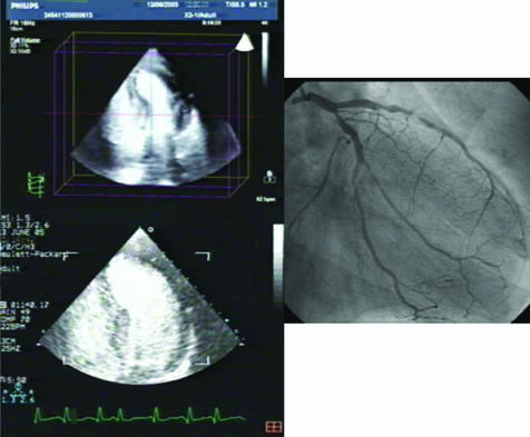
Dobutamine stress echocardiography has become a well established method of myocardial functional assessment in the diagnosis of coronary artery disease and in evaluation of its prognosis.1,2,3,4 The advent of echocardiography machines integrating all necessary systems for performing real‐time three‐dimensional echocardiography (RT3DE) holds promise as a new useful tool in cardiovascular ultrasonographic imaging. However, the clinical utility of this tool has yet to be adequately investigated. Particularly in the field of diagnosis of coronary artery disease, there is a marked lack of data regarding the usefulness of RT3DE. Our aim was to evaluate RT3DE in detecting myocardial wall‐motion abnormalities during a standard dobutamine stress protocol, in comparison to two‐dimensional echocardiography (2DE), with coronary angiography as the reference method for assessing the diagnostic power of this modality (fig 1).
Figure 1 Left top panel: three‐dimensional pyramidal dataset, following intravenous echo‐contrast administration, at peak stress, cropped in order to obtain a section similar to the two‐dimensional apical four‐chamber view. The apical interventricular septum and the apex are hypokinetic, leading to a rounded shape of the apex. Left bottom panel: two‐dimensional apical four‐chamber view in the same patient at peak stress. The same rounded shape of the apex is evident. Right panel: angiography in this patient showed significant left anterior descending artery disease.
Methods
Study population
The study population included 56 patients (mean (SD) age 64.5 (6.2) years, 38 males), referred for coronary angiography to the cardiac catheterisation laboratory of a tertiary hospital. All patients were in sinus rhythm. Exclusion criteria included the presence of symptoms of heart failure, a suspected or proven acute coronary event within the previous month, history of sustained ventricular tachycardia, moderate or severe valvular disease, uncontrolled hypertension, second or third degree atrioventricular block, cardiomyopathy and history of cerebral haemorrhage in the past 2 years. All patients provided informed consent.
Dobutamine stress protocol and echocardiography
Patients were examined in the left lateral decubitus position, using a Philips Sonos 5500 instrument with an s3 sector‐array transducer (1–3 MHz operating frequency range) for 2DE, and a Philips iE33 instrument with an X3‐1, 2400‐element matrix‐array transducer (1–3 MHz operating frequency range) for RT3DE (two machines were used in order to save time during image acquisition, by not having to make the necessary adjustments on the iE33 instrument on transition from 2DE to RT3DE imaging). An intravenous line was placed and dobutamine was infused in four 3‐min stages at dosing rates of 10–20–30–40 μg/kg/min, under continuous electrocardiographic monitoring. If the target heart rate was not achieved by the end of the fourth stage, atropine was given. Echo‐contrast agent (SonoVue, Bracco) was administered as a single‐bolus infusion at each stage before 2DE and RT3DE acquisition, in order to opacify the left ventricular cavity and achieve improved endocardial border delineation. At each stage, two‐dimensional images were acquired on the three standard apical planes: apical four chamber, apical two chamber and apical long axis (after the administration of contrast agent, parasternal images do not allow evaluation of wall motion due to shadowing by the contrast; thus, parasternal views were not interpreted). Three‐dimensional full‐volume data acquisition from the apical position followed two‐dimensional acquisition during each stage.
Image analysis
The left ventricular walls were divided into 16 segments, assigned to a vascular supply according to the American Society of Echocardiography recommendations.5,6 Each segment was scored for wall motion (1 for normal, 2 for mildly hypokinetic, 3 for severely hypokinetic, 4 for akinetic and 5 for dyskinetic). The global wall motion score index (WMSI) was calculated by dividing the sum of individual segmental scores by the number of segments.6 To compare the two modalities in the evaluation of the apex, the regional wall‐motion score of the apical four segments (apWMS) was also calculated. Ischaemia was defined as appearance of a new wall‐motion abnormality or deterioration of wall motion at stress or biphasic response. The two‐dimensional digitised images were compared on a quad‐screen (side‐by‐side viewing of electrocardiogram‐gated loops from each of the four stages of pharmacological stress). The acquired RT3DE data were cropped using the software integrated in the iE33 operating software (QLAB rev 4.2.1). RT3DE pyramidal volumetric datasets were cropped from the inferior wall (views analogous to the apical two‐dimensional four‐chamber view). Then, the cropping plane was inclined in order to get sections analogous to the two‐dimensional apical long‐axis and the apical two‐chamber views. Additional sections were obtained on the reader's choice, if deemed necessary, by rotating the pyramidal full‐volume datasets and cropping along the three rectilinear axes projected by the QLAB software. When a segmental wall motion abnormality was detected, the reader obtained more sections across this segment in order to make sure that the observed abnormality was not due to erroneous off‐axis cropping. Two‐dimensional and three‐dimensional images were interpreted by two experienced readers (CA and GR) who had no knowledge of the patients' clinical or angiographic data. They reviewed 2DE and RT3DE recordings identified by serial numbers (the reader did not know which 2DE recording corresponded to a given RT3DE study). In all, 24 studies (both 2DE and RT3DE) were read in duplicate (on different days, with different serial numbers) in order to assess intraobserver agreement. Interobserver agreement was also computed and the reports were compared in order to pinpoint discordances. Disagreements were then resolved by consultation and consensus between the two readers.
Coronary angiography
All patients underwent coronary angiography and left ventriculography by the Judkins technique, within 1 week from the echocardiography study without any intervening event. A normal arterial segment was identified immediately proximally and distally to the lesion and measured with an electronic caliper. The minimal stenosis diameter was also measured and severity was expressed as percentage reduction of normal diameter. Significant coronary artery disease was considered to be present if there was a 50% reduction in luminal diameter of at least one major epicardial vessel.
Statistical analysis
Continuous variables are presented as mean SEM and were compared using the unpaired or paired t test as appropriate. Categorical variables and proportions were compared using the χ2 test. The agreement between modalities was assessed using the κ statistic. Sensitivity, specificity, positive predictive value, negative predictive value and accuracy of the two modalities for the diagnosis of coronary artery disease were assessed and compared using the McNemar test. Analysis was performed by coronary territory. A value of p<0.05 was considered significant in all cases. Data analysis was performed using the SPSS V.10 statistical package for Windows.
Results
The distribution of major cardiovascular risk factors in the studied population (56 patients, 38 males, age 64.5 (6.2) years) was as follows: hypertension 39%, diabetes mellitus 15%, dyslipidaemia 28%, family history of coronary heart disease 22%, smoking 40%. All patients completed the stress protocol uneventfully. The mean acquisition time for all necessary views to evaluate all 16 segments was 26.3 (2.5) s for RT3DE and 58.8 (3.7) s for 2DE (p<0.001). Coronary angiography showed significant lesions (>50% stenosis) in 38 patients.
Assessment of wall‐motion abnormalities by 2DE and RT3DE
Fifteen of the patients had wall‐motion abnormalities at rest. New or worsened wall‐motion abnormalities at peak stress (sign of ischaemia) were detected in 34 patients by 2DE and in 37 patients by RT3DE. The two methods showed excellent agreement in WMSI at rest (Pearson's correlation index 0.984, 95% CI: 0.973 to 0.990, p<0.001) and very good correlation at peak stress (Pearson's correlation index 0.822, 95% CI: 0.713 to 0.892, p<0.001). The mean WMSI at rest was 1.055 (0.013) by 2DE and 1.059 (0.014) by RT3DE (mean difference 0.004, 95% CI: −0.01 to 0.001, p = 0.103). At peak stress, the mean difference in WMSI assessed by the two techniques was higher, with RT3DE reporting significantly higher WMSI (1.25 (0.032) by 2DE, 1.30 (0.037) by RT3DE; mean difference 0.053, 95% CI: 0.01 to 0.09, p = 0.014). In order to investigate whether the two modalities differ in the evaluation of the apex, which is known to present difficulties in its evaluation by 2DE,7 we compared the regional wall‐motion score for the four apical segments (apWMS) at peak stress. The mean apWMS was 1.35 (0.073) by 2DE and 1.52 (0.092) by RT3DE (p = 0.003). There was, thus, a significant difference in the evaluation of wall‐motion abnormalities by the two techniques in the apical region (mean apWMS difference 0.17, 95% CI 0.07 to 0.29) at peak stress.
Diagnostic parameters
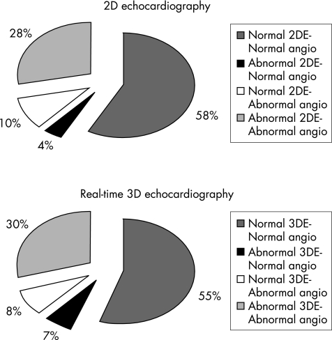
Altogether, 168 coronary territories were analysed. Of these, 64 (38%) were found to be supplied by a significantly stenosed artery on angiography. 2DE and RT3DE were in agreement in 157 of the territories (93%, κ = 0.855). The agreement of 2DE and RT3DE with the angiographic results per vascular territory was 86% and 85%, respectively (κ values 0.688 and 0.682, respectively). Figure 2 shows data regarding the concordance of echocardiographic findings to the angiographic results. Figure 3 illustrates the diagnostic parameters of the two techniques (per coronary vascular territory), using coronary angiography as the reference method. Table 1 shows the diagnostic parameters per vascular territory. It is worth noting that in the left anterior descending artery territory, in particular, where as previously mentioned RT3DE reported higher apical regional wall‐motion scores, RT3DE also showed a tendency towards higher sensitivity (85% vs 78%), although this difference did not achieve statistical significance.
Figure 2 Concordance data between the echocardiographic and the angiographic findings. The percentages express proportions to the total of 168 analysed vascular territories. Angio, angiography.
Figure 3 Diagnostic parameters for two‐dimensional (2DE) and real‐time three‐dimensional (RT3DE) dobutamine stress echocardiography in the per coronary territory analysis. Coronary angiography was used as the reference method. p>0.05 for all differences between the two methods. NPV, negative predictive value; PPV, positive predictive value.
Table 1 Diagnostic parameters of two‐dimensional and three‐dimensional dobutamine stress echocardiography for the detection of coronary artery disease and κ statistic for agreement with coronary angiography (method of reference; >50% stenoses were deemed as significant).
| Sensitivity (%) | Specificity (%) | PPV (%) | NPV (%) | Accuracy (%) | κ | |
|---|---|---|---|---|---|---|
| LAD | ||||||
| 2D echo | 78 | 92 | 95 | 82 | 85 | 0.748 |
| 3D echo | 85 | 90 | 88 | 87 | 88 | 0.749 |
| RCA | ||||||
| 2D echo | 75 | 90 | 79 | 86 | 86 | 0.646 |
| 3D echo | 80 | 86 | 76 | 89 | 84 | 0.654 |
| LCX | ||||||
| 2D echo | 65 | 96 | 85 | 86 | 86 | 0.638 |
| 3D echo | 65 | 92 | 79 | 86 | 84 | 0.600 |
LAD, left anterior descending artery; LCX, left circumflex artery; NPV, negative predictive value; PPV, positive predictive value; RCA, right coronary artery.
Differences between the two modalities were not statistically significant in all patients.
In the analysis per patient, the diagnostic parameters of 2DE versus RT3DE were: sensitivity, 82% vs 84%; specificity, 83% vs 72%; positive predictive value, 91% vs 86%; negative predictive value, 69% vs 68%; and overall accuracy, 82% vs 80%.
Interobserver variability
The agreement between the two readers (in the analysis per vascular territory) for wall‐motion assessment was 94% for 2DE and 92% for RT3DE.
Discussion
This study shows that RT3DE has significantly higher global and regional—in the apical region—wall‐motion score values than 2DE at peak stress with dobutamine infusion. Using coronary angiography as the reference method, we showed that this difference in reported regional wall‐motion score in the apex is accompanied by a tendency towards higher sensitivity of RT3DE than 2DE in the left anterior descending artery territory, without any marked difference in specificity. Moreover, we correlated RT3DE results with the anatomy of the coronary vessels, as assessed by angiography.
Real‐time three‐dimensional dobutamine echocardiography has been shown to have similar sensitivity to 2DE for the detection of myocardial ischaemia, with a first‐generation RT3DE system.8 Matsumura et al,9 using a second‐generation RT3DE system, showed a trend towards better sensitivity for RT3DE than 2DE in the left anterior descending artery territory, using single‐photon emission computed tomography as the reference standard, thus comparing two non‐invasive techniques. We proceeded further by validating the 2DE and RT3DE results with the anatomical study of coronary arteries through angiography. Our observations (also made with a second generation RT3DE system) reiterate the findings of Matsumura et al9 regarding the left anterior descending artery territory. Another point of difference in our study was the use of echo‐contrast agent for left ventricle opacification, which allowed adequate imaging in all patients—even those with poor‐quality acoustic windows. The trend of RT3DE to show higher sensitivity in the left anterior descending artery region may be interpreted as due to the increased ability of RT3DE to detect wall‐motion abnormalities in the apical segments, whereas foreshortening and poor endocardial delineation may play a negative role in the detection of wall‐motion abnormalities by 2DE.7 Moreover, the use of three‐dimensional full‐volume acquisition permits the performance of multiple sections of the apical region, essentially eliminating problems related to off‐axis image acquisition.
The above findings suggest that three‐dimensional dobutamine stress echocardiography is at least equivalent to 2DE in detecting coronary artery disease. Its true superiority, however, lies in the significantly shorter acquisition times; in our study, RT3DE acquisition times were less than half those of 2DE, comparable to those reported previously.8,9 Contemporary three‐dimensional echocardiographic instruments allow acquisition of full‐volume data of the left ventricle in as few as four cardiac beats, reducing acquisition times, which is a particularly useful characteristic for stress echocardiography, where special operating skill is needed to acquire two‐dimensional views on all imaging planes in the limited amount of time of each stress stage. Using RT3DE instead, all the operator needs to do is include the whole left ventricle in the acquisition volume. Inferior spatial resolution and frame rates compared with 2DE are the disadvantages of RT3DE; these drawbacks may offset the advantages of RT3DE (offline manipulation of real three‐dimensional images, ability to obtain multiple sections of any desired segment, virtual elimination of off‐axis acquisition, shorter acquisition time and so on) and could explain why RT3DE has not been shown to be superior to 2DE in previous reports,9 as well as in our study.
Study limitations
Our study population was selected from among those referred for angiography to the catheterisation laboratory of our hospital, and thus consisted of individuals with a high degree of suspicion for coronary artery disease. This may lead to overestimation of the accuracy of a diagnostic tool in the general population. However, since our goal was mainly to compare the two techniques (RT3DE and 2DE), rather than assess their absolute diagnostic power, this limitation should not have had an important effect on our observations.
Conclusion
RT3DE constitutes a promising development in echocardiography and its utility in various applications and patient populations needs to be evaluated. As a diagnostic approach of coronary heart disease in combination with dobutamine stress protocols, its diagnostic value is equivalent to that of 2DE, with the advantage of markedly shorter acquisition times. In this study, we validate RT3DE results using angiography findings and report that RT3DE identifies wall‐motion abnormalities more readily in the apical region than 2DE, which may underlie the tendency towards higher sensitivity in the left anterior artery territory.
Abbreviations
apWMS - apical wall‐motion score
2DE - two‐dimensional echocardiography
RT3DE - real‐time three‐dimensional echocardiography
WMSI - wall‐motion score index
Footnotes
Competing interests: None declared.
References
- 1.Mieres J H, Shaw L J, Arai A.et al Role of noninvasive testing in the clinical evaluation of women with suspected coronary artery disease: consensus statement from the Cardiac Imaging Committee, Council on Clinical Cardiology, and the Cardiovascular Imaging and Intervention Committee, Council on Cardiovascular Radiology and Intervention, American Heart Association. Circulation 2005111682–696. [DOI] [PubMed] [Google Scholar]
- 2.Becher H, Chambers J, Fox K.et al BSE procedure guidelines for the clinical application of stress echocardiography, recommendations for performance and interpretation of stress echocardiography: a report of the British Society of Echocardiography Policy Committee. Heart 200490(Suppl 6)vi23–vi30. [DOI] [PMC free article] [PubMed] [Google Scholar]
- 3.Quinones M A, Douglas P S, Foster E.et al American College of Cardiology/American Heart Association clinical competence statement on echocardiography: a report of the American College of Cardiology/American Heart Association/American College of Physicians‐American Society of Internal Medicine Task Force on Clinical Competence. Circulation 20031071068–1089. [DOI] [PubMed] [Google Scholar]
- 4.Arruda‐Olson A M, Juracan E M, Mahoney D W.et al Prognostic value of exercise echocardiography in 5,798 patients: is there a gender difference? J Am Coll Cardiol 200239625–631. [DOI] [PubMed] [Google Scholar]
- 5.Schiller N B, Shah P M, Crawford M.et al Recommendations for quantitation of the left ventricle by two‐dimensional echocardiography. American Society of Echocardiography Committee on Standards, Subcommittee on Quantitation of Two‐Dimensional Echocardiograms. J Am Soc Echocardiogr 19892358–367. [DOI] [PubMed] [Google Scholar]
- 6.Lang R M, Bierig M, Devereux R B.et al Chamber Quantification Writing Group; American Society of Echocardiography's Guidelines and Standards Committee; European Association of Echocardiography. Recommendations for chamber quantification: a report from the American Society of Echocardiography's Guidelines and Standards Committee and the Chamber Quantification Writing Group, developed in conjunction with the European Association of Echocardiography, a branch of the European Society of Cardiology, J Am Soc Echocardiogr 2005181440–1463. [DOI] [PubMed] [Google Scholar]
- 7.Marwick T H. Performance of stress echocardiography. Practical aspects of image acquisition and stress testing. In: Stress echocardiography, its role in the diagnosis and evaluation of coronary artery disease. 2nd edn. Boston: Kluwer Academic Publishers 20031–42.
- 8.Ahmad M, Xie T, McCulloch M.et al Real‐time three dimensional dobutamine stress echocardiography in assessment of ischaemia: comparison with two‐dimensional dobutamine stress echocardiography. J Am Coll Cardiol 2001371303–1309. [DOI] [PubMed] [Google Scholar]
- 9.Matsumura Y, Hozumi T, Arai K.et al Non‐invasive assessment of myocardial ischaemia using new real‐time three dimensional dobutamine stress echocardiography: comparison with conventional two dimensional methods. Eur Heart J 2005261625–1632. [DOI] [PubMed] [Google Scholar]


