Abstract
Background
Spinal cord stimulation (SCS) is an alternative treatment option for refractory angina. Controlled trials demonstrate symptom relief and improvement in functional status. Since patients experience retrosternal prickling during active SCS, there is no option for blinding patients to active treatment or for placebo control.
Objective
To examine the therapeutic effects of subthreshold SCS in patients with refractory angina in a placebo‐controlled study.
Methods
12 responders to treatment who had already been treated with SCS for refractory angina were enrolled. Patients were randomised into four consecutive treatment arms, each for 4 weeks, with various stimulation timing and output parameters: 3×2 h/day (phase A) and 24 h/day with conventional output (phase B); 3×2 h/day with a subthreshold output (phase C); and 24 h/day with 0.1 V output, which served as control (phase D). Functional status, quality of life, Canadian Cardiovascular Society classification and nitrate usage were assessed at the end of each 4‐week period.
Results
In phase D, patients showed a significant reduction in walking distance compared with phases A and C. Canadian Cardiovascular Society classification worsened in phase D compared with phases A–C. Frequency of angina attacks and the visual analogue scale were significantly worse in phase D than in phases A–C. In three patients, it was necessary to prematurely terminate phase D owing to intolerable angina attacks.
Conclusions
In this first placebo‐controlled trial to apply SCS in patients with refractory angina, improvement in functional status and symptoms was revealed in phases with conventional or subthreshold stimulation, in comparison to a low‐output (placebo) phase.
Thoracic spinal cord stimulation (SCS) has been proposed as an alternative treatment for patients who have severe disabling angina owing to coronary artery disease, and who are refractory to conventional forms of treatment.1,2,3 SCS is performed by means of an electrode installed in the epidural space (C7–Th1) and connected to a neurostimulator typically implanted subcutaneously in the upper left abdominal region. Stimulation produces a prickling sensation normally located in the dermatome, where angina pectoris is experienced. Treatment is controlled via a programmable set‐up and a handheld controller that is activated by the patient.
Randomised controlled trials4,5,6 that have compared active neurostimulation with inactive neurostimulation, as well as neurostimulation with bypass surgery, have revealed that SCS results in an enhancement of the functional status as well as a reduction in the frequency of angina and nitrate consumption. Observational studies7,8,9 and registries10 have confirmed clinical improvement, safety11 and cost effectiveness associated with SCS.12 Although numerous studies have been performed in this context, the mechanism of action is still not completely understood.13,14,15,16 Recently, a postinfarction heart failure canine model demonstrated the reduction of ischaemic ventricular arrhythmias by application of SCS.17
The lack of placebo‐controlled trials has, until now, represented a major shortcoming in this context. This lack is because of the fact that SCS is generally performed with stimulation intensity that causes a prickling sensation in the corresponding dermatome. It has consequently not proved possible, until now, to blind a patient to active treatment. The effect of thoracic SCS at a stimulation intensity below the sensory threshold had never been tested in a clinical setting, because it was thought to be ineffective.
In contrast, animal studies have revealed that SCS at intensities below motor threshold produced cutaneous vasodilatation through antidromic activation of sensory fibres.18 Gherardini et al19 tested the effect of low‐intensity stimulation (70% of motor threshold) in comparison to high‐intensity stimulation (90% of motor threshold) in a rat model. Long‐term survival of a groin flap was improved in the low‐intensity (60% survival) and in the high‐intensity (90% survival) groups in comparison to the control (0% survival). In addition, low‐voltage cervical neuromodulation improved cardiac work efficiency in a pig model.20 In humans, only observational data from single patients have been provided to date. Linderoth21 described a peripheral vasodilator response in a patient stimulated with an intensity unable to evoke paraesthesia.
We therefore argued that, in patients with refractory angina, stimulation at an intensity below the sensory threshold could well induce a therapeutic effect in comparison to stimulation intensity near 0 V (control).
Methods
Patients
This randomised, placebo‐controlled trial was performed at the Charité University Medical Centre, Berlin, Germany, between June 2003 and August 2004. Patients had chronic refractory angina pectoris (box 1) as defined by the European Society of Cardiology Joint Study Group.2 Subjects (table 1) were eligible if they had been responders to SCS treatment and had received implants at least 3 (but not >6) months before enrolment. They were classified as responders if they had shown a reduction in the number of angina episodes by at least 50%.22 No further inclusion or exclusion criteria existed. In all, 15 patients were screened for the study. Two proved to be non‐responders, and in one patient a fracture of the stimulation electrode was detected. The doctors obtained written informed consent from the patients, and the study was approved by the hospital ethics committee.
Table 1 Baseline characteristics of the study patients.
| Patient characteristics | n = 12 | % |
|---|---|---|
| Mean (SD) age, years | 65 (8) | |
| Gender (male) | 8 | 66.7 |
| Cardiac history | ||
| Coronary artery disease | 12 | 100 |
| 1 vessel | 2 | 16.7 |
| 2 vessels | 1 | 8.3 |
| 3 vessels | 9 | 75 |
| Positive myocardial stress test | ||
| Scintigraphy (pharmacological) | 12 | 91.7 |
| Scintigraphy (pharmacological) and bicycle ergometry | 5 | 41.7 |
| Angina pectoris (CCS angina classification) | ||
| CCS 3 | 7 | 58.3 |
| CCS 4 | 5 | 41.7 |
| Heart failure | ||
| Symptoms of heart failure | 7 | 58.3 |
| NYHA classification | 2±1 | |
| Mean (SD) left ventricular ejection fraction (%) | 52 (9) | |
| Previous cardiac events | ||
| Acute myocardial infarction | 11 | 91.7 |
| Percutaneous coronary intervention | 10 | 83.3 |
| Coronary artery bypass graft | 7 | 58.3 |
| Cardiac pacemaker | 4 | 33.3 |
| Extracardiac diseases | ||
| Diabetes | 4 | 33.3 |
| Obstructive pulmonary disease | 3 | 25 |
| Hypertension | 9 | 75 |
| Adiposity | 6 | 50 |
| Dyslipidaemia | 10 | 83.3 |
| History of smoking | 8 | 66.7 |
| Medication | ||
| β Blockers | 10 | 83.3 |
| Calcium antagonists | 6 | 50 |
| Long‐acting nitrates | 12 | 100 |
| Platelet inhibitors | 12 | 100 |
| Anticoagulants (warfarin) | 3 | 25 |
| ACE inhibitors/angiotensin II receptor blockers | 9 | 75 |
| Diuretics | 8 | 66.7 |
| Lipid‐lowering agents | 6 | 50 |
CCS, Canadian Cardiovascular Society; NYHA, New York Heart Association.
Box 1 Definition of chronic refractory angina pectoris
Angina pectoris >3 months
Canadian Cardiovascular Society classes III–IV
Known coronary artery disease
Reversible myocardial ischaemia
Optimal antianginal medication
No benefit from revascularisation procedures (percutaneous coronary intervention or coronary artery bypass graft)
Device selection and programming
In this study, subjects were implanted with Medtronic Itrel and Synergy neurostimulators (Medtronic, Düsseldorf, Germany). The epidural electrode used in all patients was a Pisces Quad (Medtronic).
During the regular follow‐up after implantation, stimulation parameters were optimised in each patient. Stimulation with so‐called conventional stimulation parameters elicits by definition a prickling sensation in the area in which the patient typically experiences angina pectoris. Standard parameters are stimulation amplitude 3–5.5 V, pulse width 210–300 μs and stimulation frequency 75–85 Hz. It was essential for the investigators to adjust exact stimulation ranges for each study arm in which the patients used the patient programmer.
Subthreshold output is defined as 85% (range 2.1–4 V) of the minimum stimulation output (voltage) causing paraesthesia. During programming, patients were asked to perform body movements that could modulate the effectiveness of neurostimulation: a measure undertaken to ensure that, during the subthreshold phase, the patient would under no circumstances experience any paraesthesia induced by the neurostimulator. During follow‐up, patients were questioned about paraesthesia by trained study nurses. This was particularly important to ensure that they never actually experienced paraesthesia during the subthreshold stimulation phases.
We selected 0.1 V as the maximum output in the control phase. This is the lowest output programmable while the neurostimulator is still fully functional. This is especially important, since patients use a patient‐control unit (handheld programmer with control light‐emitting diodes) to turn the device on and off and to regulate voltage output within the pre‐specified range. An output of 0.1 V is thought to have no effect on the neuronal system and accordingly served as control (placebo).
Patients were informed that they would not perceive thoracic paraesthesia in two subthreshold phases, but that improvement in functional status could nevertheless take place.
Study design
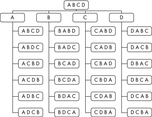
Patients were randomised (fig 1) into four consecutive treatment arms (intraindividual crossover), each lasting for 4 weeks, with various stimulation timing and output parameters: stimulation for 3×2 h/day with conventional output (phase A), 24 h/day with conventional output (phase B), 3×2 h/day with subthreshold output (phase C) and 24 h/day with 0.1 V output (phase D). Functional status—evaluated by the 6‐min walk test (6‐MWT)—and quality of life (QoL) were assessed at the end of each 4‐week treatment period. The 6‐MWT was performed according to published guidelines.23 A 35 m flat, obstacle‐free corridor was used, and patients walked unaccompanied in order that walking speed was not influenced. Angina attacks and use of short‐acting nitrates were monitored from days 8 to 28 by means of a patient diary, with the first 7 days of each treatment arm serving as a run‐in phase. Oral drugs for angina (table 1) were kept constant during the study period. We applied the Seattle Angina Questionnaire (SAQ) and the EuroQol visual analogue scale (VAS) to assess angina status and QoL at the end of each 4‐week period. The SAQ consists of five subgroups quantifying physical limitation, angina stability, angina frequency, treatment satisfaction and disease perception. The VAS, on the other hand, describes global QoL.
Figure 1 Flow chart of the randomisation procedure. Patients were initially randomised into one of the four groups (A–D); after each 4‐week study period, new randomisation took place. By the end of the study, every patient had passed through four different groups.
End point
The primary end point was the total walking distance in the 6‐MWT, which is closely related to everyday situations. Secondary end points were time to angina (6‐MWT), number of angina attacks, nitrate usage, Canadian Cardiovascular Society class, QoL according to the SAQ and the VAS, and premature termination of a study phase owing to intolerable symptoms. Assessments were performed at the same time of day at the end of each treatment arm, and in patients who received intermittent stimulation during active stimulation. All investigations were performed by specially trained study nurses who were unaware of the neurostimulator programming.
Statistical analysis
All data are shown as mean (SD). Data analysis was performed with SPSS V.11.5.1. for Windows. Since no normal distribution could be assumed, a non‐parametric test (Friedman's test) was used for global testing. In case of statistically significant differences, additional post hoc testing in pairs took place with Wilcoxon's test. Because of the exploratory character of the study, no adjustments for multiple comparisons were undertaken. For all comparisons, a value of p<0.05 was considered significant.
Results
Six‐minute walk test
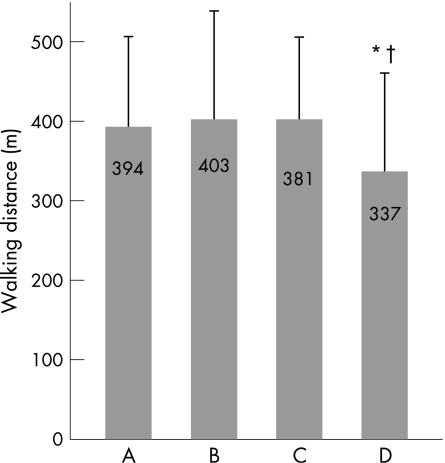
Walking distance in the 6‐MWT (fig 2) was the pre‐specified primary end point. There was no significant difference between phases A, B and C. At the end of phase D (control phase), walking distance was significantly lower than in phases A (p = 0.013) and C (p = 0.008).
Figure 2 Primary end point: physical capacity measured by total walking distance (m) in the 6‐min walk test (*p = 0.013 for A vs D; †p = 0.008 for C vs D). In all study arms, n = 12; three patients in phase D dropped out after the study end points were measured ahead of schedule.
Time to angina in the 6‐MWT (table 2) served as a secondary end point. In phases A and B, 4 (33%) patients experienced angina during the test, compared with 3 (25%) in phase C and 6 (50%) in phase D. Although there was a trend to shorter time to angina in phase D compared with phases A–C, statistical significance was not reached.
Table 2 Seattle Angina Questionnaire and visual analogue scale.
| Period A | Period B | Period C | Period D | p Value | |
|---|---|---|---|---|---|
| Structured diary, median (range) | |||||
| Angina episodes/21 days | 1.5 (1–25) | 3.5 (1–32) | 6.0 (1–28) | 11.0 (2–31) | * |
| Nitrate usage/21 days | 1.0 (0–21) | 2.0 (0–32) | 5.0 (0–27) | 9.0 (2–31) | † |
| Seattle Angina Questionnaire, mean (SD) | |||||
| Physical limitation | 52.3 (22.4) | 46.3 (26.8) | 50.2 (18.8) | 42.6 (14.9) | NS |
| Angina stability | 72.9 (29.1) | 56.3 (30.4) | 47.9 (37.6) | 25.0 (33.7) | ‡ |
| Angina frequency | 59.2 (33.7) | 54.2 (32.6) | 48.3 (37.3) | 34.2 (34.7) | § |
| Treatment satisfaction | 87.5 (13.0) | 85.9 (15.3) | 82.8 (17.5) | 82.3 (20.2) | NS |
| Disease perception | 74.3 (21.7) | 61.8 (19.3) | 60.4 (20.4) | 51.4 (24.3) | ¶ |
| VAS | 56.3 (20.4) | 57.5 (19.6) | 53.8 (21.4) | 45.9 (21.7) | ** |
| Symptom‐free walking interval | 323.3 (66.3) | 324.2 (64.2) | 323.3 (19.5) | 287.5 (98.6) | NS |
| Premature termination owing to intolerable angina pectoris (n) | 0 | 0 | 0 | 3 | |
VAS, visual analogue scale.
Score: 0–100; 0, worst and 100, best: physical limitation (NS).
*Structured diary: A–C vs D (p = 0.009; 0.015; 0.04).
†A–C vs D (p = 0.0002; 0.005; 0.015) and A vs C (p = 0.04).
‡Angina stability (A vs D (p = 0.01), B vs D (p = 0.043), C vs D (NS)).
§Angina frequency (A vs D (p = 0.007), B vs D (p = 0.044), C vs D (p = 0.034)), treatment satisfaction (NS).
¶Disease perception (A vs B–D (p = 0.03; 0.026; 0.021)).
**VAS (A and C vs D (p = 0.04), C vs D (p = 0.02)).
Quality of life
In phase D, the score obtained from the VAS was significantly lower than in phases A–C (p<0.05; table 2). Results from the SAQ were significantly different with respect to the following subgroups: angina frequency (A–C vs D) and angina stability (A and B vs D).
Angina attacks and short‐acting nitrate usage
Patients were asked to record the frequency of angina attacks and their usage of short‐acting nitrates in a diary in which 28 days were registered. Days 1–7 served as a wash‐in period. During phase D, the number of angina episodes was significantly higher than during phases A–C (p = 0.01, 0.02 and 0.04; table 2). No significant difference between phases A, B and C became apparent. This was reflected in the usage of oral short‐acting nitrates.
CCS angina classification
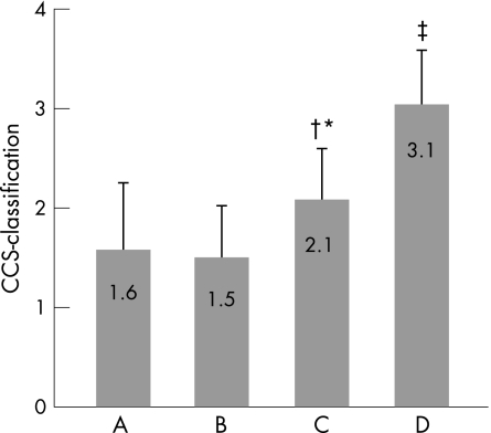
In phase D, CCS class was significantly worse than in phases A–C (p<0.05; fig 3). Owing to intolerable worsening of angina pectoris, it became necessary to terminate phase D in three patients (end points were measured completely). Two patients interrupted phase D after 5 days and one after 7 days. In phases A–C, all patients completed the 4‐week period.
Figure 3 Symptoms of angina pectoris measured by the classification of the Canadian Cardiovascular Society (CCS; *A vs C (p = 0.014); †B vs C (p = 0.012); ‡A–C vs D (p = 0.002); A vs B = 0.66). In all study arms, n = 12; three patients in phase D dropped out after the study end points were measured ahead of schedule.
Device follow‐up
During the follow‐up visits, device interrogation was performed to meter activation time and frequency. Reliable adherence to the protocol was documented in all patients.
It is important to note that during stimulation at the subthreshold level (phase C) and with 0.1 V (phase D), patients reported no perception of paraesthesia at any time.
Discussion
This is the first study that successfully tested the effect of thoracic SCS in patients with refractory angina pectoris, and in which subjects in one study phase were blinded to active treatment.
Blinding patients to active SCS
All randomised human studies reported so far had applied SCS in such a way that active treatment caused paraesthesia. Until now, it had been assumed that stimulation at a level below the sensory threshold would not lead to a therapeutic effect. It was therefore not possible to definitively rule out a placebo effect.24,25,26,27
Animal studies published in recent years have lent credence to the assumption that the stimulation effect gradually decreases with lower output parameters. In an experimental study, Gherardini et al19 investigated whether pre‐emptive SCS could increase long‐term flap survival. In 56 rats, SCS systems were implanted. After 3 days, a groin flap and the single feeding artery were occluded by a detachable clip, and after 12 h the clip was removed. In a control group, all flaps necrotised. Two groups were treated by means of neurostimulation, with stimulation amplitudes of 70% (low intensity) or 90% (high intensity) of the level that evokes muscular contractions. Flap survival was 60% in the low‐intensity group and 90% in the high‐intensity group. The authors thus concluded that the effect is dependent on stimulation intensity.
Greenwood‐Van Meerveld et al28 studied nociceptive reflexes in conscious rats with spinal cord‐stimulating electrodes implanted on the dorsal surface of the spinal cord 1 week before testing responses. The effect of SCS on nociceptive reflexes was determined by measuring abdominal contractions (visceromotor behavioural response) when the colon was distended to nociceptive levels. This study suggested that SCS did not reduce abdominal contractions until the stimulus intensity reached 80% of the motor threshold. SCS did not significantly reduce contractions between 20% and 60% of the motor threshold.
Tanaka et al18 investigated the effect of SCS below the motor threshold on cutaneous vasodilatation. In this experimental study, SCS was applied with stimulus parameters used clinically and with stimulus intensities at 30%, 60% and 90% of the motor threshold. Findings clearly showed that there was still an effect on ipsilateral blood flow at 60% and 30% of the motor threshold. Although it is obvious that animals are not able to report on paraesthesia that may or may not be induced by SCS, these cited studies clearly showed that the effect gradually decreases with lowering stimulation intensity. In addition, Linderoth21 published a case report of a patient having peripheral vascular disease, and showed that stimulation at an intensity unable to evoke paraesthesia induced vasodilatory response.
It was therefore reasonable to pose the hypothesis that, in humans, stimulation at a level not causing paraesthesia might still induce a therapeutic effect. This hypothesis was for the first time successfully tested in our patients.
Effect of SCS on walking distance and functional status
In treatment phases A–C, walking distance in the 6‐MWT did not differ significantly. By contrast, stimulation with 0.1 V was inferior compared with stimulation with conventional output, or to stimulation just below the output inducing paraesthesia. Walking distance in the 6‐MWT is an accepted outcome measure in multiple studies.29,30,31,32 It attains clinical importance owing to the fact that it reflects patients' behaviour in realistic everyday situations. A recent trial showed high concurrence between self‐reported symptoms of heart failure and 6‐MWT.33
Effect of SCS on CCS class, angina frequency and nitrate usage
There was significant worsening of CCS class and angina frequency in phase D compared with phases A–C. Most importantly, three patients in phase D (placebo) prematurely terminated this part of the study owing to intolerable angina symptoms, whereas no patients prematurely terminated phases A–C (subthreshold stimulation). In a randomised, controlled efficacy study, Hautvast et al4 studied 13 SCS and 12 control patients over a period of 6 weeks. Comparable results were disclosed with regard to reduction in angina pectoris, short‐acting nitrate usage and time to angina. It is important to note that we randomised patients who were already undergoing effective neurostimulation. This may have produced a degree of negative influence on the effects of symptom perception during subthreshold SCS in some patients. In addition, one can speculate about a trend for better results with paraesthesic compared with subthreshold stimulation (eg, angina symptoms and nitrate consumption), although the primary end point did not show significant differences. A placebo effect—as well as withdrawal of an effective treatment to which patients had become accustomed—could have contributed to this trend.
Effect of SCS on QoL
Symptom relief is one of the major goals of any treatment. It is of great importance that patients in phases A–C—in comparison to phase D—showed significant improvements in two parameters of the SAQ (angina severity and angina frequency) and in the VAS. A recent study by Diedrichs et al34 revealed a comparable improvement in QoL. In their study of 31 patients over a period of 1 year, these authors revealed that scores for angina stability and frequency improved significantly, results comparable to the findings of our study.
Comparison of stimulation duration and intensity
For almost all tested parameters, 3×2 h/day of stimulation was as effective as continuous stimulation for 24 h/day, which enables the option of using both stimulation duration set‐ups as alternatives, in accordance with patients' needs. It is necessary to note, however, that the battery lifespan will be shorter if neurostimulation is performed continuously. These results are in line with data from two registries. In the Italian registry,10 most of the patients were stimulated continuously. They experienced a reduction in CCS class from 3.4 to 2.2 after an average follow‐up of 13 months. TenVarwerk9 reported an improvement in New York Heart Association class from 3.5 to 2.1 in 517 patients.
Our results, likewise, strongly indicate that neurostimulation on a subthreshold level has the potential for replacing conventional stimulation patterns in the future. The absence of paraesthesia may lead to enhanced patient compliance owing to increased treatment comfort. Our observations support the assumption that it may be possible to eliminate the uncomfortable electrical stimulation experienced by some patients in unspecified settings.
Limitations
This is a pilot study including a limited number of patients. The results, although striking, require confirmation by a larger‐scale trial. For such a trial, a power calculation with regard to non‐inferiority (comparison between groups A, B and C) and superiority hypotheses (comparison between groups A, B, C and D) must be performed on the basis of the results of this trial.
Stimulation with 0.1 V (phase D) is generally held to be ineffective. Owing to technical constraints, it was not possible to switch off the device without unmasking inactive treatment to the patients.
Since patients turn the device on and off, the study was based on high patient compliance. We metered the number of activations via telemetry and compared the results with expected values. Our documentation confirms high adherence to the protocol.
For this study, we focused on patients who had already been identified as being responders to SCS treatment. It is known that 70–80% of patients with refractory angina (depending on the definition applied) who undergo implantation turn out to be responders. It is also important that patients become familiar with the device and with the patient remote‐control unit. In addition, we did not investigate the effect on myocardial ischaemia. Future studies should include a broader spectrum of patients and should investigate anti‐ischaemic effect in conjunction with functional status and patients' symptoms.
Conclusion
This randomised placebo‐controlled trial of SCS in patients with refractory angina pectoris reveals the possibility of improvement in functional status and in QoL, as well as of reduction in angina frequency. A placebo effect as the only mechanism of action seems unlikely. The option to use subthreshold stimulation as an active but patient‐blinded treatment facilitates attractive concepts for future studies and may extend clinical application.
Acknowledgements
We thank KD Wernecke and G Kalb for statistical analysis of the study data.
Abbreviations
6‐MWT - 6‐min walk test
QoL - quality of life
SAQ - Seattle Angina Questionnaire
SCS - spinal cord stimulation
VAS - visual analogue scale
Footnotes
Funding: This study was funded by Medtronic, Düsseldorf, Germany.
Competing interests: None.
References
- 1.Aronow W S, Frishman W H. Spinal cord stimulation for the treatment of angina pectoris. Curr Treat Options Cardiovasc Med 2004679–83. [DOI] [PubMed] [Google Scholar]
- 2.Mannheimer C, Camici P, Chester M R.et al The problem of chronic refractory angina. Report from the ESC Joint Study Group on the Treatment of Refractory Angina. Eur Heart J 200223355–370. [DOI] [PubMed] [Google Scholar]
- 3.Jessurun G A, DeJongste M J, Hautvast R W.et al Clinical follow‐up after cessation of chronic electrical neuromodulation in patients with severe coronary artery disease: a prospective randomized controlled study on putative involvement of sympathetic activity. Pacing Clin Electrophysiol 1999221432–1439. [DOI] [PubMed] [Google Scholar]
- 4.Hautvast R W, DeJongste M J, Staal M J.et al Spinal cord stimulation in chronic intractable angina pectoris: a randomized, controlled efficacy study [comment]. Am Heart J 19981361114–1120. [DOI] [PubMed] [Google Scholar]
- 5.Mannheimer C, Eliasson T, Augustinsson L E.et al Electrical stimulation versus coronary artery bypass surgery in severe angina pectoris: the ESBY Study. Circulation 1998971157–1163. [DOI] [PubMed] [Google Scholar]
- 6.De Jongste M J, Hautvast R W, Hillege H L.et al Efficacy of spinal cord stimulation as adjuvant therapy for intractable angina pectoris: a prospective, randomized clinical study. Working Group on Neurocardiology. J Am Coll Cardiol 1994231592–1597. [DOI] [PubMed] [Google Scholar]
- 7.Ekre O, Norrsell H, Wahrborg P.et al Temporary cessation of spinal cord stimulation in angina pectoris–effects on symptoms and evaluation of long‐term effect determinants. Coron Artery Dis 200314323–327. [DOI] [PubMed] [Google Scholar]
- 8.Jessurun G A, TenVaarwerk I A, DeJongste M J.et al Sequelae of spinal cord stimulation for refractory angina pectoris. Reliability and safety profile of long‐term clinical application. Coron Artery Dis 1997833–38. [DOI] [PubMed] [Google Scholar]
- 9.TenVaarwerk I A, Jessurun G A, DeJongste M J.et al Clinical outcome of patients treated with spinal cord stimulation for therapeutically refractory angina pectoris. The Working Group on Neurocardiology. Heart 19998282–88. [DOI] [PMC free article] [PubMed] [Google Scholar]
- 10.Di Pede F, Lanza G A, Zuin G.et al Immediate and long‐term clinical outcome after spinal cord stimulation for refractory stable angina pectoris. Am J Cardiol 200391951–955. [DOI] [PubMed] [Google Scholar]
- 11.Andersen C, Hole P, Oxhoj H. Does pain relief with spinal cord stimulation for angina conceal myocardial infarction? Br Heart J 199471419–421. [DOI] [PMC free article] [PubMed] [Google Scholar]
- 12.Murray S, Carson K G, Ewings P D.et al Spinal cord stimulation significantly decreases the need for acute hospital admission for chest pain in patients with refractory angina pectoris. Heart 19998289–92. [DOI] [PMC free article] [PubMed] [Google Scholar]
- 13.Armour J A, Linderoth B, Arora R C.et al Long‐term modulation of the intrinsic cardiac nervous system by spinal cord neurons in normal and ischaemic hearts. Auton Neurosci 20029571–79. [DOI] [PubMed] [Google Scholar]
- 14.Olgin J E, Takahashi T, Wilson E.et al Effects of thoracic spinal cord stimulation on cardiac autonomic regulation of the sinus and atrioventricular nodes. J Cardiovasc Electrophysiol 200213475–481. [DOI] [PubMed] [Google Scholar]
- 15.Barolat G. Spinal cord stimulation for chronic pain management. Arch Med Res 200031258–262. [DOI] [PubMed] [Google Scholar]
- 16.Foreman R D, Linderoth B, Ardell J L.et al Modulation of intrinsic cardiac neurons by spinal cord stimulation: implications for its therapeutic use in angina pectoris. Cardiovasc Res 200047367–375. [DOI] [PubMed] [Google Scholar]
- 17.Issa Z F, Zhou X, Ujhelyi M R.et al Thoracic spinal cord stimulation reduces the risk of ischemic ventricular arrhythmias in a postinfarction heart failure canine model. Circulation 20051113217–3220. [DOI] [PubMed] [Google Scholar]
- 18.Tanaka S, Barron K W, Chandler M J.et al Low intensity spinal cord stimulation may induce cutaneous vasodilation via CGRP release. Brain Res 2001896183–187. [DOI] [PubMed] [Google Scholar]
- 19.Gherardini G, Lundeberg T, Cui J G.et al Spinal cord stimulation improves survival in ischemic skin flaps: an experimental study of the possible mediation by calcitonin gene‐related peptide. Plast Reconstr Surg 19991031221–1228. [DOI] [PubMed] [Google Scholar]
- 20.Gersbach P A, Hasdemir M G, Eeckhout E.et al Spinal cord stimulation treatment for angina pectoris: more than a placebo? Ann Thorac Surg 200172S1100–S1104. [DOI] [PubMed] [Google Scholar]
- 21.Linderoth B. Spinal cord stimulation in ischemia and ischemic pain. In: Horsch S, Claeys L, eds. Spinal cord stimulation: an innovative method in the treatment of PVD and angina Darmstadt: Steinkopff, 199519–35.
- 22.Di Pede F, Zuin G, Giada F.et al Long‐term effects of spinal cord stimulation on myocardial ischemia and heart rate variability: results of a 48‐hour ambulatory electrocardiographic monitoring. Ital Heart J 20012690–695. [PubMed] [Google Scholar]
- 23.American Thoracic Society ATS statement: guidelines for the six‐minute walk test. Am J Respir Crit Care Med 2002166111–117. [DOI] [PubMed] [Google Scholar]
- 24.Boissel J P, Philippon A M, Gauthier E.et al Time course of long‐term placebo therapy effects in angina pectoris. Eur Heart J 198671030–1036. [DOI] [PubMed] [Google Scholar]
- 25.Brown W A. The placebo effect. Sci Am 199827890–95. [DOI] [PubMed] [Google Scholar]
- 26.Diedrichs H, Zobel C, Theissen P.et al Symptomatic relief precedes improvement of myocardial blood flow in patients under spinal cord stimulation. Curr Control Trials Cardiovasc Med 200567. [DOI] [PMC free article] [PubMed] [Google Scholar]
- 27.Kienle G S, Kiene H. The powerful placebo effect: fact or fiction? J Clin Epidemiol 1997501311–1318. [DOI] [PubMed] [Google Scholar]
- 28.Greenwood‐Van Meerveld B, Johnson A C, Foreman R D.et al Attenuation by spinal cord stimulation of a nociceptive reflex generated by colorectal distention in a rat model. Auton Neurosci 200310417–24. [DOI] [PubMed] [Google Scholar]
- 29.Fung J W, Yu C M, Yip G.et al Effect of beta blockade (carvedilol or metoprolol) on activation of the renin‐angiotensin‐aldosterone system and natriuretic peptides in chronic heart failure. Am J Cardiol 200392406–410. [DOI] [PubMed] [Google Scholar]
- 30.Mancini D M, Katz S D, Lang C C.et al Effect of erythropoietin on exercise capacity in patients with moderate to severe chronic heart failure. Circulation 2003107294–299. [DOI] [PubMed] [Google Scholar]
- 31.Garrigue S, Bordachar P, Reuter S.et al Comparison of permanent left ventricular and biventricular pacing in patients with heart failure and chronic atrial fibrillation: prospective haemodynamic study. Heart 200287529–534. [DOI] [PMC free article] [PubMed] [Google Scholar]
- 32.Cazeau S, Leclercq C, Lavergne T.et al Effects of multisite biventricular pacing in patients with heart failure and intraventricular conduction delay. N Engl J Med 2001344873–880. [DOI] [PubMed] [Google Scholar]
- 33.Ingle L, Shelton R J, Rigby A S.et al The reproducibility and sensitivity of the 6‐min walk test in elderly patients with chronic heart failure. Eur Heart J 2005261742–1751. [DOI] [PubMed] [Google Scholar]
- 34.Diedrichs H, Zobel C, Theissen P.et al Symptomatic relief precedes improvement of myocardial blood flow in patients under spinal cord stimulation. Curr Control Trials Cardiovasc Med 200567. [DOI] [PMC free article] [PubMed] [Google Scholar]


