Abstract
Skeletal muscle is composed of heterogeneous myofibers with distinctive rates of contraction, metabolic properties, and susceptibility to fatigue. We show that class II histone deacetylase (HDAC) proteins, which function as transcriptional repressors of the myocyte enhancer factor 2 (MEF2) transcription factor, fail to accumulate in the soleus, a slow muscle, compared with fast muscles (e.g., white vastus lateralis). Accordingly, pharmacological blockade of proteasome function specifically increases expression of class II HDAC proteins in the soleus in vivo. Using gain- and loss-of-function approaches in mice, we discovered that class II HDAC proteins suppress the formation of slow twitch, oxidative myofibers through the repression of MEF2 activity. Conversely, expression of a hyperactive form of MEF2 in skeletal muscle of transgenic mice promotes the formation of slow fibers and enhances running endurance, enabling mice to run almost twice the distance of WT littermates. Thus, the selective degradation of class II HDACs in slow skeletal muscle provides a mechanism for enhancing physical performance and resistance to fatigue by augmenting the transcriptional activity of MEF2. These findings provide what we believe are new insights into the molecular basis of skeletal muscle function and have important implications for possible therapeutic interventions into muscular diseases.
Introduction
Skeletal muscle fibers of adult vertebrates differ markedly with respect to their contractile and metabolic properties, which reflect different patterns of gene expression (1). Slow-twitch or type I myofibers exhibit an oxidative metabolism, are rich in mitochondria, heavily vascularized, and resistant to fatigue. In contrast, fast-twitch or type II fibers exhibit glycolytic metabolism, are involved in rapid bursts of contraction, and fatigue rapidly. Myofibers can be further classified as either type I, IIa, IIx/d, or IIb, depending on the type of myosin heavy chain (MHC) isoform expressed (2). The heterogeneity of skeletal myofibers is reflected at the molecular level in that almost every protein involved in contraction (MHC, myosin light chain, troponin I, troponin T, troponin C, actinin, etc.) has at least 2 isoforms expressed discretely in slow (type I) and fast (type II) fibers (3). In adult animals, specialized myofiber phenotypes remain plastic and vary in response to contractile load, hormonal milieu, and systemic diseases (4). Functional overload or exercise training results in transformation of preexisting fast fibers to a slow-twitch, oxidative phenotype (5). Conversely, decreased neuromuscular activity induced by spinal cord injury, limb immobilization, space flight, or blockade of action potential conduction causes a slow-to-fast myofiber conversion (6).
Functional demands modulate skeletal muscle phenotypes by activating signaling pathways that modify the gene expression profile of the myofiber. The signaling pathways involved in myofiber remodeling are of particular interest because of their relevance to several human disorders, including muscle dystrophy, metabolic disorders, and muscle atrophy (7). Increasing the abundance of slow, oxidative fibers in the mdx mouse model of Duchenne muscular dystrophy, for example, reduces the severity of the dystrophic phenotype (8, 9). Skeletal muscles also play an important role in whole-body metabolism, such that increasing the number of type I fibers enhances insulin-mediated glucose uptake and protects against glucose intolerance (10), which could have important therapeutic implications for diabetes and other metabolic diseases.
The myocyte enhancer factor 2 (MEF2) transcription factor, a key regulator of muscle development, is preferentially activated in slow, oxidative myofibers (11) and responds to calcium-dependent signaling pathways that promote the transformation of fast, glycolytic fibers into slow, oxidative fibers (12). The transcriptional activity of MEF2 is repressed by class II histone deacetylases (HDACs) (13–15). However, the potential involvement of MEF2 and class II HDACs in regulating myofiber identity in vivo has not been explored.
In this study, we show class II HDACs are selectively degraded by the proteasome in slow, oxidative myofibers, enabling MEF2 to activate the slow myofiber gene program. Consistent with these conclusions, forced expression of class II HDACs in skeletal muscle or genetic deletion of Mef2c or Mef2d blocks activity-dependent fast- to slow-fiber transformation whereas expression of a hyperactive MEF2 protein promotes the slow-fiber phenotype, enhancing endurance and enabling mice to run almost twice the distance of WT littermates. These findings provide new insights into the molecular basis of skeletal muscle performance and have important implications for possible therapeutic manipulation of muscle function for amelioration of muscular diseases.
Results
Reduction of class II HDAC proteins in soleus muscle.
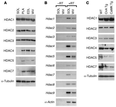
We speculated that variations in MEF2 activity patterns seen among different types of myofibers (12) might arise from differences in the extent of MEF2 repression by class II HDACs. To begin to explore this possibility, we determined the expression patterns of HDAC proteins in several skeletal muscles containing different proportions of fast and slow myofibers by Western blot analysis (Figure 1A).
Figure 1. Posttranscriptional downregulation of class II HDACs in soleus muscle.
Soleus (SOL), PLA, EDL, and WV muscles were dissected from the hind limbs of adult WT mice (8–10 weeks of age). (A) Protein expression of HDACs was assayed using antibodies specific for individual HDAC proteins. α-Tubulin level indicated equal loading. (B) RNA expression of HDACs in SOL versus WV was analyzed by RT-PCR in the presence (+) or absence (–) of reverse transcriptase (RT). Skeletal α-actin primers were used to show equivalent cDNA input. (C) Immunoblots of HDACs using WV muscle extracts from WT and 2 transgenic mouse models (10 weeks old) overexpressing active calcineurin A (CnA Tg) or CaMKIV (CaMK Tg).
Soleus muscle is composed primarily of slow, oxidative fibers with only a few fast, glycolytic fibers (16). This fiber-type composition fits the physiological functions of soleus muscle, which is used almost continuously to maintain posture and resist gravity. Three other skeletal muscles, plantaris (PLA), extensor digitorum longus (EDL), and superficial white vastus lateralis (WV), contain very few slow fibers (16). As seen in Figure 1A, class I HDACs (HDAC1, -2, and -3) were expressed at comparable levels in different muscle groups. In contrast, class II HDACs (HDAC4, -5, and -7) were expressed preferentially in the fast-fiber–dominant PLA, EDL, and WV muscles, with relatively little expression in the slow-fiber–enriched soleus (Figure 1A). MEF2 protein expression levels did not differ among muscle types (Supplemental Figure 1; supplemental material available online with this article; doi:10.1172/JCI31960DS1). The relatively low level of class II HDAC protein expression in soleus appeared to reflect a posttranscriptional mechanism since mRNA transcripts encoding class II HDACs were more abundant in soleus than in WV muscles, as revealed by both RT-PCR and Northern blot analyses (Figure 1B and Supplemental Figure 2).
Downregulation of class II HDACs in transgenic skeletal muscles transformed toward a slow, oxidative phenotype.
Forced expression of constitutively active calcineurin or calcium/calmodulin-dependent protein kinase IV (CaMKIV) in adult fast, glycolytic fibers of transgenic mice results in an increase in the number of slow fibers (17, 18). We used these transgenic mouse models to determine whether fast- to slow-fiber transformation correlated with a downregulation of class II HDACs, as might be expected if class II HDACs are involved in the fiber-type switch. The levels of class I HDACs (HDAC1, -2, and -3) were similar in WV muscles from WT and transgenic mice whereas the transformation of WV muscles toward a slow myofiber identity was associated with diminished expression of class II HDAC (HDAC4, -5, and -7) proteins (Figure 1C), consistent with the possibility that class II HDACs repress the expression of slow-fiber genes in fast myofibers.
Class II HDACs redundantly regulate slow, oxidative fiber expression.
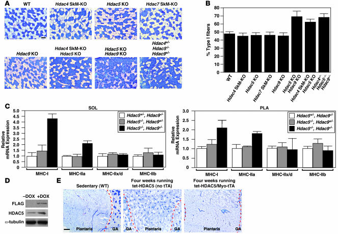
To directly examine the potential role of class II HDACs in regulating fiber-type identity, we analyzed adult skeletal muscles from mutant mice lacking 1 or more class II HDACs. Hdac5–/– and Hdac9–/– mice are viable (15, 19), so we were able to analyze the fiber-type composition of these homozygous mutants with global Hdac gene deletion in all tissues. However, because Hdac4–/– mice die at birth from skeletal defects (20) and Hdac7–/– mice die during embryogenesis from vascular defects (21), we used floxed alleles to delete these genes specifically in skeletal muscle using a skeletal muscle–specific Cre recombinase transgene (Myo-Cre) (SkM-KO mice), thereby avoiding lethality (22). Mice lacking individual class II HDACs did not display abnormalities in fiber-type switching or skeletal muscle development (Figure 2A). In contrast, soleus muscles from Hdac5–/–Hdac9–/– and Hdac4fl/-;Myo-creHdac5–/– double-knockout mice showed an increase in the percentage of slow myofibers from 48% ± 2.6% to 70% ± 3% (P < 0.003) and 63% ± 6.6% (P < 0.05), respectively (Figure 2, A and B). The fiber-type composition of the soleus of Hdac5+/–Hdac9–/– mice and Hdac4+/–Hdac5–/– mice was identical to that of WT mice (data not shown) whereas Hdac4+/–Hdac5–/–Hdac9+/– mutant mice showed an increase in slow fibers comparable to that of double-mutant mice (Figure 2A), suggesting that deletion of any combination of 4 alleles of Hdac4, -5, or -9 results in enhanced slow-fiber gene expression.
Figure 2. Class II HDACs redundantly regulate slow, oxidative fiber expression.
(A) Soleus muscles from WT, Hdac5–/– (Hdac5 KO), Hdac9–/– (Hdac9 KO), Hdac4fl/–;Myo-Cre (Hdac4 SkM-KO), Hdac7fl/–;Myo-Cre(Hdac7 SkM-KO), and class II HDAC compound mutant mice were analyzed by metachromatic ATPase staining. Type I fibers stain dark blue. Type II fibers stain light blue. Original magnification, ×10. Scale bar: 100 μm. (B) Quantification of fiber-type distribution based on fiber-type analysis in A. (C) Transcripts of MHC isoforms were determined in soleus and PLA muscles from mice of the indicated genotypes by quantitative real-time PCR. (D) Anti-FLAG M2 antibody on a Western blot analysis of proteins isolated from GP muscles of 4-week-old Myo-tTA/tet-HDAC5 mice treated with DOX or 10 days after removal of DOX. Tubulin served as a loading control. (E) Metachromatic ATPase staining of GP muscles harvested from sedentary WT, 4-week-exercised control (tet-HDAC5 [no tTA]), and 4-week-exercised HDAC5 transgenic (Myo-tTA/tet-HDAC5) mice. Original magnification, ×4. Scale bar: 300 μm. Dashed red lines delineate gastrocnemius (GA) muscle from PLA.
Analysis of the expression of transcripts encoding the individual MHC isoforms revealed an increase in expression of oxidative genes (MHC type I [MHC-I] and -IIa) in soleus and PLA muscles of Hdac5–/–Hdac9–/– mutant mice (Figure 2C) compared with Hdac5+/–Hdac9–/– or Hdac5–/–Hdac9+/– littermates. These results suggested that a reduction in expression of class II HDAC proteins below a specific threshold results in an increase in slow and oxidative fibers.
Forced expression of HDAC5 blocks fiber-type switching.
Exercise training transforms preexisting fast fibers to an oxidative phenotype (5). To determine whether class II HDACs modulated fiber-type switching in response to exercise, we constructed an inducible skeletal muscle–specific transgenic system using the myogenin promoter/MEF2 enhancer (Myo) to drive expression of the tetracycline transactivator (tTA) (Myo-tTA), which acts in trans to activate a tetracycline-responsive transgene. In this system, the transgene is not expressed in the presence of doxycycline (DOX) but is induced when the drug is removed. To verify the spatial expression of tTA in the Myo-tTA transgenic mice, we generated transgenic mice harboring a lacZ reporter gene cloned behind the tetracycline responsive (tet-responsive) expression cassette (tet-lacZ). When the Myo-tTA transgenic mice were crossed to the tet-lacZ reporter mice in the absence of DOX, lacZ expression was observed specifically in skeletal muscle without preference for adult slow or fast fibers (data not shown).
The Myo-tTA transgenic mice were bred to responder mice bearing a tet-responsive transgene encoding a signal-resistant FLAG-tagged human HDAC5 mutant protein (HDAC5S/A) (23). Expression of the FLAG-tagged HDAC5 protein was detected by Western blot analysis with an anti-FLAG antibody using total protein extracts from gastrocnemius and PLA (GP) muscles of 4-week-old tet-HDAC5/Myo-tTA transgenic mice that had DOX removed 10 days earlier (Figure 2D). Overexpression of HDAC5 in transgenic mice was confirmed by probing with an HDAC5 antibody (Figure 2D). FLAG-HDAC5 was not detected in the presence of DOX (Figure 2D).
To analyze the influence of HDAC5 on fiber-type switching, DOX was removed from 6-week-old tet-HDAC5/Myo-tTA double-transgenic mice, and these mice were provided free access to a running wheel for 4 weeks, a time period shown previously to allow the transformation of fast, glycolytic fibers to oxidative fibers (12). Tet-HDAC5/Myo-tTA transgenic mice and control mice ran voluntarily at comparable intensities (Supplemental Figure 3, A and B).
Metachromatic ATPase staining of skeletal muscles showed a pronounced increase in type I and IIa fibers within GP muscles of exercised tet-HDAC5 mice (without tTA) compared with unexercised mice (Figure 2E). In contrast, GP muscles from exercised tet-HDAC5/Myo-tTA double-transgenic mice without DOX did not show an increase in type I and type IIa fibers compared with sedentary mice (Figure 2E). Quantification of slow fibers revealed an approximate 10-fold reduction in the number of slow fibers from exercised HDAC5 transgenic mice compared with exercised WT mice (data not shown). Sedentary tet-HDAC5/Myo-tTA double-transgenic mice with and without DOX displayed normal fiber-type distributions (data not shown). We conclude that continuous repression of class II HDAC target genes in adult skeletal muscle is sufficient to inhibit exercise-induced fiber-type switching.
Requirement of MEF2 for establishing slow, oxidative myofiber identity.
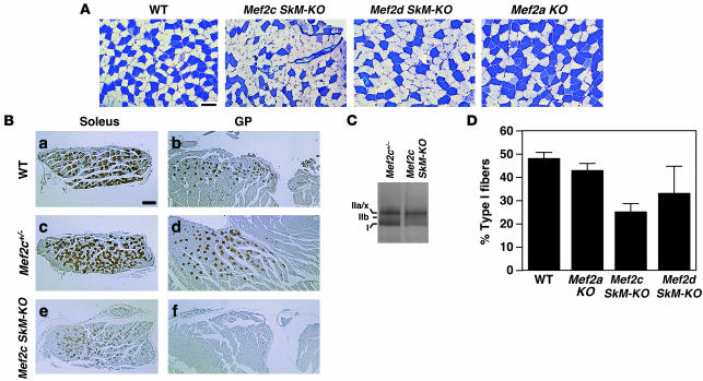
To examine whether class II HDAC regulation of fiber-type switching occurs through repression of MEF2 activity, we analyzed the skeletal muscles from individual MEF2 knockout mice. Conditional alleles of Mef2c and Mef2d (24, 25) were deleted specifically in skeletal muscle using transgenic mice that express Cre recombinase under the control of the myogenin promoter/MEF2 enhancer (Myo-Cre) (22), which is active in both fast and slow fibers (data not shown). As shown in Figure 3A, skeletal muscle–specific deletion (SkM-KO) of Mef2c or Mef2d using Myo-Cre resulted in a reduction in slow fibers within the soleus whereas the abundance of slow fibers was unaltered in Mef2a–/– mice (Figure 3A) or Mef2c+/– and Mef2d+/– mice (data not shown).
Figure 3. Requirement of MEF2 for establishing slow, oxidative myofiber identity.
Muscles from individual MEF2 knockout mice: Mef2a–/–, Mef2c SkM-KO (Mef2cfl/-;Myo-Cre), and Mef2d SkM-KO (Mef2dfl/fl;Myo-Cre) skeletal muscle conditional knockout mice were analyzed for fiber-type composition. (A) Metachromatic ATPase staining of soleus muscle. Type I fibers stain dark blue. Type II fibers stain light blue. Original magnification, ×10. Scale bar: 100 μm. (B) Immunohistochemistry of soleus and GP muscles of Mef2c SkM-KO and WT littermates using an MHC-I specific antibody. Original magnification, ×2.5. Scale bar: 300 μm. (C) Glycerol gradient silver staining of protein extracts from soleus of WT and Mef2c SkM-KO mice. MHC-I, -IIa/x, and -IIb isoforms are indicated. (D) Quantification of fiber-type distribution based on metachromatic ATPase staining of MEF2 knockout mice.
To further validate the reduction of slow fibers following skeletal muscle–specific deletion of Mef2c, we performed immunohistochemistry for type I fibers using a MHC-I specific antibody. As shown in Figure 3B, skeletal muscle lacking Mef2c displayed a loss of type I fibers in the GP and a reduction in number (and intensity) of type I fibers in the soleus. Moreover, using glycerol gradient silver staining of MHC isoforms from the Mef2c SkM-KO soleus muscle, we discovered that these muscles display a reduction in MHC-I (Figure 3C). Specifically, a decrease in the percentage of slow myofibers from 48% ± 2.6% to 25% ± 3.6% (P < 0.002) and 33% ± 11.4% (P < 0.001) was observed in the soleus of the Mef2c SkM-KO and Mef2d SkM-KO mice, respectively (Figure 3D). These findings demonstrate that MEF2C and MEF2D activate slow-fiber genes and that repression of fiber-type switching by class II HDACs is mediated, at least in part, through their repressive influence on MEF2C and MEF2D.
Activated MEF2 is sufficient to increase slow-fiber gene expression and muscle performance.
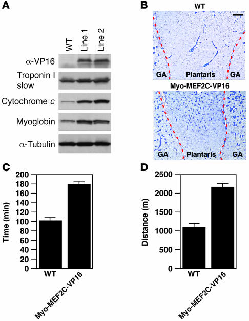
To determine whether MEF2 proteins were not only necessary, but also sufficient, for properly establishing slow, oxidative myofiber distribution, we tested whether expression of a hyperactive MEF2C-VP16 chimera, which is insensitive to HDAC repression, was sufficient to increase slow, oxidative fiber expression. Indeed, skeletal muscle expression of MEF2C-VP16 (Myo-MEF2C-VP16) was sufficient to increase the number of slow fibers in the PLA, which is normally composed primarily of fast fibers (Figure 4B and data not shown). This increase in slow-fiber abundance was remarkably similar to the fiber-type increase observed in WT mice after exercise training (compare to Figure 2F). Analysis of muscle fiber markers and mitochondrial proteins by Western blot analysis revealed an increase in the slow-fiber–specific contractile protein, Troponin I, and the type I fiber oxidative proteins myoglobin and cytochrome c (Figure 4A) in Myo-MEF2C-VP16 transgenic mice, confirming the results of metachromatic ATPase staining. In addition, an increase in expression of other metabolic genes and important metabolic transcription factors, such as peroxisome proliferator-activated receptor–γ coactivator-1α (PGC-1α), was observed in Myo-MEF2C-VP16 transgenic skeletal muscles (Supplemental Figure 4). These findings demonstrate that MEF2 is sufficient to drive fast- to slow-fiber transformation, mimicking the effect of exercise in vivo.
Figure 4. Activated MEF2 is sufficient to increase slow-fiber expression.
(A) Western blot analysis of Myo-MEF2C-VP16 transgene expression using an anti-VP16 antibody. Expression of the slow-fiber–specific troponin I and oxidative markers myoglobin and cytochrome c in protein extract of GP muscles of Myo-MEF2C-VP16 transgenic mice. (B) Metachromatic ATPase staining of gastrocnemius and PLA muscles of WT and Myo-MEF2C-VP16 transgenic mice. Original magnification, ×4. Scale bar: 300 μm. Dashed red lines delineate gastrocnemius muscle from PLA. (C and D) Exercise endurance and muscle performance showing total time running (min; C) and total distance run (m; D) of Myo-MEF2-VP16 transgenic muscles were analyzed by forced treadmill exercise. Eight-week-old Myo-MEF2C-VP16 transgenic and WT male mice with similar body weights were subjected to forced treadmill exercise (n = 5 for each group) on a 10% incline.
To examine the functional consequences of the increase in slow fibers and oxidative capacity of Myo-MEF2C-VP16 transgenic muscles, we measured the endurance of these mice by forced treadmill exercise on a 10% incline. As shown in Figure 4, C and D, Myo-MEF2C-VP16 transgenic mice displayed a 75% increase in running time and a 94% increase in distance, respectively, compared with WT mice. Thus, activation of MEF2 is sufficient to enhance skeletal muscle oxidative capacity and mitochondrial content, thereby diminishing muscle fatigability and augmenting endurance.
Class II HDACs are ubiquitinated and degraded by the proteasome.
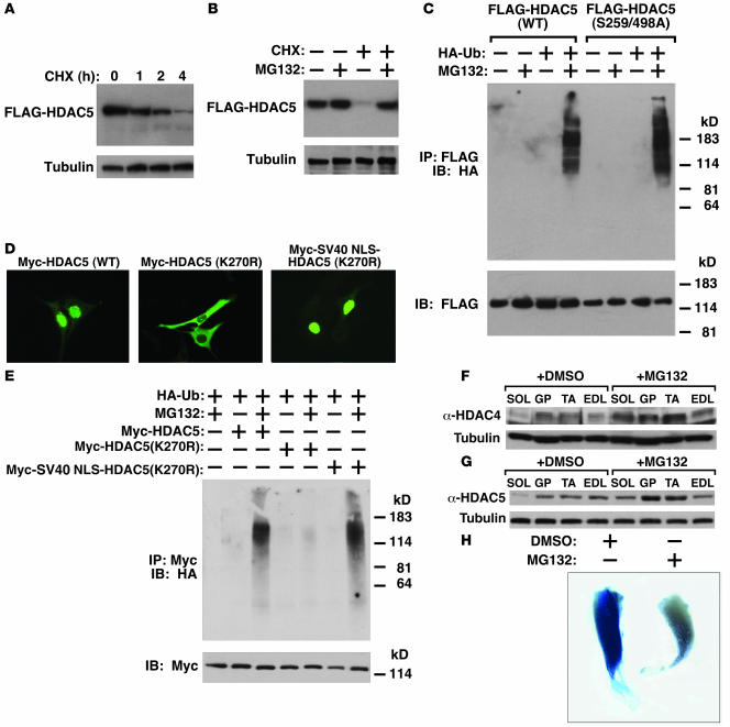
To begin to understand the mechanistic basis for the lack of accumulation of class II HDAC proteins in slow skeletal muscle fibers, we examined the half-life of HDAC5 in vitro using a stable C2C12 muscle cell line that constitutively expressed FLAG-tagged HDAC5. Inhibition of protein synthesis with cycloheximide for 4 hours resulted in a precipitous decrease in the level of HDAC5 protein in skeletal myocytes (Figure 5A). In contrast, α-tubulin was stable over this time period. The proteasome inhibitor MG132 blocked the degradation of HDAC5 (Figure 5B), suggesting that HDAC5 is degraded by the proteasome pathway.
Figure 5. Ubiquitination and degradation of class II HDACs in vitro and in vivo.
(A) C2C12 cells stably expressing FLAG-tagged HDAC5 (C2C12-HDAC5) were treated with cycloheximide (CHX, 25 μM) for 0, 1, 2, or 4 hours before cells were lysed and FLAG-HDAC5 expression was measured by Western blot analysis using anti-FLAG M2 antibody. Tubulin immunoblot showed equivalent loading of each lane. (B) C2C12-HDAC5 cells were treated with cycloheximide and MG132, singly or in combination for 4 hours, and FLAG-HDAC5 expression was analyzed. (C) C2C12-HDAC5 and C2C12 cells stably expressing CaMK-resistant HDAC5 (S259/498A) were transfected with or without HA-tagged ubiquitin (HA-Ub) and treated with or without MG-132 (25 μM) for 4 hours; the ubiquitination status of WT and mutant HDAC5 was analyzed. FLAG expression in inputs shows equal loading. (D) Subcellular localization of Myc-HDAC5 (WT), Myc-HDAC5(K270R), or Myc-SV40 NLS-HDAC5(K270R) in C2C12 cells. (E) The ubiquitination status of cytoplasmic HDAC5 [Myc-HDAC5(K270R)] or nuclear HDAC5 [Myc-SV40 NLS-HDAC5(K270R)] was analyzed. (F and G) WT C57BL/6 males (8 weeks old) were IP injected with DMSO or MG132 for 6 hours. Protein was isolated from SOL, GP, tibialis anterior (TA), and EDL muscles and analyzed for expression of (F) HDAC4 or (G) HDAC5. Tubulin shows equal loading. (H) Treatment with MG132 decreases MEF2 activation. Six hours after DesMEF-lacZ mice were injected with DMSO or MG132, mice were run for approximately 3 hours using forced treadmill exercise. Skeletal muscles were then isolated from DMSO- and MG132-treated DesMEF mice and analyzed for lacZ expression. LacZ expression was reduced in MG132-treated muscles (where class II HDAC expression was increased).
Since ubiquitination is a prerequisite for degradation by the proteasome (26), we examined whether HDAC5 was ubiquitinated. Indeed, ubiquitinated HDAC5 was readily detectable when HA-tagged ubiquitin was expressed in the HDAC5-expressing cell line in the presence of MG132 (Figure 5C). The signal-resistant HDAC5S/A mutant (13), lacking the regulatory phosphorylation sites (serines 259 and 498), was ubiquitinated to the same extent as the WT HDAC5 protein (Figure 5C), indicating that phosphorylation of HDAC5 on these residues is not a prerequisite for ubiquitination. FLAG-HDAC4, -7, and MITR (a splice variant of HDAC9) were also ubiquitinated in C2C12 cells (data not shown).
To determine whether ubiquitination of HDAC5 occurs in the nucleus or cytoplasm, we compared the ubiquitination of WT HDAC5, which is expressed primarily in the nucleus (Figure 5D), and an HDAC5 mutant with a mutation in the nuclear localization signal, HDAC5(K270R) (Figure 5D). As shown in Figure 5E, the K270R mutant was not ubiquitinated, whereas fusion of an SV40–nuclear localization signal (SV40-NLS) to the HDAC5(K270R) protein restored nuclear localization and ubiquitination. Together these results demonstrate that HDAC5 ubiquitination occurs in the nucleus.
Blockade to HDAC degradation in vivo by MG132.
To determine whether class II HDACs are degraded via the proteasome pathway in slow, oxidative myofibers in vivo, 8-week-old, WT C57BL/6 male mice were injected with either DMSO or MG132 and expression of HDAC4 and -5 was examined in the soleus, GP, tibialis anterior, and EDL skeletal muscles after 6 hours, a time period shown previously to provide proteasome inhibition in vivo (27). Treatment with MG132 in vivo increased the level of HDAC4 and HDAC5 protein expression in the soleus to that of the EDL (Figure 5, F and G, respectively), consistent with the in vitro results and demonstrating that class II HDAC proteins are specifically degraded by the proteasome in slow and oxidative myofibers.
Finally, since treatment with MG132 results in an increase in class II HDAC protein, we examined its effect on MEF2 activity in vivo using a transgene reporter line (DesMEF lacZ) that expresses lacZ under control of 3 tandem MEF2 sites (28). Prior studies showed that the expression of lacZ in these mice provides a faithful measure of MEF2 activity (12). Sedentary MEF2 reporter mice (DesMEF lacZ) do not express lacZ in skeletal muscle. However, lacZ expression is observed when DesMEF mice are exercised (12). Therefore, we injected DesMEF lacZ transgenic mice with either DMSO or MG132 and after 6 hours ran the mice for approximately 3 hours using forced treadmill exercise. Skeletal muscles were then isolated from DMSO- and MG132-treated DesMEF lacZ mice and analyzed for lacZ expression. Soleus muscle from exercised, DMSO-treated DesMEF lacZ mice expressed lacZ, while lacZ expression in exercised, MG132-treated DesMEF lacZ muscles was significantly reduced (Figure 5H). These results demonstrate that inhibition of the proteasome in vivo, which prevents HDAC degradation, reduces the activation of MEF2 and slow, oxidative fiber gene expression.
Discussion
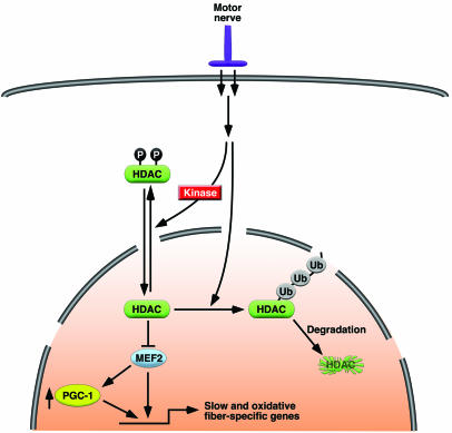
The results of this study show that slow and oxidative myofiber identity and muscle performance are governed by the balance between positive and negative signaling by MEF2 and class II HDACs, respectively. Degradation of class II HDAC proteins allows sustained activation of MEF2, which promotes the establishment of slow and oxidative myofibers and, strikingly, enhances muscle endurance and fatigue resistance (Figure 6).
Figure 6. A model for the control of slow and oxidative fibers by MEF2 and class II HDACs.
Motor nerve activity regulates MEF2 activity and myofiber identity through ubiquitination (Ub) and degradation of class II HDACs.
The contractile properties of skeletal myofibers reflect a combination of developmental and extrinsic inputs. During embryogenesis, fast- and slow-twitch fibers are patterned by specific developmental cues (3). After birth, the pattern of motor innervation plays a key role in influencing muscle fiber type. Phasic motor neuron firing at high frequency (100–150 Hz) promotes the formation of fast fibers, which display brief, high-amplitude calcium transients and low ambient calcium levels (<50 nM) whereas tonic motor neuron stimulation (10–20 Hz) favors the formation of slow fibers, which maintain higher intracellular calcium levels (100–300 nM) (29). Calcineurin and CaMK have been implicated in the transduction of calcium-dependent signals that upregulate the expression of oxidative, slow-fiber–specific genes in skeletal muscle (17, 18, 30). However, the precise mechanisms whereby these signaling pathways modulate the slow-fiber phenotype have not been defined (30).
Our results show that skeletal muscle of transgenic mice that has undergone a fast-to-slow myofiber transformation in response to activated calcineurin and CaMK displays a reduction in abundance of class II HDAC proteins, suggesting that these calcium-dependent signaling pathways act, at least in part, by enhancing degradation of class II HDAC proteins. The calcineurin and CaMK signaling pathways have also been shown to promote the phosphorylation of class II HDACs on a series of conserved serine residues, which mediate signal-dependent nuclear export and derepression of MEF2 target genes (13, 14). Thus, it seems likely that some combination of the 2 mechanisms — proteolysis and regulated nuclear export — regulates class II HDACs and thereby MEF2 activity and myofiber identity.
Nuclear factor of activated T cells (NFAT) transcription factors, which serve as transcriptional mediators of calcineurin signaling, have also been implicated in the control of slow-fiber gene expression (30, 31), as has the nuclear receptor PPARδ (32) and PGC-1α (33). Recently, PGC-1β was implicated in regulating type IIx/d fiber formation (34). MEF2 interacts with NFAT (35) and PGC-1α (36) and also regulates PGC-1α expression (23). Indeed, the MEF2-VP16 superactivator upregulated PGC-1α but not PGC-1β expression in skeletal muscle (Supplemental Figure 4). Thus, MEF2 serves as a nodal point for the control of multiple downstream transcriptional regulators of the slow-fiber phenotype and can potentially confer calcium sensitivity to other factors via its signal-dependent interaction with class II HDACs.
Our results show that ubiquitination of class II HDACs occurs in the nucleus. Nuclear ubiquitination of transcriptional activators has also been described as a mechanism to regulate the extent and duration of activation of transcriptional activators (37). The ubiquitination of class II HDACs provides another mechanism by which transcriptional activators may become activated. Class II HDACs and MEF2 are also sumoylated, which enhances the repressive activity of class II HDACs (38, 39). Whether sumoylation might be regulated during myofiber specification in vivo has not been addressed.
The mechanism that directs ubiquitination and degradation of class II HDACs in response to calcium signaling remains to be determined. It is tempting to speculate that phosphorylation of HDACs serves as a signal for ubiquitination by recruiting specific E3 ligases. Identification of the E3 ligase(s) for class II HDACs and the signals regulating their degradation are currently under investigation.
Our results show that adult skeletal muscle phenotypes are dictated by the extent of repression of MEF2 by class II HDACs and that the fast-fiber phenotype results from the absence of MEF2 activity. The fact that 4 Hdac alleles needed to be deleted to observe an increase in slow-fiber gene expression suggests that there is substantial functional redundancy among different HDACs with respect to repression of the slow-fiber gene program. Forced expression of a signal-resistant mutant of HDAC5 is sufficient to suppress the slow-fiber phenotype, whereas expression of a hyperactive MEF2-VP16 chimera is sufficient to override the repressive influence of endogenous class II HDACs and drive the slow-fiber phenotype. These findings suggest that the fast-fiber phenotype represents a “default” gene program resulting from the absence of MEF2 activity.
Therapeutic potential.
The ability of activated MEF2 to enhance oxidative capacity and endurance of skeletal muscle suggests opportunities for therapeutically enhancing muscle performance by stimulating MEF2 activity. Increasing the number of slow fibers in skeletal muscle via MEF2 also represents a potential method for treating metabolic and muscular diseases (8, 9). One could imagine augmenting MEF2 activity by interfering with the repressive activity of class II HDACs by modulating the signaling pathways that control HDAC phosphorylation, subcellular localization, or degradation. In this regard, HDAC inhibitors have recently been shown to suppress muscle pathology associated with muscular dystrophy (40, 41) in mice.
In addition to regulating skeletal muscle gene expression and function, MEF2 signaling has been shown to drive pathological cardiac growth and remodeling, which result from signal-dependent phosphorylation and nuclear export of class II HDACs in cardiac myocytes (19). These adverse consequences of MEF2 activation pose interesting challenges to the goal of enhancing MEF2 activity in skeletal muscle while avoiding possible cardiotoxicity of such strategies and, conversely, to pharmacologically preventing cardiac dysfunction without diminishing skeletal muscle function.
Methods
Plasmid constructs, tissue culture, and cell transfection.
The expression vector encoding HA-ubiquitin was described previously (42). Myc-NLS-HDAC5 was a kind gift from Tim McKinsey. The K270R point mutation was generated by site-directed mutagenesis (Stratagene). Myo-tTA and Myo-MEF2C-VP16 transgenic constructs were generated by cloning the tTA cassette from the pUDH 15-1 vector (43), fusing a VP16 activation domain in frame to the C terminus of full-length MEF2C, respectively, and cloning them into the HindIII site of a pBSIISK(+) vector containing a hGHpolyA tail. The myogenin promoter/MEF2C enhancer (22) was then cloned upstream of the cassettes into KpnI/XhoI sites.
C2C12 myoblasts were grown in DMEM supplemented with fetal bovine serum and antibiotics (100 U/ml penicillin and 100 μg/ml streptomycin). For transient transfection assays, cells were plated and transfected 12 hours later using FuGENE (Roche Applied Science) following the manufacturer’s instructions. Cells were harvested 24-48 hours after transfection. C2C12 stable lines expressing FLAG-tagged HDAC5 or FLAG-tagged HDAC5S/A were established by G418 selection of C2C12 clones transfected with plasmids encoding WT HDAC5 or a CaMK-resistant HDAC5 mutant, in which Ser259 and Ser498 were mutated into alanines (13). Cells were treated with cycloheximide (Sigma-Aldrich) and/or MG132 (Calbiochem; EMD Biosciences) at indicated concentrations.
Generation of transgenic mice and knockout mice.
Transgenic mice that express constitutively active forms of calcineurin or CaMKIV under the control of a muscle-specific enhancer from the muscle creatine kinase (MCK) gene are described elsewhere (17, 18). Tet-HDAC5S/A transgenic mice are described elsewhere (23). Myo-tTA, tet-lacZ, and Myo-MEF2C-VP16 transgenic mice were generated by injecting linearized constructs into the pronuclei of fertilized oocytes as previously described (44).
Hdac5–/– (15), Hdac9–/– (19), and Hdac7 conditional knockout mice had been generated previously (21). Hdac4 conditional mice were generated by flanking exon 6 with loxP sites, which results in an out-of-frame mutatation in the Hdac4 allele. Mef2a–/– and Mef2c and Mef2d conditional knockout mice have been described previously (24, 45). Correct gene targeting was confirmed by Southern blot analysis, genomic sequencing, and RT-PCR.
Immunoprecipitation and immunoblotting.
Immunoprecipitations were performed as previously described (20). Antibodies against HA (1:1,000; Sigma-Aldrich), FLAG M2 (1:4,000; Sigma-Aldrich), Myc (1:1,000; Santa Cruz Biotechnology Inc.), α-tubulin (1:5,000; Sigma-Aldrich), HDAC1, -2, and -3 (1:1,000 for all; Sigma-Aldrich), HDAC4 (1:500; Santa Cruz Biotechnology Inc.), HDAC5 (1:1,000; Millipore), HDAC7 (1:1,000; Cell Signaling Technology), MEF2A (1:1,000; Millipore), MEF2C (1:1,000; Santa Cruz Biotechnology Inc.), MEF2D (1:2,500; BD Biosciences), troponin I slow (1:2,500; Santa Cruz Biotechnology Inc.), cytochrome c (1:2,500; BD Biosciences — Pharmingen), and myoglobin (1:3,000; Dako) were used for immunoblot analyses. For analyzing endogenous class II HDAC proteins, the ECL Advance Western Blotting Detection Kit (Amersham Biosciences) was used.
In vivo pharmacological studies.
Myo-tTA and tet-HDAC5S/A transgenic mice were bred while receiving DOX (200 μg/ml) in water as previously described (23). Myo-tTA\tet-HDAC5S/A transgenic mice were maintained on DOX as needed. Voluntary wheel-running experiments were performed and measured as previously described (12). Animals were allowed to acclimate to running cages for 4 days prior to running recordings. DOX was removed from the mice for the days of acclimation so that the animals could begin expressing the transgene. After 4 days, wheel-running activity was measured continuously for 4 weeks.
Forced treadmill exercise experiments for analyzing Myo-MEF2C-VP16 transgenic mice were performed as follows. Prior to exercise, mice were accustomed to the treadmill (Columbus Instruments) with a 5-minute run at 7 m/min once per day for 2 days. The exercise test was performed on a 10% incline for 10 m/min for the first 60 minutes, followed by 1 m/min increment increases for 3- to 15-minute intervals, then 45 minutes at 13 m/min, followed by 1 m/min increment increases at 15-minute intervals until exhaustion. Forced exercise ended when mice were unable to avoid repeated electric shocks. MEF2 reporter mice (DesMEF lacZ) were run for approximately 3 hours at 9 m/min.
In vivo proteasome inhibition experiments were performed by intraperitoneally delivering DMSO or MG132 as previously described for 6 hours (27), except 30 μmol/kg body weight MG132 was used for injections. All experiments involving animals were reviewed and approved by the Institutional Animal Care and Research Advisory Committee of the University of Texas Southwestern Medical Center.
RNA isolation and analysis.
Total RNA was prepared from mouse tissues using TRI zol (Invitrogen) following the manufacturer’s instructions. Of total RNA, 1.5 μg was converted to cDNA using oligo dT primer and SuperScript II Reverse Transcriptase (Invitrogen). For PCR reactions, 2% of the cDNA pool was amplified. PCR cycles were optimized for each set of primers. Sequences for HDAC PCR primers have been described previously (46). Quantitative real-time PCR was performed for indicated MHC isoforms using SYBR Green (Applied Biosystems). Northern blots were performed with 20 μg of total RNA in each lane and probed in ULTRAhyb (Ambion) with labeled HDAC4, -5, or β-actin cDNA.
Fiber-type and immunohistological analysis.
Soleus and GP muscles were harvested from mice and flash frozen in embedding medium or fixed in 4% paraformaldehyde as previously described (47). Fiber-type analysis using metachromatic ATPase staining (48) and glycerol gradient silver staining were performed as previously described (47). Paraffin sections were stained with a MHC-I antibody, followed by treatment with DAB (Vector Laboratories).
Statistics.
Data are presented as mean ± SEM. Differences between groups were tested for statistical significance using the unpaired 2-tailed Student’s t test. Values of P < 0.05 were considered significant.
Supplementary Material
Acknowledgments
We thank Andrew Williams, Yuri Kim, Dillon Phan, Bryan Young, and Cheryl Nolen for technical assistance and Alisha Tizenor for assistance with graphics. This work was supported by grants from the NIH, the D.W. Reynolds Clinical Cardiovascular Research Center, the Texas Advanced Technology Program, the Muscular Dystrophy Association (to E.N. Olson), and the Robert A. Welch Foundation.
Footnotes
Nonstandard abbreviations used: CaMK, calcium/calmodulin-dependent protein kinase; DOX, doxycycline; EDL, extensor digitorum longus; GP, gastrocnemius and plantaris; HDAC, histone deacetylase; MEF2, myocyte enhancer factor 2; MHC, myosin heavy chain; Myo, myogenin promoter/MEF2 enhancer; Myo-Cre, Myo expressing Cre recombinase; Myo-MEF2C-VP16, Myo expressing a MEF2C-VP16 fusion protein; Myo-tTA, Myo expressing tTA; NLS, nuclear localization signal; PGC-1α, peroxisome proliferator-activated receptor–γ coactivator-1α; PLA, plantaris; SkM-KO mice, mice with a skeletal muscle–specific knockout; tet, tetracycline responsive; tTA, tetracycline transactivator; WV, white vastus lateralis.
Conflict of interest: The authors have declared that no conflict of interest exists.
Citation for this article: J. Clin. Invest. 117:2459–2467 (2007). doi:10.1172/JCI31960
See the related Commentary beginning on page 2468.
References
- 1.
- 2.Pette D., Staron R.S. Myosin isoforms, muscle fiber types, and transitions. Microsc. Res. Tech. 2000;50:500–509. doi: 10.1002/1097-0029(20000915)50:6<500::AID-JEMT7>3.0.CO;2-7. [DOI] [PubMed] [Google Scholar]
- 3.Schiaffino S., Reggiani C. Molecular diversity of myofibrillar proteins: gene regulation and functional significance. Physiol. Rev. 1996;76:371–423. doi: 10.1152/physrev.1996.76.2.371. [DOI] [PubMed] [Google Scholar]
- 4.Baldwin K.M., Haddad F. Effects of different activity and inactivity paradigms on myosin heavy chain gene expression in striated muscle. J. Appl. Physiol. 2001;90:345–357. doi: 10.1152/jappl.2001.90.1.345. [DOI] [PubMed] [Google Scholar]
- 5.Sugiura T., Miyata H., Kawai Y., Matoba H., Murakami N. Changes in myosin heavy chain isoform expression of overloaded rat skeletal muscles. Int. J. Biochem. 1993;25:1609–1613. doi: 10.1016/0020-711x(93)90519-k. [DOI] [PubMed] [Google Scholar]
- 6.Talmadge R.J. Myosin heavy chain isoform expression following reduced neuromuscular activity: potential regulatory mechanisms. Muscle Nerve. 2000;23:661–679. doi: 10.1002/(sici)1097-4598(200005)23:5<661::aid-mus3>3.0.co;2-j. [DOI] [PubMed] [Google Scholar]
- 7.Bassel-Duby R., Olson E.N. Signaling pathways in skeletal muscle remodeling. Annu. Rev. Biochem. 2006;75:19–37. doi: 10.1146/annurev.biochem.75.103004.142622. [DOI] [PubMed] [Google Scholar]
- 8.Chakkalakal J.V., et al. Stimulation of calcineurin signaling attenuates the dystrophic pathology in mdx mice. Hum. Mol. Genet. 2004;13:379–388. doi: 10.1093/hmg/ddh037. [DOI] [PubMed] [Google Scholar]
- 9.Stupka N., et al. Activated calcineurin ameliorates contraction-induced injury to skeletal muscles of mdx dystrophic mice. J. Physiol. 2006;575:645–656. doi: 10.1113/jphysiol.2006.108472. [DOI] [PMC free article] [PubMed] [Google Scholar]
- 10.Ryder J.W., Bassel-Duby R., Olson E.N., Zierath J.R. Skeletal muscle reprogramming by activation of calcineurin improves insulin action on metabolic pathways. J. Biol. Chem. 2003;278:44298–44304. doi: 10.1074/jbc.M304510200. [DOI] [PubMed] [Google Scholar]
- 11.Wu H., et al. MEF2 responds to multiple calcium-regulated signals in the control of skeletal muscle fiber type. EMBO J. 2000;19:1963–1973. doi: 10.1093/emboj/19.9.1963. [DOI] [PMC free article] [PubMed] [Google Scholar]
- 12.Wu H., et al. Activation of MEF2 by muscle activity is mediated through a calcineurin-dependent pathway. EMBO J. 2001;20:6414–6423. doi: 10.1093/emboj/20.22.6414. [DOI] [PMC free article] [PubMed] [Google Scholar]
- 13.McKinsey T.A., Zhang C.L., Lu J., Olson E.N. Signal-dependent nuclear export of a histone deacetylase regulates muscle differentiation. Nature. 2000;408:106–111. doi: 10.1038/35040593. [DOI] [PMC free article] [PubMed] [Google Scholar]
- 14.McKinsey T.A., Zhang C.L., Olson E.N. MEF2: a calcium-dependent regulator of cell division, differentiation and death. Trends Biochem. Sci. 2002;27:40–47. doi: 10.1016/s0968-0004(01)02031-x. [DOI] [PubMed] [Google Scholar]
- 15.Chang S., et al. Histone deacetylases 5 and 9 govern responsiveness of the heart to a subset of stress signals and play redundant roles in heart development. Mol. Cell. Biol. 2004;24:8467–8476. doi: 10.1128/MCB.24.19.8467-8476.2004. [DOI] [PMC free article] [PubMed] [Google Scholar]
- 16.Burkholder T.J., Fingado B., Baron S., Lieber R.L. Relationship between muscle fiber types and sizes and muscle architectural properties in the mouse hindlimb. J. Morphol. 1994;221:177–190. doi: 10.1002/jmor.1052210207. [DOI] [PubMed] [Google Scholar]
- 17.Naya F.J., et al. Stimulation of slow skeletal muscle fiber gene expression by calcineurin in vivo. J. Biol. Chem. 2000;275:4545–4548. doi: 10.1074/jbc.275.7.4545. [DOI] [PubMed] [Google Scholar]
- 18.Wu H., et al. Regulation of mitochondrial biogenesis in skeletal muscle by CaMK. Science. 2002;296:349–352. doi: 10.1126/science.1071163. [DOI] [PubMed] [Google Scholar]
- 19.Zhang C.L., et al. Class II histone deacetylases act as signal-responsive repressors of cardiac hypertrophy. Cell. 2002;110:479–488. doi: 10.1016/s0092-8674(02)00861-9. [DOI] [PMC free article] [PubMed] [Google Scholar]
- 20.Vega R.B., et al. Histone deacetylase 4 controls chondrocyte hypertrophy during skeletogenesis. Cell. 2004;119:555–566. doi: 10.1016/j.cell.2004.10.024. [DOI] [PubMed] [Google Scholar]
- 21.Chang S., et al. Histone deacetylase 7 maintains vascular integrity by repressing matrix metalloproteinase 10. Cell. 2006;126:321–334. doi: 10.1016/j.cell.2006.05.040. [DOI] [PubMed] [Google Scholar]
- 22.Li S., et al. Requirement for serum response factor for skeletal muscle growth and maturation revealed by tissue-specific gene deletion in mice. Proc. Natl. Acad. Sci. U. S. A. 2005;102:1082–1087. doi: 10.1073/pnas.0409103102. [DOI] [PMC free article] [PubMed] [Google Scholar]
- 23.Czubryt M.P., McAnally J., Fishman G.I., Olson E.N. Regulation of peroxisome proliferator-activated receptor gamma coactivator 1 alpha (PGC-1 alpha) and mitochondrial function by MEF2 and HDAC5. Proc. Natl. Acad. Sci. U. S. A. 2003;100:1711–1716. doi: 10.1073/pnas.0337639100. [DOI] [PMC free article] [PubMed] [Google Scholar]
- 24.Arnold M.A., et al. MEF2C transcription factor controls chondrocyte hypertrophy and bone development. Dev. Cell. 2007;12:377–389. doi: 10.1016/j.devcel.2007.02.004. [DOI] [PubMed] [Google Scholar]
- 25.Haberland M., et al. Regulation of HDAC9 gene expression by MEF2 establishes a negative feedback loop in the transcriptional circuitry of muscle differentiation. Mol. Cell. Biol. 2006;27:518–525. doi: 10.1128/MCB.01415-06. [DOI] [PMC free article] [PubMed] [Google Scholar]
- 26.Pickart C.M. Mechanisms underlying ubiquitination. Annu. Rev. Biochem. 2001;70:503–533. doi: 10.1146/annurev.biochem.70.1.503. [DOI] [PubMed] [Google Scholar]
- 27.Luker G.D., Pica C.M., Song J., Luker K.E., Piwnica-Worms D. Imaging 26S proteasome activity and inhibition in living mice. Nat. Med. 2003;9:969–973. doi: 10.1038/nm894. [DOI] [PubMed] [Google Scholar]
- 28.Naya F.J., Wu C., Richardson J.A., Overbeck P., Olson E.N. Transcriptional activity of MEF2 during mouse embryogenesis monitored with a MEF2-dependent transgene. Development. 1999;126:2045–2052. doi: 10.1242/dev.126.10.2045. [DOI] [PubMed] [Google Scholar]
- 29.Olson E.N., Williams R.S. Calcineurin signaling and muscle remodeling. Cell. 2000;101:689–692. doi: 10.1016/s0092-8674(00)80880-6. [DOI] [PubMed] [Google Scholar]
- 30.Chin E.R., et al. A calcineurin-dependent transcriptional pathway controls skeletal muscle fiber type. Genes Dev. 1998;12:2499–2509. doi: 10.1101/gad.12.16.2499. [DOI] [PMC free article] [PubMed] [Google Scholar]
- 31.Delling U., et al. A calcineurin-NFATc3-dependent pathway regulates skeletal muscle differentiation and slow myosin heavy-chain expression. Mol. Cell. Biol. 2000;20:6600–6611. doi: 10.1128/mcb.20.17.6600-6611.2000. [DOI] [PMC free article] [PubMed] [Google Scholar]
- 32.Wang Y.X., et al. Regulation of muscle fiber type and running endurance by PPARdelta. PLoS Biol. 2004;2:e294. doi: 10.1371/journal.pbio.0020294. [DOI] [PMC free article] [PubMed] [Google Scholar]
- 33.Lin J., et al. Transcriptional co-activator PGC-1 alpha drives the formation of slow-twitch muscle fibres. Nature. 2002;418:797–801. doi: 10.1038/nature00904. [DOI] [PubMed] [Google Scholar]
- 34.Arany Z., et al. The transcriptional coactivator PGC-1beta drives the formation of oxidative type IIX fibers in skeletal muscle. Cell Metab. 2007;5:35–46. doi: 10.1016/j.cmet.2006.12.003. [DOI] [PubMed] [Google Scholar]
- 35.Blaeser F., Ho N., Prywes R., Chatila T.A. Ca(2+)-dependent gene expression mediated by MEF2 transcription factors. J. Biol. Chem. 2000;275:197–209. doi: 10.1074/jbc.275.1.197. [DOI] [PubMed] [Google Scholar]
- 36.Moore M.L., Park E.A., McMillin J.B. Upstream stimulatory factor represses the induction of carnitine palmitoyltransferase-Ibeta expression by PGC-1. J. Biol. Chem. 2003;278:17263–17268. doi: 10.1074/jbc.M210486200. [DOI] [PubMed] [Google Scholar]
- 37.Kodadek T., Sikder D., Nalley K. Keeping transcriptional activators under control. Cell. 2006;127:261–264. doi: 10.1016/j.cell.2006.10.002. [DOI] [PubMed] [Google Scholar]
- 38.Zhao X., Sternsdorf T., Bolger T.A., Evans R.M., Yao T.P. Regulation of MEF2 by histone deacetylase 4- and SIRT1 deacetylase-mediated lysine modifications. Mol. Cell. Biol. 2005;25:8456–8464. doi: 10.1128/MCB.25.19.8456-8464.2005. [DOI] [PMC free article] [PubMed] [Google Scholar]
- 39.Gregoire S., Yang X.J. Association with class IIa histone deacetylases upregulates the sumoylation of MEF2 transcription factors. Mol. Cell. Biol. 2005;25:2273–2287. doi: 10.1128/MCB.25.6.2273-2287.2005. [DOI] [PMC free article] [PubMed] [Google Scholar]
- 40.Minetti G.C., et al. Functional and morphological recovery of dystrophic muscles in mice treated with deacetylase inhibitors. Nat. Med. 2006;12:1147–1150. doi: 10.1038/nm1479. [DOI] [PubMed] [Google Scholar]
- 41.Avila A.M., et al. Trichostatin A increases SMN expression and survival in a mouse model of spinal muscular atrophy. J. Clin. Invest. 2007;117:659–671. doi: 10.1172/JCI29562. [DOI] [PMC free article] [PubMed] [Google Scholar]
- 42.Hakak Y., Martin G.S. Ubiquitin-dependent degradation of active Src. Curr. Biol. 1999;9:1039–1042. doi: 10.1016/s0960-9822(99)80453-9. [DOI] [PubMed] [Google Scholar]
- 43.Resnitzky D., Gossen M., Bujard H., Reed S.I. Acceleration of the G1/S phase transition by expression of cyclins D1 and E with an inducible system. Mol. Cell. Biol. 1994;14:1669–1679. doi: 10.1128/mcb.14.3.1669. [DOI] [PMC free article] [PubMed] [Google Scholar]
- 44.Cheng T.C., Wallace M.C., Merlie J.P., Olson E.N. Separable regulatory elements governing myogenin transcription in mouse embryogenesis. Science. 1993;261:215–218. doi: 10.1126/science.8392225. [DOI] [PubMed] [Google Scholar]
- 45.Naya F.J., et al. Mitochondrial deficiency and cardiac sudden death in mice lacking the MEF2A transcription factor. Nat. Med. 2002;8:1303–1309. doi: 10.1038/nm789. [DOI] [PubMed] [Google Scholar]
- 46.Wu H., Olson E.N. Activation of the MEF2 transcription factor in skeletal muscles from myotonic mice. J. Clin. Invest. 2002;109:1327–1333. doi: 10.1172/JCI200215417. [DOI] [PMC free article] [PubMed] [Google Scholar]
- 47.Oh M., et al. Calcineurin is necessary for the maintenance but not embryonic development of slow muscle fibers. Mol. Cell. Biol. 2005;25:6629–6638. doi: 10.1128/MCB.25.15.6629-6638.2005. [DOI] [PMC free article] [PubMed] [Google Scholar]
- 48.Ogilvie R.W., Feeback D.L. A metachromatic dye-ATPase method for the simultaneous identification of skeletal muscle fiber types I, IIA, IIB and IIC. Stain Technol. 1990;65:231–241. doi: 10.3109/10520299009105613. [DOI] [PubMed] [Google Scholar]
Associated Data
This section collects any data citations, data availability statements, or supplementary materials included in this article.


