Abstract
INTRODUCTION
Fine needle aspiration cytology (FNAC) is a well-established investigation in thyroid disease. Fine needle sampling without aspiration (FNS) is less commonly used but often easier to perform. Both methods have advantages and disadvantages but, as yet, there is no agreement on which method produces better specimens for cytological diagnosis.
MATERIALS AND METHODS
We undertook a review of the literature and performed a meta-analysis of the results of four crossover trials.
RESULTS
The resulting odds ratio favoured FNS (OR = 0.99; 95% CI 0.88–1.11) but was not statistically significant. A fifth paper not included in the meta-analysis reported results in favour of FNS (P = 0.003).
CONCLUSIONS
There is no evidence from the meta-analysis that one method is superior to the other; however, taking into consideration all available evidence, it seems that FNS may be easier to perform and may produce better samples.
Keywords: Cytology, Fine needle biopsy, Needle biopsy, Thyroid gland, Non-aspiration
Fine needle aspiration is a commonly used technique to obtain material for cytological diagnosis. It is often the firstline investigation for tissue diagnosis of soft-tissue swellings in many sites in the body. In the field of otolaryngology, it is most often used for the investigation of neck swellings, particularly swellings of the thyroid gland.
Although cytology is less reliable than histopathology,1 it remains a very sensitive and specific test2 for thyroid lesions and is minimally invasive investigation. Although there are a number of methods for performing this investigation, the most commonly performed methods are fine needle aspiration cytology (FNAC) and fine needle sampling (FNS).
Fine needle aspiration cytology (FNAC)
This is usually performed by the method described by Lowhagen et al.3 A hollow needle of a fine gauge (usually 21–23) is attached to a 10-ml or 20-ml syringe. The needle is then inserted into the lesion and suction is applied by pulling back the on the plunger of the syringe. The needle is then passed back and forth through the lesion several times. The plunger of the syringe is then slowly released and the needle withdrawn. The contents of the needle and syringe are then sprayed onto a glass slide for examination by a cytopathologist.
Fine needle non-aspiration cytology (fine needle capillary cytology, non-aspiration cytology, cytopuncture; FNS)
This technique was first described for use in the investigation of thyroid lesions by Santos and Leiman4 in 1988. A similarly sized needle is used. The needle is usually used with no syringe attached but may have a syringe attached containing air. The needle is passed through the lesion in the same way as FNAC, but no suction is applied. The contents of the needle are then sprayed onto a slide.
Both methods have theoretical advantages and drawbacks. FNAC may yield more material because of the suction effect, but may aspirate more blood which will affect the quality of the sample. FNS may produce less material, but may cause less bleeding and produce a specimen of higher quality.
Owing to the fact that fine needle cytology is performed regularly in otolaryngological practice and that it is used primarily for conditions where malignancy is on the list of differential diagnoses, it is important that best practice be used when performing this procedure.
The objective of this study was to determine which method of fine needle biopsy is most effective for diagnosis in thyroid lesions, fine needle aspiration cytology (FNAC) or fine needle sampling (FNS).
Materials and Methods
Design
A literature search and systematic review was undertaken, looking for prospective trials to compare these methods.
Data sources
The following databases were searched: Medline/PubMed, Cochrane database of systematic reviews + randomised controlled trials, and EMBASE.
The following terms were used in combination: cytology, fine needle, aspiration, capillary, non-aspiration, suction, and thyroid.
Reference lists of the identified papers were checked for further work on the subject.
Review methods
Criteria for inclusion of studies into meta-analysis were: (i) randomised controlled trials or cross-over trials; (ii) blinded randomisation allocation; and (iii) blinded cytopathologist. The outcome measures were: (i) adequacy of sample for diagnosis; and (ii) reliability of diagnosis made.
The following papers fulfilled the inclusion criteria: Haddad-Nezhad et al.,5 Ghosh et al.,6 Mair et al.,7 Raghuveer et al.,8 and Santos and Leiman.4 All used the same method of double sampling each thyroid lesion by FNAC and FNS.
Assessment of aspirate
The first four studies all used the same technique for assessing the aspirate. This was achieved by the use of a point-scoring system devised by Mair et al.7 Each aspirate was scored according to background blood or clot, amount of cellular material, degree of cellular degeneration, degree of cellular trauma and retention of appropriate architecture (Table 1). Each category was scored from 0 to 2 to give a total for each sample of 10. Santos and Leiman4 used a different system where each sample was categorised into diagnostic superior, diagnostic or unsuitable. This study was not included in the meta-analysis.
Table 1.
Scoring system developed by Mair et al.7 to classify quality of cytological aspirate
| Criterion | Qualitative description | Point score |
|---|---|---|
| Background blood or clot | Large amount; great compromise to diagnosis | 0 |
| Moderate amount; diagnosis possible | 1 | |
| Minimal diagnosis easy; specimen of ‘textbook’ quality | 2 | |
| Amount of cellular material | Minimal to absent; diagnosis not possible | 0 |
| Sufficient for diagnosis | 1 | |
| Abundant; diagnosis simple | 2 | |
| Degree of cellular degeneration | Marked; diagnosis impossible | 0 |
| Moderate; diagnosis possible | 1 | |
| Minimal; good preservation; diagnosis easy | 2 | |
| Degree of cellular trauma | Marked; diagnosis impossible | 0 |
| Moderate; diagnosis possible | 1 | |
| Minimal; diagnosis obvious | 2 | |
| Retention of appropriate architecture | Minimal to absent; non-diagnostic | 0 |
| Moderate; some preservation of, for example, follicles | 1 | |
| Excellent architectural display closely reflecting histology; diagnosis obvious | 2 |
Data included in meta-analysis
Although the system used was similar for Ghosh et al.,6 Mair et al.,7 and Raghuveer et al.,8 Haddad-Nezhad et al.5 used the same system but did not score the sample on cellular trauma and thus had a scoring system out of eight for each sample. To allow inclusion of this study, we removed the data concerning that criterion from the other studies to produce a uniform scoring system out of eight for each sample.
For each study, a total maximum score was calculated for the samples taken by each method and an average score for each method was calculated from the average score from the objective scoring system. These data were entered into the meta-analysis. The software used was Revman® v.4.2.
Results
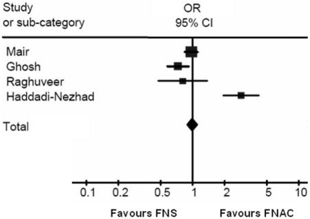
Figure 1 shows a Forest plot of the four included studies. The odds ratio produced is 0.99 in favour of FNS (95% CI 0.88–1.11) which is not statistically significant.
Figure 1.
A Forest plot of studies included in the meta-analysis.
Discussion
The fact that four of the five cross-over studies used a similar scoring system made a meta-analysis of the data fairly easy. It was unfortunate that one of the criteria had to be removed to achieve uniformity. The Santos and Leiman4 study was excluded from the meta-analysis as it did not provide comparable data on the scoring of the aspirate material. The outcome measures of this study were more crude and did not classify the aspirate into categories impacting analysis. The results of this study were, however, significant. Of the 22 cases in their study on which both FNAC and FNS were performed, 22 from the FNS group were in the diagnostically superior group whereas only 4 from the FNAC group were in this category. This difference is highly significant (P = 0.003).
Owing to the fact that this important study could not be included, the results of the meta-analysis should be viewed with caution.
Conclusions
Although there have been five high-quality trials on the subject, there is no evidence from a meta-analysis that one method of collection of cytological material is better than another in the investigation of thyroid lesions. Taking into consideration data not entered into the meta-analysis, there seems to be some evidence favouring FNS.
References
- 1.Carrillo JF, Frias-Mendivil M, Ochoa-Carrillo FJ, Ibarra M. Accuracy of fine-needle aspiration biopsy of the thyroid combined with an evaluation of clinical and radiologic factors. Otolaryngol Head Neck Surg. 2000;122:917–21. doi: 10.1016/S0194-59980070025-8. [DOI] [PubMed] [Google Scholar]
- 2.Mamoon N, Mushtaq S, Muzaffar M, Khan AH. The use of fine needle aspiration biopsy in the management of thyroid disease. J Pak Med Assoc. 1997;47:255–8. [PubMed] [Google Scholar]
- 3.Lowhagen T, Granberg PO, Lundell G, Skinnari P, Sundblad R, Willems JS. Aspiration biopsy cytology (ABC) in nodules of the thyroid gland suspected to be malignant. Surg Clin North Am. 1979;59:3–18. doi: 10.1016/s0039-6109(16)41729-9. [DOI] [PubMed] [Google Scholar]
- 4.Santos JE, Leiman G. Nonaspiration fine needle cytology. Application of a new technique to nodular thyroid disease. Acta Cytol. 1988;32:353–6. [PubMed] [Google Scholar]
- 5.Haddadi-Nezhad S, Larijani B, Tavangar SM, Nouraei SM. Comparison of fineneedle-nonaspiration with fine-needle-aspiration technique in the cytologic studies of thyroid nodules. Endocr Pathol. 2003;14:369–73. doi: 10.1385/ep:14:4:369. [DOI] [PubMed] [Google Scholar]
- 6.Ghosh A, Misra RK, Sharma SP, Singh HN, Chaturvedi AK. Aspiration vs nonaspiration technique of cytodiagnosis – a critical evaluation in 160 cases. Indian J Pathol Microbiol. 2000;43:107–12. [PubMed] [Google Scholar]
- 7.Mair S, Dunbar F, Becker PJ, Du PW. Fine needle cytology – is aspiration suction necessary? A study of 100 masses in various sites. Acta Cytol. 1989;33:809–13. [PubMed] [Google Scholar]
- 8.Raghuveer CV, Leekha I, Pai MR, Adhikari P. Fine needle aspiration cytology versus fine needle sampling without aspiration. A prospective study of 200 cases. Indian J Med Sci. 2002;56:431–9. [PubMed] [Google Scholar]


