Abstract
INTRODUCTION
There are a wide variety of different lesions which present as lumps of the foot. There have been very few studies which look at the presenting characteristics or the differential diagnosis of such lesions.
PATIENTS AND METHODS
All patients who underwent excision or biopsy of a foot lump over a period of 4 years were studied in order to determine patient demographics, presenting characteristics, diagnoses encountered and to assess the diagnostic accuracy of the surgeon.
RESULTS
In total, 101 patients were identified. Average age was 47.3 years (range, 14–79 years); there was a marked female preponderance with 73 females and 28 males. Thirty different histological types were identified; ganglion cysts were the most commonly encountered lesions and there was only one malignant lesion encountered in this study. Only 58 out of the 101 lumps were correctly diagnosed prior to surgery. Certain lesions were more commonly encountered in specific zones of the foot.
CONCLUSIONS
We have shown that there are a wide variety of potential diagnoses which have to be considered when examining a patient with a foot lump. There is a low diagnostic accuracy for foot lumps and, therefore, surgical excision and histological diagnosis should be sought if there is any uncertainty.
Keywords: Foot diseases, Orthopaedics, Biopsy, Differential diagnosis
Lumps of the foot present relatively infrequently to the orthopaedic service. There have been very few studies which look at the presenting characteristics or the differential diagnosis of such lesions. The World Health Organization (WHO) classification system which was modified by Enzinger and Weiss recognises 82 different benign and malignant soft tissue lesions of 10 histiogenic types which occur in the foot and ankle.1 This means the clinician has a wide choice of diagnoses to consider and this, coupled with the low incidence, may make their diagnostic accuracy low.
We studied our experience of foot lumps treated surgically in order to determine patient demographics, presenting characteristics, diagnoses encountered and to assess the diagnostic accuracy of the surgeon.
Patients and Methods
All patients who underwent excision or biopsy of a foot lump in the foot and ankle units of North Glasgow Hospitals NHS Trust (NGT) between 1 January 2001 to 31 December 2004 were included in the study. NGT has a catchment area of 550,000 of mixed socio-economic population. During the study period, staffing in the foot and ankle units consisted of 2 permanent consultants and 16 rotational specialist registrars. There was also an orthopaedic oncology surgeon who treated malignant lesions of the foot and ankle.
Patients were identified from theatre records and all lumps excised during the study period were sent to the pathology department as per the units' usual practice. Case records and the trust's pathology database were retrospectively reviewed. Subjects were excluded if their lesion did not present as a palpable lump; Morton's neuromas were, therefore, excluded. Patients were also excluded if they were referred from outwith the NGT catchment area, children under the age of 13 years were also not included as the hospitals involved did not treat children under this age.
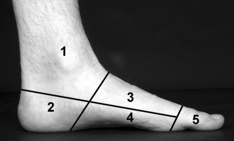
The data assessed included patient demographics, presenting symptoms (pain, footwear problems, cosmesis or neurological), the duration of the symptoms, the location of the lump, the presumed clinical diagnosis and the confirmed histological diagnosis. For the purpose of recording the site of the lesion, the foot was divided into 5 zones (Fig. 1) as described by Kirby et al.2
Figure 1.
The zones of the foot used to analyse the data as described by Kirby et al.2 The lines correspond to an oblique coronal plane, drawn from the mid-tarsal joint to the posterior margin of the longitudinal arch; a transverse plane, drawn from the mid-point of the metatarsal heads to the level of insertion of the Achilles tendon into the calcaneus; and a coronal plane, drawn through the metatarsophalangeal joints. These regions were numbered 1–5, to correspond to the ankle, heel, dorsum of the foot, plantar surface of the foot, and toes.
An SPSS database was created and the data analysed using descriptive statistics; the differences between the groups were assessed using Student's t-test. A difference was considered significant if P < 0.05.
Results
Age and sex
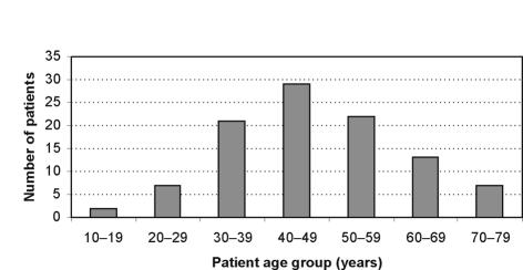
A total of 101 patients were identified. Average age was 47.3 years (range, 14–79 years; median, 45 years) and 71.3% of patients were in their 4th, 5th or 6th decade (Fig. 2). There was a marked female preponderance with 73 females and 28 males.
Figure 2.
Number of patients by age.
Presenting symptoms
Mean duration of symptoms was 23.6 months (range, 5–60 months; median, 24 months) which was not significantly different between males and females. Pain was the single most common presenting complaint followed by footwear problems. Only three patients attended because of cosmetic reasons and neurological symptoms were very rare with only one patient complaining of paraesthesia (Table 1).
Table 1.
Frequency of presenting symptoms
| Symptom | Number of patients |
|---|---|
| Pain | 49 |
| Footwear problems | 22 |
| Pain and footwear problems | 10 |
| Cosmesis | 3 |
| Paraesthesia | 1 |
| Unspecified | 16 |
Zones of the foot
Of the lesions, 60% were found on the toes (zone 5) and on the dorsum of the foot (zone 3) whereas only 7% were present on the heel (zone 2). Certain lesions were more commonly encountered in specific zones (Table 2). All cases of plantar fibromatosis (n = 6) were found in zone 4, 56% of ganglion cysts (22/39) were found in zone 3 and 83% of simple lipomas were found in zone 1 (5/6).
Table 2.
Distribution of lesions by zones of the foot
| Zone | Histological diagnosis |
|---|---|
| 1 | Lipoma (5) |
| Ganglion cyst (8) | |
| Neuroma (2) | |
| Connective tissue histiocyte reaction (1) | |
| 2 | Rheumatoid nodule (2) |
| Schwannoma (1) | |
| Viral wart (1) | |
| Cavernous haemangioma (1) | |
| Calcific tendonitis (1) | |
| Angioleiomyoma (1) | |
| 3 | Ganglion cyst (22) |
| Angioleiomyoma (1) | |
| Chondroma (1) | |
| Fibroma (1) | |
| Inclusion cyst (1) | |
| Lipoma (1) | |
| Fibrous histiocytoma (1) | |
| 4 | Plantar fibromatosis (6) |
| Neuroma (6) [3 with adventitious bursa] | |
| Rheumatoid nodule (2) [1 with adventitious bursa] | |
| Cavernous haemangioma (1) | |
| Fibrovascular tissue + fat (1) | |
| Adventitious bursa (1) | |
| 5 | Ganglion cyst (9) |
| Fibroma (3) | |
| Keratinous horn (2) | |
| Mucoid cyst (2) | |
| Neuroma (2) | |
| Subungal exostosis (2) | |
| Epidermal cyst (1) | |
| Fibrin fibrous tissue (1) | |
| Neurofibroma (1) | |
| Osteoid osteoma (1) | |
| Osteophyte (1) | |
| Plexiform fibrohistiocytic tumour (1) | |
| Tenosynovial giant cell tumour (1) | |
| Tophaceous gout (2) | |
| Intradermal naevus (1) | |
| Spindle lipoma (1) | |
| Chondroma (2) |
Pain was the presenting symptom in many lumps on the sole of the foot (88.2%) but only 46.9% of lesions on the toes present with this problem.
Type of lesion
Thirty different types of lesion were identified in 101 patients (Table 3). Ganglion cysts were the most commonly encountered lesion, followed by neuroma, lipoma and plantar fibromatosis. Only one malignant lesion was encountered – plexiform fibrohistiocytic tumour; this had not been suspected pre-operatively and further surgery was required. There were many other lesions which occurred only once or twice. Only 58 (57.4%) of the lumps were correctly diagnosed prior to surgery.
Table 3.
Distribution of lesions by histiogenic type
| Tissue precursor | Lesion | No. of lesions |
|---|---|---|
| Adipose | Lipoma | 6 |
| Spindle lipoma | 1 | |
| Cartilage/bone | Chondroma | 3 |
| Subungual exostosis | 2 | |
| Osteophyte | 1 | |
| Osteoid osteoma | 1 | |
| Fibrous | Plantar fibromatosis | 6 |
| Fibroma | 4 | |
| Fibrin fibrous tissue | 1 | |
| Fibrohistiocytic | Plexiform fibrohistiocytic | 1* |
| Fibrous histiocytoma | 1 | |
| Neural | Neuroma | 10 |
| Neurofibroma | 1 | |
| Schwannoma | 1 | |
| Smooth muscle | Angioleiomyoma | 2 |
| Vascular | Haemangioma | 2 |
| Fibrovascular tissue | 1 | |
| Synovial | Giant cell tumour | 1 |
| Miscellaneous | Ganglion cyst | 39 |
| Adventitious bursa | 1# | |
| Gouty tophus | 2 | |
| Calcific tendonitis | 1 | |
| Connective tissue histiocytic reaction | 1 | |
| Tumour-like | Benign intradermal naevus | 1 |
| Rheumatoid nodule | 4 | |
| Mucoid cyst | 2 | |
| Epidermal inclusion cyst | 2 | |
| Viral wart | 1 | |
| Keratinous horn | 2 |
Denotes malignant tumour.
Four other specimens of adventitious bursa were found in association with other lumps.
Ganglion cyst
Ganglions were the largest single lesions present in 39 out of the 101 patients. The average age for patients with a ganglion was 43.5 years which was significantly lower than the average age of patients with other lumps (49.7 years; P = 0.039). The majority of patients with ganglions were female (85%) and ganglions represented the diagnosis for 45% of females compared to only 21% of males. Ganglions were more commonly encountered in zone 3 (56%) but also occurred in zone 5 (23%) and zone 1 (21%). There were no ganglions on the plantar surface of the foot or at the heel. Thirty-five (89.7%) of the ganglions were correctly identified pre-operatively. Eleven other lesions were incorrectly diagnosed as ganglions.
Discussion
This is the first study in the literature to examine lesions which specifically present as foot lumps. Other studies have looked at benign and malignant lesions of the foot and ankle or individual diagnoses such as ganglia.2,3 We found a marked female preponderance which is consistent with previous work.2 The distribution of lumps by age suggests that, in adults, foot lumps are most common in 30–60-year-olds with a peak in the 5th decade.
There was a wide variation in duration of symptoms with a mean of 2 years which probably represents the patients' lack of concern about the nature of the lump. This is consistent with a case series reported by Alvi et al.,4 who reported late presentation leading to delay in treatment for tumours on the sole of the foot. The commonest symptoms were pain followed by footwear problems. We were surprised that cosmesis was rarely of concern to patients as the toes and dorsum of the foot were the most frequent location for lumps. These lesions are most likely to be seen by the patients and may deter them from wearing footwear which exposes their feet.
The diagnostic accuracy in our series was low and this is probably not surprising when 30 different diagnoses were encountered in 101 patients. Previous studies of soft tissue tumours of the foot have demonstrated a higher percentage of malignant tumours. Synovial sarcoma is believed to represent about 6% of all soft tissue tumours about the foot and ankle; in one retrospective review of sarcomas of the foot and ankle, 43% were given initial diagnoses of benign entities.5 In a recent review of the tumour workload presenting to a foot and ankle clinic in the UK, 12 malignant lesions were encountered in 80 consecutive cases.6 The low number of malignant lesions in our study may be due to the different inclusion criteria used as we excluded lesions which did not present as a lump and those which were referred from out with our hospitals' catchment area.
Ganglion cyst was the commonest single diagnosis. Patients with a ganglion were generally female and more likely to be younger than patients with other foot lumps. No ganglia were found on the sole of the foot or heel a finding which replicates that of Kirby et al.2 Although lumps in these areas may be ganglia, the surgeon should probably consider other diagnoses in the first instance.
Lumps on the sole of the foot were more likely to produce pain and this is probably due to localised pressure when walking.
This study is retrospective and, therefore, its main weakness is that it is dependent on the accuracy and completeness of documentation of the surgeon. We looked only at patients who underwent surgical treatment for their lump and this introduces some bias as it is likely that some patients will have been seen with foot lumps and were then discharged without surgery. A prospective study of foot and ankle lumps may allow better understanding of the differences in presentation and clinical findings between lesions.
Conclusions
We have shown that there is a wide variety of potential diagnoses which have to be considered when examining a patient with a foot lump. The majority of these lesions are benign and ganglion cyst is the single most common diagnosis with a marked female preponderance. Some lesions demonstrate a tendency to occur in a particular region of the foot; nevertheless, there is a low diagnostic accuracy for foot lumps and a histological diagnosis should be sought if there is any uncertainty.
References
- 1.Enzinger FM, Weis SW. Soft tissue tumours. St Louis, MO: Mosby; 1983. [Google Scholar]
- 2.Kirby EJ, Shereff MJ, Lewis MM. Soft-tissue tumors and tumor-like lesions of the foot. An analysis of eighty-three cases. J Bone Joint Surg Am. 1989;71:621–6. [PubMed] [Google Scholar]
- 3.Rozbrch SR, Chang V, Bohne WH, Deland JT. Ganglion cysts of the lower extremity: an analysis of 54 cases and review of the literature. Orthopaedics. 1998;21:141–8. doi: 10.3928/0147-7447-19980201-07. [DOI] [PubMed] [Google Scholar]
- 4.Alvi F, Rafee A, Khan T. Tumours on the sole of the foot: a case series. J Bone Joint Surg Br. 2005;87(Suppl 1):80. [Google Scholar]
- 5.Scully SP, Temple HT, Harrelson JM. Synovial sarcoma of the foot and ankle. Clin Orthop. 1999;364:220–6. doi: 10.1097/00003086-199907000-00028. [DOI] [PubMed] [Google Scholar]
- 6.Hart WJ, Hemmady M, Cool WP. A review of the tumour workload presenting to a foot and ankle clinic over an eighteen month period. J Bone Joint Surg Br. 2005;87(Suppl 1):2. [Google Scholar]


