

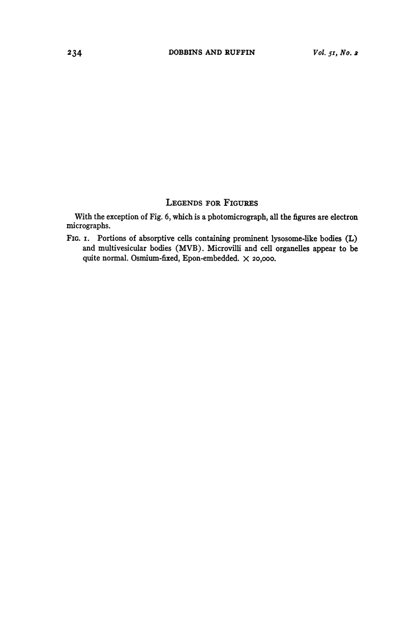
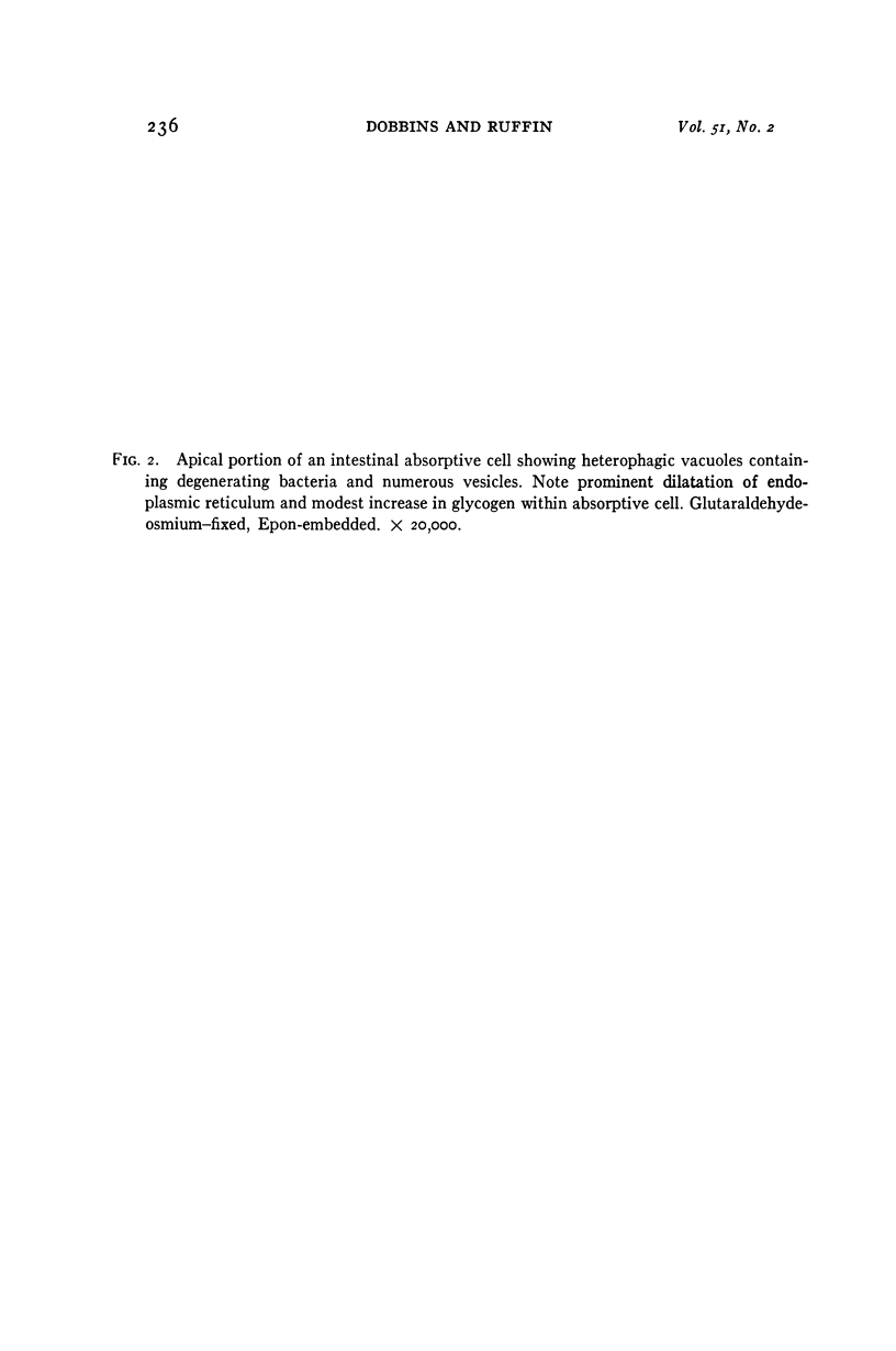
Full text
PDF
Images in this article
Selected References
These references are in PubMed. This may not be the complete list of references from this article.
- ADAMS W. R., WOLFSOHN A. W., SPIRO H. M. Some morphologic characteristics of Whipple's disease. Am J Pathol. 1963 Apr;42:415–429. [PMC free article] [PubMed] [Google Scholar]
- ASHWORTH C. T., DOUGLAS F. C., REYNOLDS R. C., THOMAS P. J. BACILLUS-LIKE BODIES IN WHIPPLE'S DISEASE; DISAPPEARANCE WITH CLINICAL REMISSION AFTER ANTIBIOTIC THERAPY. Am J Med. 1964 Sep;37:481–490. doi: 10.1016/0002-9343(64)90203-7. [DOI] [PubMed] [Google Scholar]
- BLACK-SCHAFFER B. The tinctoral demonstration of a glycoprotein in Whipple's disease. Proc Soc Exp Biol Med. 1949 Oct;72(1):225–227. doi: 10.3181/00379727-72-17388. [DOI] [PubMed] [Google Scholar]
- BRICERS J. R., OWEN E. E., TYOR M. P. AMINO ACID UPTAKE AND FATTY ACID ESTERIFICATION BY INTESTINAL MUCOSA FROM PATIENTS WITH WHIPPLE'S DISEASE AND NONTROPICAL SPRUE. Gastroenterology. 1965 May;48:584–592. [PubMed] [Google Scholar]
- CHEARS W. C., Jr, ASHWORTH C. T. Electron microscopic study of the intestinal mucosa in Whipple's disease. Demonstration of encapsulated bacilliform bodies in the lesion. Gastroenterology. 1961 Aug;41:129–138. [PubMed] [Google Scholar]
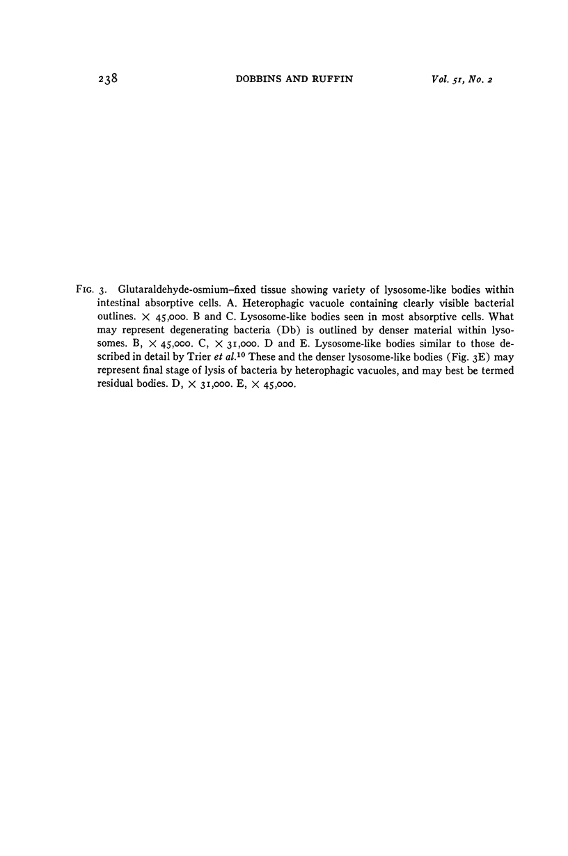
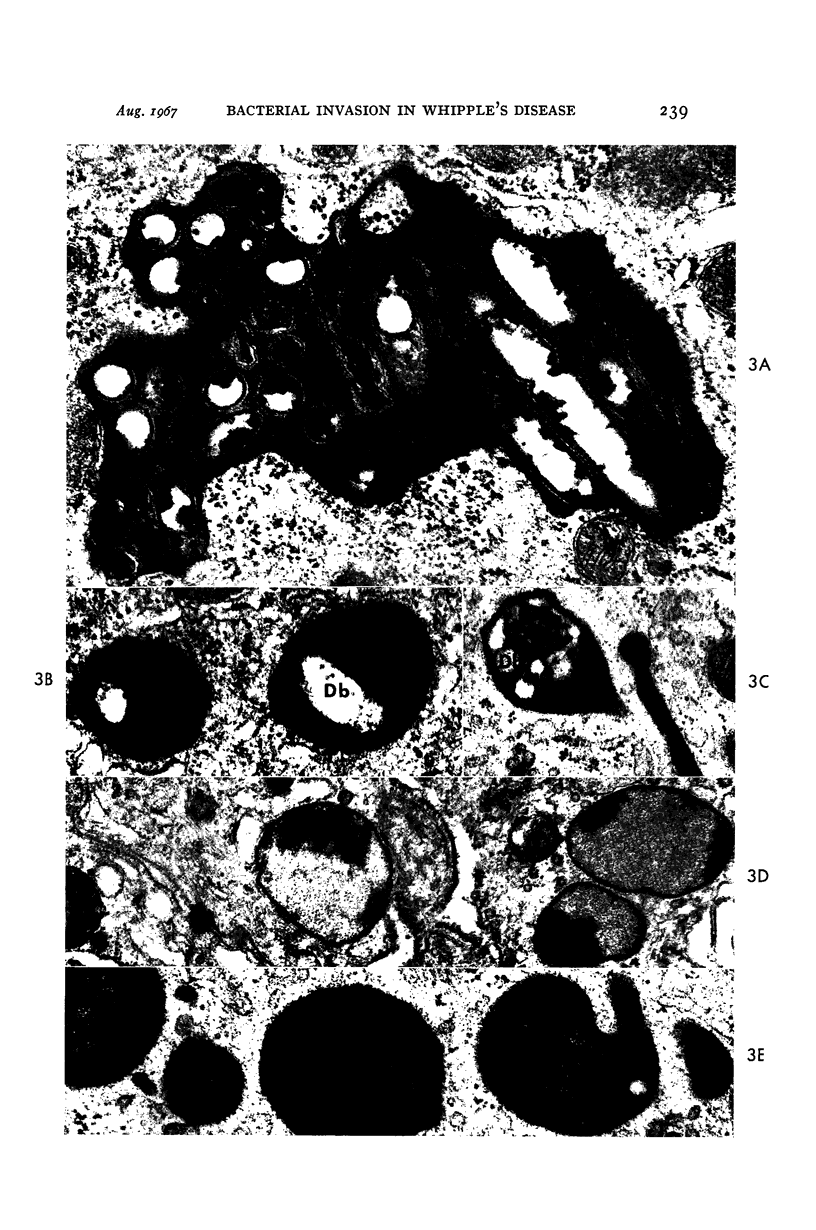
- De Duve C., Wattiaux R. Functions of lysosomes. Annu Rev Physiol. 1966;28:435–492. doi: 10.1146/annurev.ph.28.030166.002251. [DOI] [PubMed] [Google Scholar]
- Dobbins W. O., 3rd Electron microscopic study of the intestinal mucosa in intestinal lymphangiectasia. Gastroenterology. 1966 Dec;51(6):1004–1017. [PubMed] [Google Scholar]
- Dobbins W. O., 3rd The intestinal mucosal lymphatic in man. A light and electron microscopic study. Gastroenterology. 1966 Dec;51(6):994–1003. [PubMed] [Google Scholar]
- FISHER E. R. Whipple's disease: pathogenetic considerations. Electron microscopic and histochemical observations. JAMA. 1962 Aug 4;181:396–403. [PubMed] [Google Scholar]
- FRASCA J. M., PARKS V. R. A ROUTINE TECHNIQUE FOR DOUBLE-STAINING ULTRATHIN SECTIONS USING URANYL AND LEAD SALTS. J Cell Biol. 1965 Apr;25:157–161. doi: 10.1083/jcb.25.1.157. [DOI] [PMC free article] [PubMed] [Google Scholar]
- HAUBRICH W. S., WATSON J. H., SIERACKI J. C. Unique morphologic features of Whipple's disease. A study by light and electron microscopy. Gastroenterology. 1960 Oct;39:454–468. [PubMed] [Google Scholar]
- KOK N., DYBKAER R., ROSTGAARD J. BACTERIA IN WHIPPLE'S DISEASE. RESULTS OF CULTIVATION FROM REPEATED JEJUNAL BIOPSIES PRIOR TO, DURING, AND AFTER EFFECTIVE ANTIBIOTIC TREATMENT. Acta Pathol Microbiol Scand. 1964;60:431–449. doi: 10.1111/apm.1964.60.3.431. [DOI] [PubMed] [Google Scholar]
- LUFT J. H. Improvements in epoxy resin embedding methods. J Biophys Biochem Cytol. 1961 Feb;9:409–414. doi: 10.1083/jcb.9.2.409. [DOI] [PMC free article] [PubMed] [Google Scholar]
- Laster L., Waldmann T. A., Fenster L. F., Singleton J. W. Albumin metabolism in patients with Whipple's disease. J Clin Invest. 1966 May;45(5):637–644. doi: 10.1172/JCI105379. [DOI] [PMC free article] [PubMed] [Google Scholar]
- MACDONALD W. C., TRIER J. S., EVERETT N. B. CELL PROLIFERATION AND MIGRATION IN THE STOMACH, DUODENUM, AND RECTUM OF MAN: RADIOAUTOGRAPHIC STUDIES. Gastroenterology. 1964 Apr;46:405–417. [PubMed] [Google Scholar]
- PEREZ V., SCHAPIRA A., DE PELLEGRINO A. I., RYBAK B. J., DE LARRECHEA I. LIGHT- AND ELECTRON-MICROSCOPE FINDINGS ON JEJUNAL BIOPSY IN WHIPPLE'S DISEASE: STUDIES BEFORE AND AFTER ANTIBIOTIC THERAPY. Am J Dig Dis. 1963 Sep;8:718–728. doi: 10.1007/BF02236080. [DOI] [PubMed] [Google Scholar]
- Pittman F. E., Smith W. T., Mizrahi A., Blanc W. A., Pittman J. C. Clinical, histochemical, and electron microscopic study of colonic histiocytosis. Gut. 1966 Oct;7(5):458–467. doi: 10.1136/gut.7.5.458. [DOI] [PMC free article] [PubMed] [Google Scholar]
- TRIER J. S., PHELPS P. C., EIDELMAN S., RUBIN C. E. WHIPPLE'S DISEASE: LIGHT AND ELECTRON MICROSCOPE CORRELATION OF JEJUNAL MUCOSAL HISTOLOGY WITH ANTIBIOTIC TREATMENT AND CLINICAL STATUS. Gastroenterology. 1965 Jun;48:684–707. [PubMed] [Google Scholar]
- Takeuchi A. Electron microscope studies of experimental Salmonella infection. I. Penetration into the intestinal epithelium by Salmonella typhimurium. Am J Pathol. 1967 Jan;50(1):109–136. [PMC free article] [PubMed] [Google Scholar]
- Takeuchi A., Sprinz H., LaBrec E. H., Formal S. B. Experimental bacillary dysentery. An electron microscopic study of the response of the intestinal mucosa to bacterial invasion. Am J Pathol. 1965 Dec;47(6):1011–1044. [PMC free article] [PubMed] [Google Scholar]
- VENABLE J. H., COGGESHALL R. A SIMPLIFIED LEAD CITRATE STAIN FOR USE IN ELECTRON MICROSCOPY. J Cell Biol. 1965 May;25:407–408. doi: 10.1083/jcb.25.2.407. [DOI] [PMC free article] [PubMed] [Google Scholar]