Full text
PDF

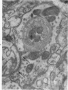
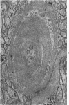
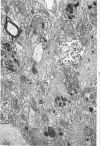
Images in this article
Selected References
These references are in PubMed. This may not be the complete list of references from this article.
- COLONNIER M. EXPERIMENTAL DEGENERATION IN THE CEREBRAL CORTEX. J Anat. 1964 Jan;98:47–53. [PMC free article] [PubMed] [Google Scholar]
- COLONNIER M., GUILLERY R. W. SYNAPTIC ORGANIZATION IN THE LATERAL GENICULATE NUCLEUS OF THE MONKEY. Z Zellforsch Mikrosk Anat. 1964 Apr 9;62:333–355. doi: 10.1007/BF00339284. [DOI] [PubMed] [Google Scholar]
- Cancilla P. A., Zimmerman H. M. The fine structure of a cerebellar hemangioblastoma. J Neuropathol Exp Neurol. 1965 Oct;24(4):621–628. doi: 10.1097/00005072-196510000-00005. [DOI] [PubMed] [Google Scholar]
- DE ROBERTIS E. Submicroscopic changes of the synapse after nerve section in the acoustic ganglion of the guinea pig; an electron microscope study. J Biophys Biochem Cytol. 1956 Sep 25;2(5):503–512. doi: 10.1083/jcb.2.5.503. [DOI] [PMC free article] [PubMed] [Google Scholar]
- GRAY E. G., GUILLERY R. W. The basis for silver staining of synapses of the mammalian spinal cord: a light and electron microscope study. J Physiol. 1961 Aug;157:581–588. doi: 10.1113/jphysiol.1961.sp006744. [DOI] [PMC free article] [PubMed] [Google Scholar]
- Gonatas N. K., Anderson W., Evangelista I. The contribution of altered synapses in the senile plaque: an electron microscopic study in Alzheimer's dementia. J Neuropathol Exp Neurol. 1967 Jan;26(1):25–39. doi: 10.1097/00005072-196701000-00003. [DOI] [PubMed] [Google Scholar]
- Gonatas N. K., Evangelista I., Walsh G. O. Axonic and synaptic changes in a case of psychomotor retardation: an electron microscopic study. J Neuropathol Exp Neurol. 1967 Apr;26(2):179–199. doi: 10.1097/00005072-196704000-00001. [DOI] [PubMed] [Google Scholar]
- Gonatas N. K., Goldehsohn E. S. Unusual neocortical presynaptic terminals in a patient with convulsions, mental retardation and cortical blindness: an electron microscopic study. J Neuropathol Exp Neurol. 1965 Oct;24(4):539–562. [PubMed] [Google Scholar]
- Gray E. G., Guillery R. W. Synaptic morphology in the normal and degenerating nervous system. Int Rev Cytol. 1966;19:111–182. doi: 10.1016/s0074-7696(08)60566-5. [DOI] [PubMed] [Google Scholar]
- HAMA K. Some observations on the fine structure of the giant nerve fibers of the earthworm, Eisenia foetida. J Biophys Biochem Cytol. 1959 Aug;6(1):61–66. doi: 10.1083/jcb.6.1.61. [DOI] [PMC free article] [PubMed] [Google Scholar]
- KIDD M. ALZHEIMER'S DISEASE--AN ELECTRON MICROSCOPICAL STUDY. Brain. 1964 Jun;87:307–320. doi: 10.1093/brain/87.2.307. [DOI] [PubMed] [Google Scholar]
- LOCKE M. THE STRUCTURE OF SEPTATE DESMOSOMES. J Cell Biol. 1965 Apr;25:166–169. doi: 10.1083/jcb.25.1.166. [DOI] [PMC free article] [PubMed] [Google Scholar]
- Luse S. A., Smith K. R., Jr The ultrastructure of senile plaques. Am J Pathol. 1964 Apr;44(4):553–563. [PMC free article] [PubMed] [Google Scholar]
- Smith C. A., Rasmussen G. L. Degeneration in the efferent nerve endings in the cochlea after axonal section. J Cell Biol. 1965 Jul;26(1):63–77. doi: 10.1083/jcb.26.1.63. [DOI] [PMC free article] [PubMed] [Google Scholar]
- TERRY R. D., GONATAS N. K., WEISS M. ULTRASTRUCTURAL STUDIES IN ALZHEIMER'S PRESENILE DEMENTIA. Am J Pathol. 1964 Feb;44:269–297. [PMC free article] [PubMed] [Google Scholar]
- WOOD R. L. Intercellular attachment in the epithelium of Hydra as revealed by electron microscopy. J Biophys Biochem Cytol. 1959 Dec;6:343–352. doi: 10.1083/jcb.6.3.343. [DOI] [PMC free article] [PubMed] [Google Scholar]
- Walberg F. An electron microscopic study of terminal degenration in the inferior olive of the cat. J Comp Neurol. 1965 Oct;125(2):205–222. doi: 10.1002/cne.901250205. [DOI] [PubMed] [Google Scholar]


