Abstract
Several brittle culm mutations of rice (Oryza sativa) causing fragility of plant tissues have been identified genetically but not characterized at a molecular level. We show here that the genes responsible for three distinct brittle mutations of rice, induced by the insertion of the endogenous retrotransposon Tos17, correspond to CesA (cellulose synthase catalytic subunit) genes, OsCesA4, OsCesA7 and OsCesA9. Three CesA genes were expressed in seedlings, culms, premature panicles, and roots but not in mature leaves, and the expression profiles were almost identical among the three genes. Cellulose contents were dramatically decreased (8.9%-25.5% of the wild-type level) in the culms of null mutants of the three genes, indicating that these genes are not functionally redundant. Consistent with these results, cell walls in the cortical fiber cells were shown to be thinner in all the mutants than in wild-type plants. Based on these observations, the structure of a cellulose-synthesizing complex involved in the synthesis of the secondary cell wall is discussed.
Cellulose is a homogenous polymer of β-1,4-glucan synthesized from UDP-Glc (Delmer and Amor, 1995). Plant CesA (cellulose synthase catalytic subunit) genes were first isolated by random sequencing of cotton (Gossypium hirsutum) expressed sequence tags (Pear et al., 1996). The first evidence that the CesA gene encodes the enzyme responsible for cellulose synthesis was obtained from analysis of a CesA mutant of Arabidopsis (Arioli et al., 1998). Another important piece of evidence rests on the structural analysis of cellulose synthesis complexes. The rosette terminal cellulose-synthesizing complexes, displaying 6-fold symmetry, are known to associate with cellulose microfibril impressions in the plasma membrane (Mueller and Brown, 1980), and CESA protein has been immunolocalized to the rosette complexes (Kimura et al., 1999a). In addition, the korrigan gene encoding a endo-1,4-β-glucanase has been shown to be required for cellulose synthesis (Zuo et al., 2000; Lane et al., 2001; Sato et al., 2001) and proposed to function in the cleavage of sitosterol-β-glucoside from the growing cellulose polymer chain (Peng et al., 2002).
Ten CesA genes have been found in the completed genomic sequence of Arabidopsis (http://cellwall.stanford.edu). Based on mutant analyses, characterization of six CesA genes of Arabidopsis has been reported. The rsw1 mutation in AtCesA1 causes a reduction of cellulose synthesis, when grown at the nonpermissive temperature, resulting in disassembly of the rosette complexes, widespread morphological abnormalities, and the accumulation of noncrystalline β-1,4-glucan (Arioli et al., 1998). The irx3 mutant of AtCesA7 shows a reduction of cellulose content in the stem and a defect in the secondary cell wall formation in xylem that causes collapse of xylem elements (Taylor et al., 1999). These results indicate that AtCesA1 and AtCesA7 contribute to cellulose syntheses in the properly developed primary and secondary cell wall, respectively. The PRC1 encodes AtCESA6, and, like the rsw1 mutant of AtCesA1, its mutant exhibits decreased cell elongation, especially in roots and dark-grown hypocotyls, because of a cellulose deficiency in the primary wall (Fagard et al., 2000). In addition to their similar mutant phenotypes, both AtCesA6 and AtCesA1 also show similar expression profiles in various organs and growth conditions, although embryonic expression of these CesA genes is different (Beeckman et al., 2002). The IRX1 and IRX5 genes, whose mutants exhibit a similar phenotype to that of the irx3 mutant of AtCesA7, encode AtCESA8 and AtCESA4, respectively, and three proteins of AtCESA4, -7, and -8 have been shown to be co-expressed in the same cells of stems (Taylor et al., 2000, 2003). In addition, interaction of all three proteins of AtCESA4, -7, and -8 has been shown. These findings suggest that AtCESA1 and -6 or AtCESA4, -7, and -8 form the functional CESA units that assemble into a rosette complex. Scheible et al. (2001) and Desprez et al. (2002) also show that the ixr1 and ixr2 genes, which confer resistance to cellulose synthesis inhibitors (isoxban and thiazolidione) encode AtCESA3 and AtCESA6, respectively.
The CesA gene family in monocot crop plants such as maize (Zea mays), barley (Hordeum vulgare), and rice (Oryza sativa) was also identified by analyses of cDNA, expressed sequence tags, and genome sequencing (Holland et al., 2000; see cell wall Web site, above). In rice, at least 10 genes (OsCesA1-10) have been identified. As clearly demonstrated in Arabidopsis, analysis of mutants is a powerful strategy for defining the function of CesA genes. Brittle mutants of monocots have been identified in barley (Takahashi et al., 1953, 1966), maize (Briggs and Robert, 1966), and rice (Jones, 1933; Nagano and Takahashi, 1963; Takahashi et al., 1968). In the barley mutants, the physiological, morphological, and biochemical properties of the brittle culm phenotype have been well characterized (Kokubo et al., 1989, 1991; Kimura et al., 1999b). The maximum bending stress of culms of barley brittle culm mutants is less than one-half that of non-brittle strains, and the cell walls of epidermal, collenchyma, and parenchyma tissues of the mutants are thinner than those of the corresponding non-brittle strains. In addition, the cellulose content is lower in the cell walls of the barley mutants than in the wild type, but there are no significant differences in the amounts of lignin, pectin, and noncellulosic polysaccharides in the cell wall of mutants and wild-type plants (Kokubo et al., 1989, 1991). In one of the barley mutants, a decrease in the number of rosette complex also has been reported (Kimura et al., 1999b). Therefore, the brittle phenotype of these barley mutants is thought to be caused by the decreased cellulose synthesis. To further characterize the brittle mutations, isolation of the affected genes is needed. In addition, information of these genes will be useful in improving the mechanical strength of tissues in monocot crop plants, such as rice, barley, maize, and wheat (Triticum aestivum), thereby overcoming abiotic problems such as lodging.
We produced about 50,000 mutant lines of rice induced by the insertion of the endogenous retrotransposon Tos17 (A. Miyao, K. Tanaka, K. Murata, H. Sawaki, S. Takeda, K. Abe, Y. Shinozuka, K. Onosato, and H. Hirochika, unpublished data). Tos17 is inactive in normally propagated plants but becomes active in tissue culture (Hirochika et al., 1996; Hirochika, 2001). In regenerated plants, transposed and original copies of Tos17 become silent, and these copies are germinally inherited in the next generation. We have shown the utility of Tos17-induced mutations for forward (Agrawal et al., 2001) and reverse (Sato et al., 1999; Takano et al., 2001) genetic studies of genefunctions. Here, we report the isolation and characterization of mutants of three CesA genes induced by insertion of Tos17.
RESULTS
Screening of the Brittle Culm Mutants Induced by the Endogenous Retrotransposon Tos17
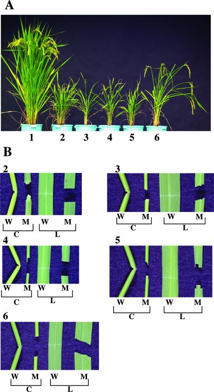
To screen for the mutants displaying the brittle phenotype, the next generations (R1) of about 3,500 regenerated rice lines were observed in the paddy field. In these populations, four regenerated lines, called NC0259, ND2395, ND8759, and NE1031, exhibited similar mutant phenotypes; a dwarfed growth habit (Fig. 1A, 2-5) and easy fracturing of leaf and culm by stressing between fingers, the so-called brittle culm phenotype (Fig. 1B, 2-5). These four mutants also exhibited other similar characteristic phenotypes, such as small leaves, thin culms, and withering of leaf apex (data not shown). In addition, fertility of the four mutants was low (0%-32.3%). The semidwarfed mutant of NF1011 (Fig. 1A, 6) also showed a brittle culm phenotype (Fig. 1B, 6). Although this mutant had normal-sized leaves, most of the mature leaves of the mutant plants growing in the paddy field were fractured in the middle region by wind pressure (data not shown). The semidwarfed mutant of NF1011 also had a thin culm, but it was thicker than those of the other four mutants (data not shown).
Figure 1.
Phenotypes of the five rice brittle culm mutants. A, Plant heights of the wild-type plant (1) and the mutants of NC0259 (2), ND8759 (3), NE1031 (4), ND2395 (5), and NF1011 (6). B, Brittleness of culm (C) and mature leaf (L) from wild-type plant (W) and the mutant (M) as demonstrated by the damage caused by stressing between fingers. Numbers indicate mutant lines as described above. All culms were prepared from the second internodes.
To examine whether these mutations were induced by the insertion of Tos17 and to identify the specific transposed Tos17 copies responsible for the mutations, cosegregation of Tos17 with the observed phenotypes was examined by genomic Southern analysis of the R1 populations of the five lines as described previously (Agrawal et al., 2001). Five to 10 copies of newly transposed Tos17 were found in each line, and one of the copies was shown to cosegregate with the observed brittle phenotype in each line (data not shown), strongly suggesting that all of the five mutant phenotypes were caused by insertion of Tos17. In the following section, this result was confirmed. This perfect tagging with Tos17 is quite unusual because tagging efficiency for various mutations has been shown to be 5% to 10% (Hirochika, 2001). In the previous study, the existence of preferred target sites for Tos17 insertion has been shown (Yamazaki et al., 2001). One possible explanation for the perfect tagging in the present study is that the causative genes for the brittle mutations are preferred targets. This explanation was supported by the finding that, among the five mutants examined, two allelic pairs were found, representing two independent mutations each in two of the three genes identified (see the following section).
Identification of Three Brittle Culm Genes Encoding Cellulose Synthase Catalytic Subunits
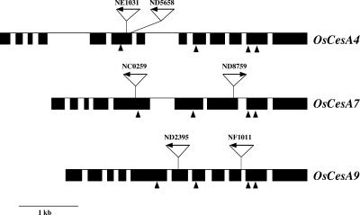
To identify the causative brittle culm genes, the genomic fragments from the region flanking Tos17 were amplified by thermal asymmetric interlaced (TAIL)-PCR and suppression PCR methods. Using genomic DNA from the mutant of NC0259, TAIL-PCR was performed with a Tos17-specific primer and the degenerate primer called AD7. Several DNA fragments were amplified, but genomic Southern analysis with these fragments as probes showed cosegregation of the fragment NC0259_14_701_1A with brittle culm phenotype (data not shown). The amino acid sequence deduced from the DNA sequence of NC0259_14_701_1A showed high similarity to those of plant CESAs (data not shown). Because the brittle phenotype can be explained by a CesA mutation, it is likely that the CesA gene is the gene responsible. This was further confirmed by the reduction of cellulose content in the mutant (see the following section). Using NC0259_14_701_1A as a probe, the locus was mapped onto the rice linkage map. The disrupted CesA gene of NC0259 was mapped at 44.0 cM on chromosome 10 (data not shown), and the RFLP marker clone R2825, which shows high similarity to plant CESAs (data not shown), was located at the identical position. The clone R2825 was shown to be a partial cDNA clone of the disrupted gene. The genomic sequence of the locus at 44.0 cM is published by the International Rice Genome Sequencing Project, and the disrupted gene of NC0259, encoding CESA, is annotated as OsCesA7 (CESA sequence database at http://cellwall.stanford.edu). OsCesA7 consists of nine exons and eight introns (Fig. 2) and encodes a protein of 1,063 amino acids (Fig. 3). In NC0259, Tos17 was inserted in the fifth exon of OsCesA7 (Fig. 2). In ND8759, Tos17 was inserted into the seventh exon of OsCesA7 (Fig. 2), and the homozygous mutant of ND8759 also exhibited a similar brittle culm phenotype, as shown in Figure 1B, 3. Based on these results, it is concluded that the OsC-esA7 gene is a causative gene for the brittle culm mutation.
Figure 2.
Genomic structure of three rice CesA genes, OsCesA4, -7, and -9. The black boxes and lines indicate exon and intron sequences, respectively. Scale bar = 1 kb. Arrows and numbers indicate the Tos17 insertion sites and mutant lines, respectively. Arrowheads indicate the positions of the four UDP-Glc-binding motifs shown in Figure 3.
Figure 3.
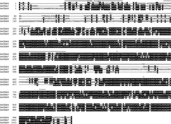
Alignment of the deduced amino acid sequences of OsCesA4, -7, and -9. Numbers indicate the position of the amino acid residues of each protein. The residues that are identical in at least two CesA genes are marked in reverse contrast letters. The three Asp (D) residues and the QxxRW motif that are critical for the function of CESA (see text) are marked by asterisks. The plant conserved region (P-CR), variable regions (VR1 and -2), and RING finger motif are also shown by overhead double-dashed lines.
The DNA fragments carrying putative brittle culm genes of NE1031, ND2395, and NF1011 were amplified by the suppression PCR method. The amplified DNA fragments T12748T, ND2395_30, and NF1011-7_2 also showed high similarity to CesA genes and cosegregated with the brittle culm phenotype (data not shown). The disrupted gene of NE1031, encoding CESA, was mapped at 129.6 cM on chromosome 1 (data not shown), and this locus corresponds to RFLP marker R2417, whose clone also shows high similarity to plant CESAs (data not shown). R2417 was shown to be a partial cDNA of the disrupted gene of NE1031, whose genomic sequence was determined by International Rice Genome Sequencing Project and shown to be OsCesA4. OsCesA4 consists of 13 exons and 12 introns (Fig. 2) and encodes a protein of 990 amino acids (Fig. 3). The insertion site of Tos17 in NE1031 was found in the sixth exon (Fig. 2). To isolate allelic mutants of OsCesA4, PCR screening was carried out using DNA pools from mutant lines induced by the insertion of Tos17 (Hirochika, 2001; H. Hirochika and A. Miyao, unpublished data). The mutant line of ND5658 with an insertion of Tos17 in the sixth exon of OsCesA4 was found (Fig. 2). As expected, the mutant of ND5658 also exhibited the brittle culm phenotype (data not shown).
The disrupted gene of ND2395 was mapped at 158.9 cM on chromosome 1 (data not shown), but the corresponding sequence was not found in the published genomic sequence. Therefore, using the Monsanto Rice Genome Sequence Database (http://www.rice-research.org/), the genomic sequence of the brittle culm gene of ND2395, encoding CESA, was determined. This CesA gene was shown to be OsCesA9, which consists of 11 exons and 10 introns (Fig. 2) and encodes a protein of 1,055 amino acids (Fig. 3). Part of the genomic sequence of OsCesA9 was identical to that of NF1011-7_2 and shown to cosegregate with the mutant phenotype of NF1011 by the genomic Southern analysis (data not shown). Tos17 was inserted into the 6th exon in ND2395 and the ninth intron in NF1011 (Fig. 2).
Based on the results described in this section, it is concluded that the genes responsible for the three brittle mutations are three different CesA genes.
Comparison of Gene Structures and Deduced Amino Acid Sequences of OsCesA4, OsCesA7, and OsCesA9
To characterize the three rice CesA genes, the deduced amino acid sequences were compared (Fig. 3). There is a high degree of sequence similarity among the deduced CESA proteins. Furthermore, the proteins possess four motifs that have been identified as being conserved in CESAs and all processive glucosyl transferase (Saxena et al. 1995). These are motifs surrounding three Asp residues and a QxxRW motif downstream of the third Asp residue that are essential for binding for the UDP-Glc. A sequence highly conserved in the plant CESAs, so-called P-CR (Pear et al., 1996), and two variable regions called VR1 and VR2 (Taylor et al., 1999), also exist in the rice CESAs. In addition to these common features, there is a structural feature unique to one of three rice CESAs, OsCESA4. The sequence of Cys-9 to Cys-47 at the N terminus of OsCESA4 shows a low similarity to those of OsCESA7 and OsCESA9 (Fig. 3). This region corresponds to a Cys-rich region conserved in plant CesA genes that has been suggested to form two zinc fingers with high homology to the RING finger motif, probably involved in protein-protein interactions (Delmer, 1999; Kurek et al. 2002). Although OsCESA4 possesses eight Cys residues required to form two zinc fingers (Fig. 3), eight amino acid residues are missing in the first RING finger motif.
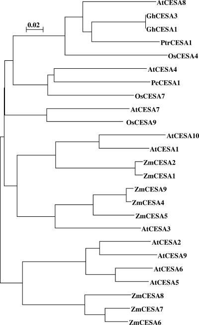
Based on the sequences shown in Figure 3, phylogenetic relationships of CesA genes from higher plants were examined (Fig. 4). Three rice CesA genes were considered to function to synthesize cellulose in the secondary cell walls responsible for the overall strength of the plant, judging from the brittle culm phenotype (Fig. 1B) exhibited by their respective mutants (Fig. 2). In Arabidopsis, three CesA genes contributing to cellulose synthesis in the secondary cell walls, AtCesA4, -7, and -8, have been found by mutant analyses (Taylor et al., 1999, 2000, 2003). The phylogenetic relationships between the three rice CesA genes and all of the Arabidopsis genes indicated that OsCesA9, OsCesA4, and OsCesA7 are the functional analogues for AtCesA7, AtCesA8, and AtCesA4, respectively. This result strongly suggests that these three rice CESA proteins, like the three Arabidopsis CESA proteins, make a complex essential for cellulose synthesis in secondary cell wall. Two cotton CesA genes (GhCesA1 and -3) that are considered to be members of secondary wall-forming genes (Pear et al., 1996; Peng et al., 2002) belong to the same group including OsCesA4 and AtCesA8. GhCesA1 and -3 may have been evolved by recent gene duplication. In maize, only eight complete amino acid sequences have been determined, and no CesA genes analogous to the three rice/Arabidopsis genes have been isolated, probably due to low abundance of the cDNAs.
Figure 4.
Phylogenetic relationships of CesA genes from higher plants. Phylogenetic trees based on the complete amino acid sequences were generated by using ClustalX (version 1.8), then bootstrapping at random number generator seed = 1,000 and number of bootstrap trials = 10,000. Plant species are as follows: At, Arabidopsis; Gc, cotton; Os, rice; Pc, Populus × canescens; Ptr, Populus tremuloides; and Zm, maize.
Expression of OsCesA4, OsCesA7, and OsCesA9
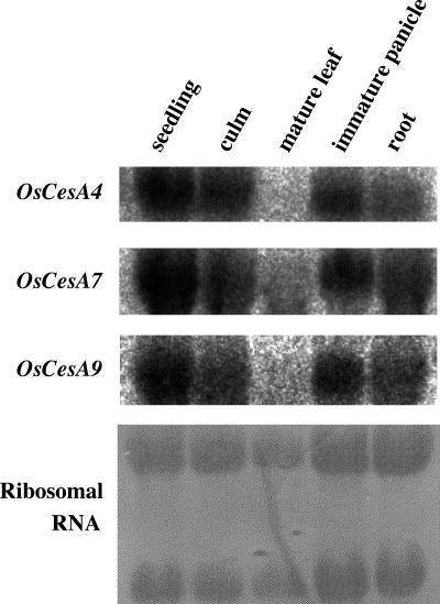
The expression patterns for OsCesA4, OsCesA7, and OsCesA9 were investigated in culms, mature leaves, roots and immature panicles, and 2-week old seedlings. Signals corresponding to these genes could be detected by northern-blot analyses (Fig. 5). Transcripts of the three CesA genes were found in seedlings, culms, immature panicles and roots, but not in mature leaves, and the expression patterns were almost identical among three genes. Therefore, it is concluded that the three rice CesA genes are expressed coordinately in seedlings and three organs, although it is uncertain whether these genes are co-expressed in the same cell.
Figure 5.
Expression profiles of OsCesA4, -7, and -9 in wild-type plant. Total RNAs were prepared from 2-week-old seedlings, culms, mature leaves, immature panicles, and roots and subjected to northern-blot analysis with probes for OsCesA4, -7, and -9. Equal loading of the gel was confirmed by staining of ribosomal RNA with methylene blue.
Cellulose Contents in the Culms of the Brittle Culm Mutants
Cellulose contents in the second culm internodes of the mutants andthe wild-type plant were investigated. As summarized in Table I, cellulose contents were decreased in all the mutants, but some differences were observed among the mutants. The brittle culm phenotype of the mutants can be readily explained by these reductions in cellulose. The OsCesA4 and -7 mutants exhibiting the same phenotype showed almost the same reductions, and these Tos17 insertion sites are found in the exon of the gene (Fig. 2). This result indicates that the mutations of OsCesA4 and -7 affected the gene function, possibly leading to complete loss of function. On the other hand, the cellulose contents were different in two OsCesA9 mutants exhibiting different phenotypes, and the Tos17 insertion sites are one in an exon and the other in an intron (Fig. 2). The mild phenotype (semidwarf, Fig. 1A) exhibited by the NF1011 line could be explained by its higher cellulose content. One possible explanation for the partial loss-of-function phenotype of NF1011 line is incomplete splicing out of the Tos17-containing intron. Analysis of reverse transcription (RT)-PCR products showed that incomplete splicing out did occur (data not shown). One important finding here is that complete loss of function of any one of the three CesA genes leads to a dramatic reduction in cellulose contents (8.9%-25.5% of the wild-type level), suggesting that these genes are not functionally redundant to each other.
Table I.
Measurement of cellulose content in the culms of the wild-type plant and five brittle culm mutants
Culm materials were prepared from the second internode of the wild-type plant and five mutants. The samples were freeze dried, enzymatically and chemically treated, and cellulose content was determined by the phenol-sulfuric acid method. Numbers in parentheses indicate percentage of wild type.
| Line | Cellulose/Dry Wt |
|---|---|
| % | |
| Wild-type Nipponbare | 24.23 ± 3.40 (100) |
| CesA4 disruption | |
| NE1031 | 4.95 ± 2.01 (20.4) |
| CesA7 disruption | |
| NC0259 | 5.81 ± 3.13 (24.0) |
| ND8759 | 6.17 ± 1.87 (25.5) |
| CesA9 disruption | |
| ND2395 | 2.16 ± 1.26 (8.9) |
| NF1011 | 13.03 ± 3.91 (53.8) |
Scanning Electron Micrographs of Cross Sections of the Culms of the Five Brittle Culm Mutants
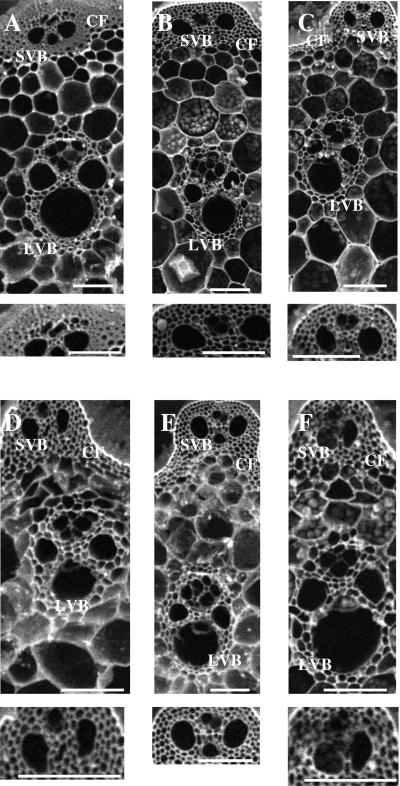
To investigate structural changes in the culms of the five brittle culm mutants, cross sections of the second culm internodes from the wild-type plant and five mutants were observed by scanning electron micrograph (Fig. 6). Notably thinner cell walls were found in the cortical fiber cells in all the mutants. Similar results were obtained by the analysis of the third internodes (data not shown). The significant differences in the cell walls between the wild-type plant and five mutants can be explained by a decrease in cellulose content of the cell walls of the mutants (Table I). In three Arabidopsis mutants of the CesA genes required for cellulose synthesis in the secondary cell walls, collapsed xylem cells have been found (Taylor et al., 1999, 2000, 2003). In all the rice mutants, the large and small vascular bundle cells in the second internodes were normal in shape (Fig. 6), and no collapsed xylem cells were detected in mature leaves (data not shown).
Figure 6.
Scanning electron micrographs of cross sections of the culms of the wild-type plant and five brittle culm mutants. The cross sections were prepared from the second internode of wild-type plant (A) and the mutants of NC0259 (B), ND8759 (C), ND2395 (D), NF1011 (E), and NE1031 (F). White scale bars = 100 μm. CF, LVB, and SVB, Cortical fiber, large vascular bundle, and small vascular bundle, respectively. Lower panels show tissues of CF and SVB corresponding to upper panels of the wild-type plant and five mutants.
DISCUSSION
Brittle culm mutants have been isolated in several monocot species (Jones, 1933; Takahashi et al., 1953, 1966, 1968; Nagano and Takahashi, 1963; Briggs and Robert, 1966), and some of the mutants have been studied in relation to cellulose synthesis (Kimura et al., 1999b; Kokubo et al., 1989, 1991). However, the genes affected in these mutations were still not known. Here, isolation of the causative genes for three brittle culm mutations induced by the retrotransposon Tos17 is reported. These genes are shown to encode three distinct CESAs, OsCESA4, OsCESA7, and OsCESA9.
The plant CESAs are generally considered to be associated with rosette complexes, the cellulose-synthesizing units in the plasma membrane (Kimura et al., 1999a). The discovery of a family of CesA genes raised the important question of why plants have many genes for cellulose synthesis. One explanation is that each rosette complex is composed of two or more distinct CESA proteins to form a functional CESA unit (Fagard et al., 2000; Scheible et al., 2001; Taylor et al., 2000, 2003; Peng et al., 2002). This proposal is based on the following observations. Two Arabidopsis mutants of AtCesA1 and -6 genes encoding the enzymes synthesizing cellulose in the primary cell wall are phenotypically indistinguishable, and both genes are expressed in the same tissues (Fagard et al., 2000). Mutants of AtCesA4, -7, and -8, genes involved in the biosynthesis of secondary walls, also exhibited almost identical phenotype, including collapse of xylem elements (Taylor et al., 2003). Three genes are co-expressed in the same cells, and proteins encoded by these genes interact with each other. Therefore, at least two or three distinct CESAs seem to be essential to form a functional CESA unit. To clarify the combination of CesA genes forming a functional CESA unit in maize, the expression patterns of eight CesA genes in 10 different tissues have been examined (Dhugga, 2001). Although the expression profiles of four CesA genes (ZmCesA3-6) are different, coordinate expression of the other four CesA genes (ZmCesA1, -2, -7, and -8) has been suggested. By in situ RT-PCR analysis, ZmCesA1 has been shown to be highly expressed in the pith and cortex of the cell elongation region and ZmCesA8 in the region of developing vascular tissue. This observation suggests that ZmCESA1 and ZmCESA8 are primary wall-forming and secondary wall-forming enzymes, respectively (Holland et al., 2000). The phylogenetic relationships of ZmCesA1, -2, -7, and -8 (Holland et al., 2000; Fig. 5), together with expression profiles, suggest ZmCesA2 and -7 are the enzymes synthesizing primary and secondary cell walls, respectively (Dhugga, 2001). Further studies are needed to determine whether ZmCESA1 and -2 or ZmCESA7 and -8 assemble into a functional CESA unit. The expression profiles of the three rice CesA genes (OsCesA4, OsCesA7, and OsCesA9) were indistinguishable in four different organs in the wild-type plant (Fig. 5), and the complete loss-of-function mutants of these genes exhibited almost identical phenotypes with respect to the cellulose content (Table I), brittleness of culm (Fig. 1B, 2-5), the growth habit (Fig. 1A, 2-5), and cell wall thickness in the cortical fiber cells of the culm (Fig. 6). These results strongly suggest that the three CESAs (OsCESA4, -7, and -9) are not functionally redundant to each other, and these proteins form a functional CESA unit. Here, we propose that in rice at least three distinct CesA genes are required to form functional CESA units for the secondary wall, as in the case of Arabidopsis (Taylor et al., 2003).
The phylogenetic analysis shows that the secondary wall-forming enzymes in Arabidopsis are classified into three subgroups (Fig. 4). Interestingly, the secondary wall-forming enzymes in rice are also classified into these three subgroups. This suggests that a functional CESA unit would consist of three different CESA subunits in higher plants. This suggestion agrees with the model of a functional CESA unit (Doblin et al., 2002), proposing that three different types of subunits might be required for rosette assembly.
All the plant CESA proteins possess not only the conserved three Asp residues and QxxRW motif that are critical for the function of CESA but also the RING finger motif in the N-terminal region of the proteins (Delmer, 1999). Here, it was found that one of three CESA proteins, OsCESA4, shows a low similarity to the other two proteins and lacks eight amino acid residues in the first zinc finger (Fig. 3). The mutant of OsCesA4 exhibited similar cellulose deficiency to those of other two CesA genes, OsCesA7 and -9 (Table I). This result indicates that OsCESA4, although lacking eight amino acid residues in the first zinc finger, has an essential role for cellulose production in the secondary wall. In cotton, a RING finger motif has been shown to be involved in dimerization of CESAs via intermolecular disulfide bridges in the oxidized state. Based on this result, it was proposed that dimerization between the domains of the RING finger motifs in CESA subunits is necessary for the assembly of a cellulose-synthesizing unit (Kurek et al., 2002). The spacing of the Cys residues in the RING-finger motifs in CESA proteins is CxxC-x(15)-CxxC-x(4)-CxxC-x(11)-CxxC, and two rice CESA proteins of OsCESA7 and -9 also show the identical spacing (Fig. 3). Therefore, OsCESA4 possesses a unique RING finger motif of CxxC-x(7)CxxC(4)-CxxC-x(11)-CxxC to interact with OsCESA7 or -9.
Two OsCesA9 allelic mutants, ND2395 and NF1011, although both showing the brittle culm phenotype (Fig. 1B, 5 and 6) and thinning of cortical fiber cell walls in the culm (Fig. 6), exhibited different growth habits (Fig. 1A, 5 and 6) and different cellulose contents (Table I). The mild mutant phenotype exhibited by the NF1011 line is likely due to partial loss of function. The partial loss of function is considered to result from partial inhibition of splicing of the Tos17-containing intron because the Tos17 insertion site is found in the intron (Fig. 2). Data supporting this hypothesis were obtained by RT-PCR analysis.
The complete loss-of-function mutants of OsCesA4, -7, and -9 exhibited morphological abnormality such as dwarf (Fig. 1A). In Arabidopsis, the collapsed xylem elements have been observed in the mutants of AtCesA4, -7, and -8, the members of the secondary wall-forming genes, but these mutants did not exhibit morphological abnormalities (Turner and Somerville, 1997). In the rice mutants, thinner walls in the cortical fiber cells were found instead of the collapsed xylem cells (Fig. 6). The composition of cell wall is considerably different between rice and Arabidopsis. For example, the mixed-linked (1→3)(1→4)β-d-glucan in the primary wall is a well-known wall polysaccharide found only in grasses including rice (Carpita, 1996). The difference in the mutant phenotype between Arabidopsis and rice, especially topology of xylem element, may have been caused by that of the cell wall composition. In the Arabidopsis mutants of CesA genes required for cellulose synthesis in the primary wall, various mutant phenotypes such as root swelling, stunted growth, and reduced length of root and hypocotyl have been observed (Reiter, 2002). The abnormal phenotype of three rice CesA mutants (Fig. 1) may be explained by alteration in the primary wall composition.
The mutant of AtCesA3, one of the members of the primary wall-forming genes, has been reported to overproduce jasmonate and ethylene, which are induced by a number of biotic and abiotic stresses such as wounding and pathogen attack (Ellis et al., 2002). In addition, constitutive expression of the jasmonate-responsive genes or the jasmonate- and ethylene-responsive genes in the mutants of two primary-wall-forming genes, AtCesA1 and -3, has been shown. Based on these observations, it was proposed that cell walls with altered composition caused by mutations in CesA genes might contain high level of elicitors inducing jasmonate and ethylene, or the altered wall architecture might allow the untimely release of such molecules. In the rice mutants, the reduction in cellulose content in the secondary wall may lead to alteration in the primary wall composition and accumulation of jasmonate and ethylene, thus resulting in a dwarf phenotype.
Several brittle culm mutations have been genetically identified in rice and mapped onto the linkage map as follows; bc1 on chromosome 3, bc2 on chromosome 5, bc3 on chromosome 2, bc4 on chromosome 6, and bc5 on chromosome 2 (Nagato and Yoshimura, 1998). These mutants showed three different growth habits: a dwarf phenotype like that of three CesA mutants observed in the present study (Fig. 1), a normal morphology with growth retardation, and a normal growth like the wild-type plant (Jones, 1933; Nagano and Takahashi, 1963; Takahashi et al., 1968). However, the genes associated with these mutations have not been isolated. We showed here isolation of the causative genes for three brittle culm mutations, OsCesA4, -7, and -9. These CesA genes were mapped on chromosomes 1 and 10 (data not shown) and, thus, do not correspond to the causative genes for the brittle culm mutations found previously. The brittle culm phenotype is probably caused by the defects in the secondary cell walls responsible for the overall strength of the plant. The defective cell walls are considered to be caused by loss or reduction of one of the wall components including cellulose, lignin, pectin, and noncellulosic polysaccharides. In the lignin-deficient mutant irx4 in Arabidopsis, both strength and stiffness of the stems are severely reduced (Jones et al., 2001). In an Arabidopsis mutant deficient in Fuc, one of the noncellulosic polysaccharides, the cell walls are fragile (Reiter et al., 1993). Therefore, the causative genes for rice brittle culm mutations might be involved in the synthesis of cellulose or noncellulosic components in the secondary cell walls. Isolation of these genes is important not only for further understanding of the mechanism of cell wall synthesis but also for improving the mechanical strength of tissues in monocot crop plants to provide lodging tolerance.
In the present study, three causative genes for brittle culm mutations were isolated after screening only 3,500 mutant lines induced by the Tos17. Further screening should contribute to the isolation of new genes involved in cell wall synthesis.
MATERIALS AND METHODS
Plant Materials
Rice (Oryza sativa) mutant lines induced by insertion of the endogenous retrotransposon Tos17 were grown in the paddy field. For various analyses, such as observation by scanning electron microscopy, the culm internode was collected after harvesting and fixed in Nawaschin Claf type III (3:2:1:4 [v/v] 1% chromic acid:10% acetic acid:35% formalin:water; Miksche, 1976).
Southern Analysis
Rice genomic DNA was prepared from mature leaves by the cetyltrimethyl-ammonium bromide method (Murray and Thompson, 1980). Five hundred nanograms of the prepared DNA was digested with restriction enzyme XbaI and electrophoresed on 0.8% (w/v) agarose gels with the Multiblotter MB24 system (Labimap, Plaisir, France). Hybridization was performed at 65°C with Church-Gilbert buffer (Church and Gilbert, 1984) containing a 32P-labeled probe.
TAIL-PCR and Suppression PCR
TAIL-PCR (Liu and Whittier, 1995) was carried out with a Tos17-specific primer and a degenerate one. The Tos17-specific primers and thermal conditions were described by Miyao et al. (1998). Here, we used four kinds of degenerate primer called AD1, 4, 5, and 7 to amplify several genomic fragments from the region flanking Tos17, as described elsewhere (A. Miyao, K. Tanaka, K. Murata, H. Sawaki, S. Takeda, K. Abe, Y. Shinozuka, K. Onosato, and H. Hirochika, unpublished data). The target fragment NC0259_14_701_1A in this study was isolated by PCR with the AD7, 5′-CGTGNAG(A/T) ANCNAAG-3′, and Tos17-specific primers.
A modified method of suppression PCR was used, as described elsewhere (Miyao et al., 1998). The target fragment T12748T was amplified by PCR using genomic DNA digested with DraI as a template. The fragments ND2395_30 and NF1011-7_2 were prepared from genomic DNA digested with NruI.
Northern-Blot Analysis
Total RNA was isolated from elongating culms, mature leaves, panicles at the meiotic stage, roots, and 2-week-old seedlings with the Isogen kit (Nippon Gene, Toyama, Japan), and 10 μg of total RNA was electrophoresed on 0.8% (w/v) denatured agarose gels. The gel was blotted on Hybond N+ membrane (Amersham, Little Chalfont, UK), and the membrane was hybridized with the same buffer as Southern analysis. The probes for OsCesA4, -7, and -9 were prepared with the DNA fragments re-amplified from the RT-PCR products by PCR with the gene-specific primers designed from the nested region of the sequence of the primers used in the first PCR. RT-PCR was carried out using a ReverTra Dash kit (Toyobo, Osaka) with the gene-specific primers according to the manufacturer's recommendation. The forward and reverse primers used for primary and secondary reactions were as follows: for OsCesA4, 5′-GACAGATGCATATGAGCCAC-3′ and 5′-CTTTCTTGTGGTGTTGGTAC-3′, and 5′-CTCGGATCATTCCTATATCG-3′ and 5′-CTCGAAACATAGACTAGCCG-3′; for OsCesA7, 5′-GCGGCTTCCCGGTGTGCAAG-3′ and 5′-GCTGGACCTTGTCCTTGAGG-3′, and 5′-TGCTACGAGTACGAGCGCAG-3′ and 5′-GTCGAAGAGCAGCATGGACGCGC-3′; and for OsCesA9, 5′-CATGCTCAACTTGGATTGCG-3′ and 5′-TCCACTTGAAGGCGTAGAGC-3′, and 5′-TCAACAACAGCAAGGCCATC-3′ and 5′-CGGTGAAGTTGGTGTCGATC-3′.
DNA Sequencing
The DNA sequencing analysis was performed using the dideoxy chain termination method (Sanger et al., 1977) and a BigDyeTM Terminator Cycle Sequencing Ready Reaction (Applied Biosystems, Foster City, CA). The sequence was determined by ABI377, ABI310, and ABI3100 automatic DNA sequencers (Applied Biosystems).
Mapping
The loci of the rice brittle genes were mapped on the Nipponbare/Kasalath high-density linkage map constructed by Harushima et al. (1998) according to the procedures described by Kurata et al. (1994).
Measurement of Cellulose Content
The second culm internodes of rice were freeze dried at -20°C for 1 d and 4°C for 2 d with Unitop HL and Freezemobile 12EL systems (Virtis, Gardiner, NY). The freeze-dried materials were homogenized at 2,400 rpm for 20 min with Multi-Beads Shocker MB301 (Yasui Kikai, Osaka). The homogenate was suspended in 50 mm Tris-HCl (pH 7.0), incubated for 5 min in boiled water, and then cooled at room temperature. The suspension was treated for 2 h at 37°C with 20 units mL-1 porcine pancreatic α-amylase (Sigma, St. Louis) and centrifuged at 1,000g for 10 min. The resulting precipitate was resuspended in 17.5% (w/v) NaOH/0.02% (w/v) NaBH4. The suspension was centrifuged, and the precipitate was resuspended in the alkaline solution as described above and incubated at room temperature for 24 h. The incubated suspension was centrifuged, and the precipitate was washed once with the same alkaline solution. After alkaline treatment, the precipitate was washed three times with 0.03 m acetic acid, three times with ethanol, three times with ethanol/ether (1:1 [v/v]), and three times with ether. These procedures were modified from the method of Kokubo et al. (1991). The dried material was hydrolyzed with 67% (v/v) H2SO4 for 1 h at room temperature, and cellulose content was determined on the appropriate dilution of the hydrolyzed sample by the phenol-sulfic acid method, as described by Dubois et al. (1956).
Observation under Scanning Electron Microscope
The fixed culm internodes were substituted in water overnight. The substituted material was cut perpendicularly to culm axis with a microtome blade. The specimens were examined directly with a scanning electron microscope (S-2380N, Hitachi, Tokyo).
Acknowledgments
We thank M. Harada, N. Kojima, Y. Machida, M. Tanabe, and Y. Yamashita (Institute of Society for Techno-Innovation of Agriculture, Forestry and Fisheries, Tsukuba, Japan) for experimental and technical assistance during this study. We thank T. Ando, A. Shomura (Institute of Society for Techno-Innovation of Agriculture, Forestry, and Fisheries, Tsukuba, Japan), and Dr. M. Yano (National Institute of Society for Techno-Innovation of Agriculture, Forestry, and Fisheries, and National Institute of Agrobiological Sciences, Ibaraki, Japan) for supporting the experimental procedure of gene mapping. We thank Drs. H. Nakagawa and H. Takatsuji (National Institute of Agrobiological Sciences) for performing the scanning electron microscopy. We thank Prof. N. Sakurai (Hiroshima University, Japan) for insightful advice regarding the cellulose content measurement.
This work was supported by the Program for Promotion of Basic Research Activities for Innovation Biosciences.
References
- Agrawal GK, Yamazaki M, Kobayashi M, Hirochika R, Miyao A, Hirochika H (2001) Screening of the rice viviparous mutants generated by endogenous retrotransposon Tos17 insertion: tagging of a zeaxanthin epoxidase gene and a novel OsTATC gene. Plant Physiol 125: 1248-1257 [DOI] [PMC free article] [PubMed] [Google Scholar]
- Arioli T, Peng L, Betzner AS, Burn J, Wittke W, Herth W, Camilleri C, Hofte H, Plazinski J, Birch R et al. (1998) Molecular analysis of cellulose biosynthesis in Arabidopsis. Science 279: 717-720 [DOI] [PubMed] [Google Scholar]
- Beeckman T, Przemeck GKD, Stamatiou G, Lau R, Terryn N, Rycke RD, Inze D, Berleth T (2002) Genetic complexity of cellulose synthase A gene function in Arabidopsis embryogenesis. Plant Physiol 130: 1883-1893 [DOI] [PMC free article] [PubMed] [Google Scholar]
- Briggs RW, Robert W (1966) Recognition and classification of some genetic traits in maize. J Hered 57: 35-42 [Google Scholar]
- Carpita NC (1996) Structure and biogenesis of the cell walls of grasses. Annu Rev Plant Physiol Plant Mol Biol 47: 445-476 [DOI] [PubMed] [Google Scholar]
- Church GM, Gilbert W (1984) Genomic sequencing. Proc Natl Acad Sci USA 81: 1991-1995 [DOI] [PMC free article] [PubMed] [Google Scholar]
- Delmer DP (1999) Cellulose biosynthesis: exciting times for a difficult field of study. Annu Rev Plant Physiol Plant Mol Biol 50: 245-276 [DOI] [PubMed] [Google Scholar]
- Delmer DP, Amor Y (1995) Cellulose biosynthesis. Plant Cell 7: 987-1000 [DOI] [PMC free article] [PubMed] [Google Scholar]
- Desprez T, Vernhettes S, Fagard M, Refregier G, Desnos T, Aletti E, Py N, Pelletier S, Hofte H (2002) Resistance against herbicide isoxaben and cellulose deficiency caused by distinct mutations in same cellulose synthase isoform CESA6. Plant Physiol 128: 482-490 [DOI] [PMC free article] [PubMed] [Google Scholar]
- Dhugga KS (2001) Building the wall: genes and enzyme complexes for polysaccharide synthases. Curr Opin Plant Biol 4: 488-493 [DOI] [PubMed] [Google Scholar]
- Dublin MS, Kurek I, Jacob-Wilk D, Delmer DP (2002) Cellulose biosynthesis in Plants: from genes to rosettes. Plant Cell Physiol 43: 1407-1420 [DOI] [PubMed] [Google Scholar]
- Dubois M, Gilles KA, Hamilton JK, Rebers PA, Smith F (1956) Colorimetric method for determination of sugars and related substances. Anal Chem 28: 350-356 [Google Scholar]
- Ellis C, Karafyllidis I, Wasternack C, Turner JG (2002) The Arabidopsis mutant cev1 links cell wall signaling to jasmonate and ethylene responses. Plant Cell 14: 1557-1566 [DOI] [PMC free article] [PubMed] [Google Scholar]
- Fagard M, Desnos T, Desprez T, Goubet F, Refregier G, Mouille G, McCann M, Rayon C, Vernhettes S (2000) Hofte H PROCUSTE1 encodes a cellulose synthase required for normal cell elongation specifically in roots and dark-grown hypocotyls of Arabidopsis. Plant Cell 12: 2409-2423 [DOI] [PMC free article] [PubMed] [Google Scholar]
- Harushima Y, Yano M, Shomura A. Sato M, Shimano T, Kuboki Y, Yamamoto T, Lin SY, Antonio BA, Parco A et al. (1998) A high-density rice genetic linkage map with 2275 markers using a single F2 population. Genetics 148: 479-494 [DOI] [PMC free article] [PubMed] [Google Scholar]
- Hirochika H (2001) Contribution of the Tos17 retrotransposon to rice functional genomics. Curr Opin Plant Biol 4: 118-122 [DOI] [PubMed] [Google Scholar]
- Hirochika H, Sugimoto K, Otsuki Y, Tsugawa H, Kanda M (1996) Retrotransposon of rice involved in mutations induced by tissue culture. Proc Natl Acad Sci USA 93: 7783-7788 [DOI] [PMC free article] [PubMed] [Google Scholar]
- Holland N, Holland D, Helentjaris T, Dhugga KS, Xoconostle-Cazares B, Delmer DP (2000) A comparative analysis of the plant cellulose synthase (CesA) gene family. Plant Physiol 123: 1313-1323 [DOI] [PMC free article] [PubMed] [Google Scholar]
- Jones JW (1933) Inheritance of characters in rice. J Agric Res 47: 771-782 [Google Scholar]
- Jones L, Ennos AR, Turner SR (2001) Cloning and characterization of irregular xylem4 (irx4): a severely lignin-deficient mutant of Arabidopsis. Plant J 26: 205-216 [DOI] [PubMed] [Google Scholar]
- Kimura S, Laosinchai W, Itoh T, Cui X, Linder CR, Brown RMJ (1999a) Immunogold labeling of rosette terminal cellulose-synthesizing complexes in the vascular plant Vigna angularis. Plant Cell 11: 2075-2085 [DOI] [PMC free article] [PubMed] [Google Scholar]
- Kimura S, Sakurai N, Itoh T (1999b) Different distribution of cellulose synthesizing complexes in brittle and non-brittle strains of barley. Plant Cell Physiol 40: 335-338 [Google Scholar]
- Kokubo A, Kuraishi S, Sakurai N (1989) Culm strength of barley: correlation among maximum bending stress, cell wall dimensions, and cellulose content. Plant Physiol 91: 876-882 [DOI] [PMC free article] [PubMed] [Google Scholar]
- Kokubo A, Sakurai N, Kuraishi S, Takeda K (1991) Culm brittleness of barley (Hordeum vulgare L.) mutants is caused by small number of cellulose molecules in cell wall. Plant Physiol 97: 509-514 [DOI] [PMC free article] [PubMed] [Google Scholar]
- Kurata N, Nagamura Y, Yamamoto K, Harushima Y, Sue N, Wu J, Antonio BA, Shomura A, Shimizu T, Lin SY et al. (1994) A 300 kilobase interval genetic map of rice including 883 expressed sequences. Nat Genet 8: 365-372 [DOI] [PubMed] [Google Scholar]
- Kurek I, Kawagoe Y, Jacob-Wilk D, Doblin M, Delmer D (2002) Dimerization of cotton fiber cellulose synthase catalytic subunits occurs via oxidation of the zinc-binding domains. Proc Natl Acad Sci USA 99: 11109-11114 [DOI] [PMC free article] [PubMed] [Google Scholar]
- Lane DR, Wiedemeier A, Peng L, Hofte H, Vernhettes S, Desprez T, Hocart CH, Birch RJ, Baskin TI, Burn JE et al. (2001) Temperature-sensitive alleles of RSW2 link the KORRIGAN endo-1, 4-β-glucanase to cellulose synthesis and cytokinesis in Arabidopsis. Plant Physiol 126: 278-288 [DOI] [PMC free article] [PubMed] [Google Scholar]
- Liu YG, Whittier RF (1995) Thermal asymmetric interlaced PCR: automatable amplification and sequencing of insert end fragments from P1 and YAC clones for chromosome walking. Genomics 25: 674-681 [DOI] [PubMed] [Google Scholar]
- Miksche JP (1976) Botanical microtechnique and cytochemistry. Iowa State University Press, Ames
- Miyao A, Yamazaki M, Hirochika H (1998) Systematic screening of mutants of rice by sequencing retrotransposon-insertion sites. Plant Biotechnol 15: 253-256 [Google Scholar]
- Mueller SC, Brown RMJ (1980) Evidence for an intramembranous component associated with a cellulose microfibril synthesizing complex in higher plants. J Cell Biol 84: 315-326 [DOI] [PMC free article] [PubMed] [Google Scholar]
- Murray MG, Thompson WF (1980) Rapid isolation of high molecular weight plant DNA. Nucleic Acids Res 8: 4321. [DOI] [PMC free article] [PubMed] [Google Scholar]
- Nagano S, Takahashi M (1963) Trial construction of twelve linkage groups in Japanese rice: XXVII. Genetical studies on rice plant. J Fac Agr, Hokkaido Univ, Sapporo 53: 72-130 [Google Scholar]
- Nagato Y, Yoshimura A (1998) B. Report of the committee on gene symbolization, nomenclature and linkage groups. Rice Genet Newslett 15: 13-74 [Google Scholar]
- Pear JR, Kawagoe Y, Schreckengost WE, Delmer DP, Stalker DM (1996) Higher plants contain homologs of the bacterial celA genes encoding the catalytic subunit of cellulose synthase. Proc Natl Acad Sci USA 93: 12637-12642 [DOI] [PMC free article] [PubMed] [Google Scholar]
- Peng L, Kawagoe Y, Hogan P, Delmer D (2002) Sitosterol-β-glucoside as primer for cellulose synthesis in plants. Science 295: 147-150 [DOI] [PubMed] [Google Scholar]
- Reiter W-D (2002) Biosynthesis and properties of the plant cell wall. Curr Opin Plant Biol 5: 536-542 [DOI] [PubMed] [Google Scholar]
- Reiter W-D, Chapple CCS, Somerville CR (1993) Altered growth and cell walls in a fucose-deficient mutant of Arabidopsis. Science 261: 1032-1035 [DOI] [PubMed] [Google Scholar]
- Sanger F, Nicklen S, Coulson AR (1977) DNA sequencing with chain-terminating inhibitors. Proc Natl Acad Sci USA 74: 5463-5467 [DOI] [PMC free article] [PubMed] [Google Scholar]
- Sato S, Kato T, Kakegawa K, Ishii T, Liu YG, Awano T, Takabe K, Nishiyama Y, Kuga S, Sato S et al. (2001) Role of the putative membrane-bound endo-1, 4-β-glucanase KORRIGAN in cell elongation and cellulose synthesis in Arabidopsis thaliana. Plant Cell Physiol 42: 251-263 [DOI] [PubMed] [Google Scholar]
- Sato Y, Sentoku N, Miura Y, Hirochika H, Kitano H, Matsuoka M (1999) Loss-of-function mutations in the rice homeobox gene OSH15 affect the architecture of internodes resulting in dwarf plants. EMBO J 18: 992-1002 [DOI] [PMC free article] [PubMed] [Google Scholar]
- Saxena IM, Brown RM Jr, Fevrc M, Geremia RA, Henrissat B (1995) Multidomain architecture of β-glucosyl transferases: implications for mechanism of action. J Bacteriol 177: 1419-1424 [DOI] [PMC free article] [PubMed] [Google Scholar]
- Scheible W-R, Eshed R, Richmond T, Delmer D, Somerville C (2001) Modifications of cellulose synthase confer resistance to isoxaben and thiazolidinone herbicides in Arabidopsis Ixr1 mutants. Proc Natl Acad Sci USA 98: 10079-10084 [DOI] [PMC free article] [PubMed] [Google Scholar]
- Takahashi M, Kinoshita T, Takeda K (1968) Character expressions and casual genes of some mutants in rice plant: XXXIII. Genetical studies on rice plant. J Fac Agr, Hokkaido Univ, Sapporo 55: 496-512 [Google Scholar]
- Takahashi R, Hayashi J, Hiura U (1966) Inheritance and linkage studies in barley: III. Linkage studies of the gene for fragile stem-2 and orientation of linkage map on barley chromosome 5. Ber Ohara Inst landw Biol Okayama Univ 13: 199-212 [Google Scholar]
- Takahashi R, Yamamoto J, Yasuda S, Itano Y (1953) Inheritance and linkage studies in barley. Ber Ohara Inst landw Forsch 10: 29-52 [Google Scholar]
- Takano M, Kanegae H, Shinomura T, Miyao A, Hirochika H, Furuya M (2001) Isolation and characterization of rice phytochrome A mutants. Plant Cell 13: 521-534 [DOI] [PMC free article] [PubMed] [Google Scholar]
- Taylor NG, Howells RM, Huttly AK, Vickers K, Turner SR (2003) Interactions among three distinct CesA proteins essential for cellulose synthesis. Proc Natl Acad Sci USA 100: 1450-1455 [DOI] [PMC free article] [PubMed] [Google Scholar]
- Taylor NG, Laurie S, Turner SR (2000) Multiple cellulose synthase catalytic subunits are required for cellulose synthesis in Arabidopsis. Plant Cell 12: 2529-2539 [DOI] [PMC free article] [PubMed] [Google Scholar]
- Taylor NG, Scheible W-R, Cutler S, Somerville CR, Turner SR (1999) The irregular xylem3 locus of Arabidopsis encodes a cellulose synthase required for secondary cell wall synthesis. Plant Cell 11: 769-779 [DOI] [PMC free article] [PubMed] [Google Scholar]
- Turner SR, Somerville CR (1997) Collapsed xylem phenotype of Arabidopsis identifies mutants deficient in cellulose deposition in the secondary cell wall. Plant Cell 9: 689-701 [DOI] [PMC free article] [PubMed] [Google Scholar]
- Yamazaki M, Tsugawa H, Miyao A, Yano M, Wu J, Yamamoto S, Matsumoto T, Sasaki T, Hirochika H (2001) The rice retrotransposon Tos17 prefers low-copy-number sequences as integration targets. Mol Genet Genomics 265: 336-344 [DOI] [PubMed] [Google Scholar]
- Zuo J, Niu Q-W, Nishizawa N, Wu Y, Kost B, Chua N-H (2000) KORRIGAN, an Arabidopsis endo-1,4-β-glucanase, localizes to the cell plate by polarized targeting and is essential for cytokinesis. Plant Cell 12: 1137-1152 [DOI] [PMC free article] [PubMed] [Google Scholar]


