
Fig. 4. Methylation-specific PCR (MSP) analysis of methylated and unmethylated DNA in cervical precursor lesions and tumors.
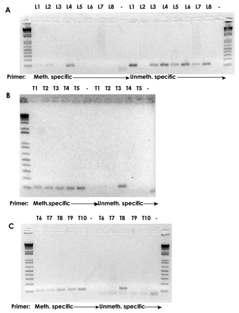
MSP analysis of 18 HPV-18 samples with known methylation patterns (Fig. 3). Analysis of the eight DNAs from precursor lesions (L) with methylation-specific primers gave signals only with the clones L2 and L4, primer pairs specific for unmethylated gave signals for seven samples. A mutation overlapping with the reverse primer likely explains the unsatisfactory outcome with clone L2. Analysis of the tumors (T) with methylation specific primers gave positive signals for all ten samples. Only T3 and T8 gave showed for unmethylated DNA as expected from Fig.3. The DNA of L8 was exhausted and could not be included in this analysis.