Abstract
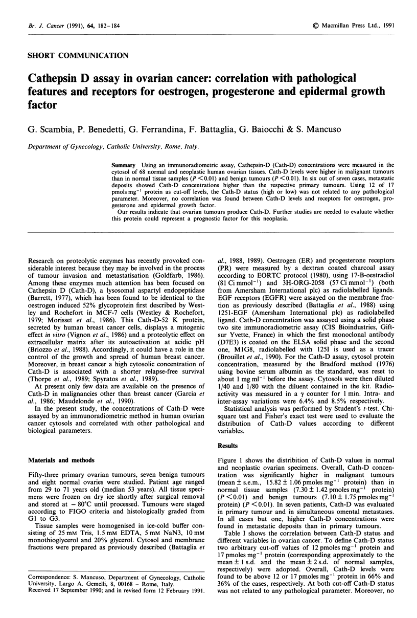
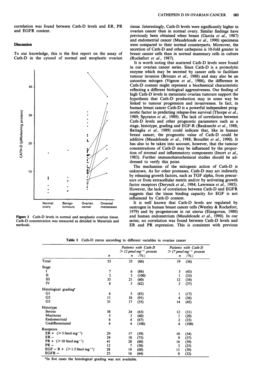
Using an immunoradiometric assay, Cathepsin-D (Cath-D) concentrations were measured in the cytosol of 68 normal and neoplastic human ovarian tissues. Cath-D levels were higher in malignant tumours than in normal tissue samples (P less than 0.01) and benign tumours (P less than 0.01). In six out of seven cases, metastatic deposits showed Cath-D concentrations higher than the respective primary tumours. Using 12 of 17 pmols.mg-1 protein as cut-off levels, the Cath-D status (high or low) was not related to any pathological parameter. Moreover, no correlation was found between Cath-D levels and receptors for oestrogen, progesterone and epidermal growth factor. Our results indicate that ovarian tumours produce Cath-D. Further studies are needed to evaluate whether this protein could represent a prognostic factor for this neoplasia.
Full text
PDF


Selected References
These references are in PubMed. This may not be the complete list of references from this article.
- Battaglia F., Scambia G., Rossi S., Panici P. B., Bellantone R., Polizzi G., Querzoli P., Negrini R., Iacobelli S., Crucitti F. Epidermal growth factor receptor in human breast cancer: correlation with steroid hormone receptors and axillary lymph node involvement. Eur J Cancer Clin Oncol. 1988 Nov;24(11):1685–1690. doi: 10.1016/0277-5379(88)90068-5. [DOI] [PubMed] [Google Scholar]
- Bauknecht T., Runge M., Schwall M., Pfleiderer A. Occurrence of epidermal growth factor receptors in human adnexal tumors and their prognostic value in advanced ovarian carcinomas. Gynecol Oncol. 1988 Feb;29(2):147–157. doi: 10.1016/0090-8258(88)90209-0. [DOI] [PubMed] [Google Scholar]
- Bradford M. M. A rapid and sensitive method for the quantitation of microgram quantities of protein utilizing the principle of protein-dye binding. Anal Biochem. 1976 May 7;72:248–254. doi: 10.1006/abio.1976.9999. [DOI] [PubMed] [Google Scholar]
- Briozzo P., Morisset M., Capony F., Rougeot C., Rochefort H. In vitro degradation of extracellular matrix with Mr 52,000 cathepsin D secreted by breast cancer cells. Cancer Res. 1988 Jul 1;48(13):3688–3692. [PubMed] [Google Scholar]
- Brouillet J. P., Theillet C., Maudelonde T., Defrenne A., Simony-Lafontaine J., Sertour J., Pujol H., Jeanteur P., Rochefort H. Cathepsin D assay in primary breast cancer and lymph nodes: relationship with c-myc, c-erb-B-2 and int-2 oncogene amplification and node invasiveness. Eur J Cancer. 1990 Apr;26(4):437–441. doi: 10.1016/0277-5379(90)90012-i. [DOI] [PubMed] [Google Scholar]
- Derynck R., Roberts A. B., Winkler M. E., Chen E. Y., Goeddel D. V. Human transforming growth factor-alpha: precursor structure and expression in E. coli. Cell. 1984 Aug;38(1):287–297. doi: 10.1016/0092-8674(84)90550-6. [DOI] [PubMed] [Google Scholar]
- Elangovan S., Moulton B. C. Progesterone and estrogen control of rates of synthesis of uterine cathepsin D. J Biol Chem. 1980 Aug 10;255(15):7474–7479. [PubMed] [Google Scholar]
- Garcia M., Lacombe M. J., Duplay H., Cavailles V., Derocq D., Delarue J. C., Krebs B., Contesso G., Sancho-Garnier H., Richer G. Immunohistochemical distribution of the 52-kDa protein in mammary tumors: a marker associated with cell proliferation rather than with hormone responsiveness. J Steroid Biochem. 1987;27(1-3):439–445. doi: 10.1016/0022-4731(87)90338-4. [DOI] [PubMed] [Google Scholar]
- Garcia M., Salazar-Retana G., Pages A., Richer G., Domergue J., Pages A. M., Cavalie G., Martin J. M., Lamarque J. L., Pau B. Distribution of the Mr 52,000 estrogen-regulated protein in benign breast diseases and other tissues by immunohistochemistry. Cancer Res. 1986 Jul;46(7):3734–3738. [PubMed] [Google Scholar]
- Imort M., Zühlsdorf M., Feige U., Hasilik A., von Figura K. Biosynthesis and transport of lysosomal enzymes in human monocytes and macrophages. Effects of ammonium chloride, zymosan and tunicamycin. Biochem J. 1983 Sep 15;214(3):671–678. doi: 10.1042/bj2140671. [DOI] [PMC free article] [PubMed] [Google Scholar]
- Lawrence D. A., Pircher R., Jullien P. Conversion of a high molecular weight latent beta-TGF from chicken embryo fibroblasts into a low molecular weight active beta-TGF under acidic conditions. Biochem Biophys Res Commun. 1985 Dec 31;133(3):1026–1034. doi: 10.1016/0006-291x(85)91239-2. [DOI] [PubMed] [Google Scholar]
- Maudelonde T., Khalaf S., Garcia M., Freiss G., Duporte J., Benatia M., Rogier H., Paolucci F., Simony J., Pujol H. Immunoenzymatic assay of Mr 52,000 cathepsin D in 182 breast cancer cytosols: low correlation with other prognostic parameters. Cancer Res. 1988 Jan 15;48(2):462–466. [PubMed] [Google Scholar]
- Maudelonde T., Martinez P., Brouillet J. P., Laffargue F., Pages A., Rochefort H. Cathepsin-D in human endometrium: induction by progesterone and potential value as a tumor marker. J Clin Endocrinol Metab. 1990 Jan;70(1):115–121. doi: 10.1210/jcem-70-1-115. [DOI] [PubMed] [Google Scholar]
- Morisset M., Capony F., Rochefort H. The 52-kDa estrogen-induced protein secreted by MCF7 cells is a lysosomal acidic protease. Biochem Biophys Res Commun. 1986 Jul 16;138(1):102–109. doi: 10.1016/0006-291x(86)90252-4. [DOI] [PubMed] [Google Scholar]
- Spyratos F., Maudelonde T., Brouillet J. P., Brunet M., Defrenne A., Andrieu C., Hacene K., Desplaces A., Rouëssé J., Rochefort H. Cathepsin D: an independent prognostic factor for metastasis of breast cancer. Lancet. 1989 Nov 11;2(8672):1115–1118. doi: 10.1016/s0140-6736(89)91487-6. [DOI] [PubMed] [Google Scholar]
- Thorpe S. M., Rochefort H., Garcia M., Freiss G., Christensen I. J., Khalaf S., Paolucci F., Pau B., Rasmussen B. B., Rose C. Association between high concentrations of Mr 52,000 cathepsin D and poor prognosis in primary human breast cancer. Cancer Res. 1989 Nov 1;49(21):6008–6014. [PubMed] [Google Scholar]
- Vignon F., Capony F., Chambon M., Freiss G., Garcia M., Rochefort H. Autocrine growth stimulation of the MCF 7 breast cancer cells by the estrogen-regulated 52 K protein. Endocrinology. 1986 Apr;118(4):1537–1545. doi: 10.1210/endo-118-4-1537. [DOI] [PubMed] [Google Scholar]
- Westley B., Rochefort H. Estradiol induced proteins in the MCF7 human breast cancer cell line. Biochem Biophys Res Commun. 1979 Sep 27;90(2):410–416. doi: 10.1016/0006-291x(79)91250-6. [DOI] [PubMed] [Google Scholar]


