Abstract
We have developed an automated method to assess bone age of children using a digital hand atlas. The hand Atlas consists of two components. The first component is a database which is comprised of a collection of 1,400 digitized left hand radiographs from evenly distributed normally developed children of Caucasian (CA), Asian (AS), African-American (AA) and Hispanic (HI) origin, male (M) and female (F), ranged from 1 to 18 year old; and relevant patient demographic data along with pediatric radiologists' readings of each radiograph. This data is separate into eight categories: CAM, CAF, AAM, AAF, HIM, HIF, ASM, and ASF. In addition, CAM, AAM, HIM, and ASM are combined as one male category; and CAF, AAF, HIF, and ASF are combined as one female category. The male and female are further combined as the F & M category. The second component is a computer-assisted diagnosis (CAD) module to assess a child bone age based on the collected data. The CAD method is derived from features extracted from seven regions of interest (ROIs): the carpal bone ROI, and six phanlangeal PROIs. The PROIs are six areas including the distal and middle regions of three middle fingers. These features were used to train the eleven category fuzzy classifiers: one for each race and gender, one for the female, one male, and one F & M, to assess the bone age of a child. The digital hand atlas is being integrated with a PACS for validation of clinical use.
Keywords: bone age assessment of children, digital hand atlas, computer aided diagnosis, feature extraction, fuzzy logic
1. Introduction
Bone age assessment (BAA) is a common radiological examination used in pediatrics to determine any discrepancy between a child's skeletal age (the developmental age of their bones) and their chronological age (in years, taken from birth date). The examination is straightforward to perform, involving a single view of the left hand which includes all relevant regions of interest within the hand and wrist (Fig. 1). A difference between chronological age and skeletal age may suggest abnormalities in skeletal development. Delayed or accelerated appearance of ossification centers caused by an illness may serve as an example. Assessment of skeletal age is helpful in the monitoring of growth hormone therapy and diagnosis of endocrine disorders. BAA is also performed when surgery for correcting deformities of the long bones or the vertebral column is planned. Bone age determinations are also commonly used to predict individual's final height [1].
Figure 1.
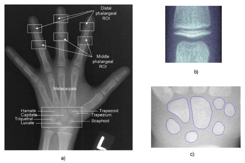
An example of hand image radiograph with superimposed regions of interest: a) a hand image with seven ROIs, b) a phalangeal region of interest (PROI), c) the carpal bones region
The classical method of skeletal bone age assessment (BAA) utilizes the recognition of changes in the radiographic appearance of the maturity indicators in a hand-wrist radiograph by comparison with a reference data set which consists of series of radiographs grouped according to sex and age. The most commonly used reference standard is the atlas published by Greulich and Pyle (G&P) [1]. They were derived from the population of the middle socioeconomic class of Caucasian children from Midwest, USA from 1931-1942. The atlas remains unchanged from its initial publication and is commonly used in clinical practice to assess bone age of children of Caucasian, African American, Hispanic, Asian, and other descent. The examination is subjective because the radiologist analyzes each individual bone of the hand and wrist, determines an overall bone age, and finally fits the amalgamated results into a closest match to the reference radiographs in the atlas. Using the G&P atlas, inter-observer reading differences ranging from 0.37 to 0.6 years and intra-observer reading differences from 0.25 to 0.47, even up to 0.96 years have been reported [2, 6]. The average time of a single case reading depends on radiologist's clinical experience and falls within 2 - 5 minutes.
More objective methods are also available [3-5], (TW1, TW2, TW3). The radiological patterns of ossification centers were derived from the population of 3000 normal British boys and girls. An overall bone age is derived from the sum of the developmental scores from all of the individual ossification centers. Because this approach is both complicated and time consuming, it is seldom used in clinical practice, particularly in the United States.
In this paper, we describe an automated method to assess bone age of children using a digital hand atlas. The hand Atlas consists of two components: a database which is a collection of 1,400 digitized left hand radiographs from evenly distributed normally developed children of Caucasian, African-American, Hispanic and Asian origin, male and female, ranged from 1 to 18 year old. Relevant patient demographic data along with pediatric radiologists' readings of each radiograph were also included. Currently, 1,390 are included in the database. The second component is a computer-assisted diagnosis (CAD) module to assess a child bone age based on the collected data. The CAD method is derived from features extracted from seven regions of interest (ROIs), the six phanlangeal and one carpal bone ROIs.
First, the concept of the Digital Hand Atlas and the data collection is described. Next, a hand image processing work flow of the CAD is introduced. Quantitative features extraction from ROIs is followed by the preprocessing of the hand image. The final bone age assessment based on extracted quantitative features is obtained by means of an array of fuzzy classifiers. The integration of the digital hand Atlas with a PACS for validation of clinical use is presented.
2. Data Collection of the Digital Hand Atlas
Rationale
During past years, numerous results have demonstrated that variations in skeletal maturation in prepubertal children are greater than those reflected in the Greulich and Pyle atlas. More precisely, prepubertal American children of European (EA) descent have significantly delayed skeletal maturation when compared with those of African descent. Also, postpubertal EA males have significantly advanced skeletal maturation when compared with postpubertal African American males [7]. Therefore, it is apparent that multiethnic pediatric population normal images are needed in order to more accurately assess today's children bone age. We started to collect normal children hand images in the late 90s through the support of grants supported by the National Institutes of Health. The process of data collection was conducted at the Childrens Hospital of Los Angeles (CHLA). Candidates for this study underwent a protocol approved by the institutional review board for clinical investigations. A physical examination by a pediatric endocrinologist was performed to determine health and Tanner stage of sexual development of all subjects. According to the clinical examinations, their skeletal development had been confirmed as normal. Measurements of height, trunk height and weight were also obtained. The bone age of each normal was evaluated by at least two pediatric radiologists from the left hand radiograph according to the method of G & P atlas. Each radiograph was digitized to a 2K×2K image using a laser film scanner (Array, Tokyo, Japan); and the patient demographic records were manually entered via the scanner GUI (graphical user interface) and saved as a DICOM file. An example of data stored in the DICOM file is presented in Table 1.
Table 1.
An example of image header fields in a hand image DICOM file.
| DICOM Field Name | Date of Birth | Date of Examination | Chronological Age | Patient Race | Patient Sex | Tanner Index | Trunk Height | Body Height | Body Weight | Reading R1 | Reading R2 |
|---|---|---|---|---|---|---|---|---|---|---|---|
| Unit | dd/mm/yy | dd/mm/yy | years | - | - | - | cm | cm | kg | years | years |
| Field value | 26/02/89 | 25/06/98 | 9.33 | CAU | M | 1.0 | 71.12 | 137.8 | 39.1 | 9.0 | 9.5 |
Data Collection
The data collection was scheduled in two separate cycles. In the first cycle a total number of 1103 left hand radiographs of normally developed children of four races: Caucasian (CA), African American (AA), Hispanic (HI), and Asian (AS) for both male and female [8, 9] have been collected. The data collection protocol is given in Appendix A, with the hand positioned as presented in Fig. 1.
For each race and gender, five images per age group for children aging from one to nine years, and ten images per age group for older have been collected respectively. During an almost 10 year period of collecting the data in this cycle, groundwork of the methodology of developing the CAD for bone age assessment has also been established [10-13].
The data was evaluated as normal comparing to: (1) the body mass index (BMI) for children based on the growth chart from National Health and Nutrition Examination Survey [14], (2) Tanner maturity index, and (3) chronological age vs. skeletal age based on the 1940 Brush Foundation Study. Comparison to (1) and (2) shown that the data is normal. According to comparison with (3), bone age assessment with first cycle data was consistently lower between ages 5-14 for boys and girls in every ethnic origin. A difference of approximately ten months was observed between item (3) and data collected.
Because of the difference in item (3), in order to increase the statistical power of the first cycle data set we started a second cycle data collection with additional 287 images for the rapid maturation stage of children from ages 5 to 14 year old. A summary of first and second cycle data from the Digital Hand Atlas as of today is presented in Table 2 (a-b).
Table 2a.
Digital Hand Atlas: the summary of the first cycle data collection.
| First cycle of data collection: each case is with two readings | ||||||||
|---|---|---|---|---|---|---|---|---|
| Age group/Category | ASF | ASM | AAF | AAM | CAF | CAM | HIF | HIM |
| 00 | 1 | 2 | 4 | 5 | 3 | 3 | 1 | 4 |
| 01 | 5 | 5 | 5 | 5 | 5 | 5 | 5 | 5 |
| 02 | 5 | 5 | 5 | 5 | 5 | 5 | 5 | 5 |
| 03 | 5 | 5 | 5 | 5 | 5 | 5 | 5 | 5 |
| 04 | 5 | 5 | 5 | 5 | 5 | 5 | 5 | 5 |
| 05 | 5 | 5 | 5 | 5 | 5 | 5 | 5 | 5 |
| 06 | 5 | 5 | 5 | 5 | 5 | 5 | 5 | 5 |
| 07 | 5 | 5 | 5 | 5 | 5 | 5 | 5 | 5 |
| 08 | 5 | 5 | 5 | 5 | 5 | 5 | 5 | 5 |
| 09 | 5 | 5 | 5 | 5 | 5 | 5 | 5 | 5 |
| 10 | 10 | 10 | 10 | 10 | 10 | 10 | 10 | 10 |
| 11 | 10 | 10 | 10 | 10 | 10 | 10 | 10 | 10 |
| 12 | 10 | 10 | 10 | 10 | 10 | 10 | 10 | 10 |
| 13 | 10 | 10 | 10 | 10 | 10 | 10 | 10 | 10 |
| 14 | 10 | 10 | 10 | 10 | 10 | 10 | 10 | 10 |
| 15 | 10 | 10 | 10 | 10 | 10 | 10 | 10 | 10 |
| 16 | 10 | 10 | 10 | 10 | 10 | 10 | 10 | 10 |
| 17 | 10 | 10 | 10 | 10 | 10 | 10 | 10 | 10 |
| 18 | 10 | 10 | 10 | 10 | 10 | 10 | 10 | 10 |
| Images total / category | 136 | 137 | 139 | 140 | 138 | 138 | 136 | 139 |
| Images total | 1,103 | |||||||
Table 2b.
Digital Hand Atlas: the summary of the second cycle data collection.
| Second cycle of data collection: each case is with four readings | ||||||||
|---|---|---|---|---|---|---|---|---|
| Age group/Category | ASF | ASM | AAF | AAM | CAF | CAM | HIF | HIM |
| 05 | 3 | 4 | 4 | 4 | 2 | 5 | 5 | 4 |
| 06 | 1 | 1 | 4 | 2 | 2 | 3 | 5 | 4 |
| 07 | 2 | 2 | 4 | 4 | 3 | 4 | 5 | 5 |
| 08 | 4 | 0 | 5 | 5 | 4 | 5 | 4 | 5 |
| 09 | 2 | 2 | 4 | 5 | 3 | 2 | 5 | 5 |
| 10 | 5 | 4 | 2 | 5 | 2 | 1 | 4 | 2 |
| 11 | 2 | 5 | 0 | 5 | 3 | 4 | 5 | 4 |
| 12 | 4 | 5 | 5 | 5 | 4 | 3 | 5 | 5 |
| 13 | 5 | 5 | 5 | 5 | 4 | 2 | 5 | 5 |
| 14 | 3 | 2 | 2 | 4 | 1 | 0 | 4 | 4 |
| Images total / category | 31 | 30 | 35 | 44 | 28 | 29 | 47 | 43 |
| Images total | 287 | |||||||
Hand Image Database
A Hand Image Database containing the image data collected along with patient demographic data and radiologist readings can be browsed using the following link: http://www.ipilab.org/BAAweb/. Fig. 2 shows a screen shot of a page retrieved from the eleven year old Asian female data.
Figure 2.
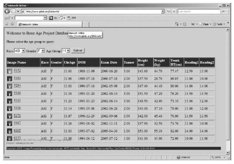
A GUI window of the bone age assessment web site (http://www.ipilab.org/BAAweb/) with hand image database. The website provides hand images, information about subjects' demographic data and radiologists' readings.
3. The CAD Module
CAD using the data collected described in Section 2 for the BAA has been designed and implemented to provide an objective assessment of a child's skeletal age as an aid to the pediatric radiologist's reading. The collected data together with features extracted related to skeletal development from phalangeal ROIs [15] and the Carpal bone ROI [17] were transformed into knowledge rules of fuzzy classifiers [16] for bone age assessment. The Digital Hand Atlas is a combination of the collected data and the CAD module. A work flow of the CAD is presented in Fig. 3 which consists of five steps: 1) image preprocessing, 2) ROIs determination, 3) phalangeal and carpal bones features extraction, 4) fuzzy classifiers, and 5) aggregation of fuzzy classifier results to obtain the bone age assessment.
Figure 3.
Five steps of the CAD workflow. Located seven regions of interest is subjected to features extraction and category classifiers provide fuzzy information about boner age derived from the regions of interest. Final bone age is calculated after the aggregation procedure.
Image Pre-Processing, ROI Determination, Feature Extraction
Image preprocessing includes background suppression and radiological markers removal (see Fig. 1 stripes and background nonuniformitiy may not be well visible in this example, except for the “L” marker). [12, 13] Next, six distal and middle phalangeal ROIs (PROIs) of three middle fingers [13] and the carpal bone ROI are located [11]. Fig. 1 left shows six PROIs and the carpal bone ROI, and the right is the magnification of a PROI and the carpal bone ROI, [17]. Considering first the six PROIs, eleven features were extracted from each ROI. [16,18,19] Two feature sets, those features related to size and shape of the epiphysis, and the wavelet features derived from stage of fusion advancement of the epiphysis, describe the degree of bone development in this single PROI. Similar procedures were performed for each PROI. As a result, a total of twelve feature sets were obtained, two from each PROI. Methods of feature extraction have been discussed in details in previous publications [10-19]. These twelve feature sets were used to train twelve PROI fuzzy classifiers for bone age assessment contributed by PROIs features.
The Category Fuzzy Classifier and Aggregator
The data from 1 – 18 years shown in Table 2 was separated into eight categories (CAM, CAF, AAM, AAF, HIM, HIF, ASM, and ASF) for race and gender comparison study. In addition, CAM, AAM, HIM and ASM were combined as one male category; and CAF, AAF, HIF, and ASF were combined as one female category. The male and female were further combined as one universal F & M category. Therefore we developed one category fuzzy classifier for each of the eleven categories.
For each category, the category fuzzy classifier using the PROIs feature sets was trained as follows. For each PROI, there were two classifiers, one was based on the shape and size features, and the other was based on wavelet features. The design and training of these two classifiers were based on Mamdani's original concept [21], and applied to phalangeal ROIs discussed in details in Ref [16]. The design and training of each category fuzzy classifier using the carpal bone ROI is given in [17]. Fifty percents of the collected images were used for training, and the other 50% for evaluation.
The design of the category fuzzy classifier is an open architecture model meaning that if fuzzy results from other ossification centers are provided they can be appended to the existing structure before the defuzzification (or aggregation) step, for example in each category classifier, the carpal bone ROI classifier could be appended to the phanlangeal ROI classifiers for bone age assessment [17].
The final BAA was derived from the aggregation of BAA results from the phalangeal ROIs and carpal ROI. In general, carpal bone ROIs determines the bone age of boys from 1 to 7, and girls from 1 to 5; phalangeal ROIs determine the bone age for both sexes above age 13. For girls from 6 – 12, and boys from 8 -12, both carpal ROI and phalangeal ROIs contribute. The final bone age value was obtained by defuzzication with the center of gravity method. Fig. 6 middle left illustrates the concept of the aggregation of 12 PROI classifiers and the carpal ROI classifier to form the combined bone age value in a category fuzzy classifier.
Figure 6.
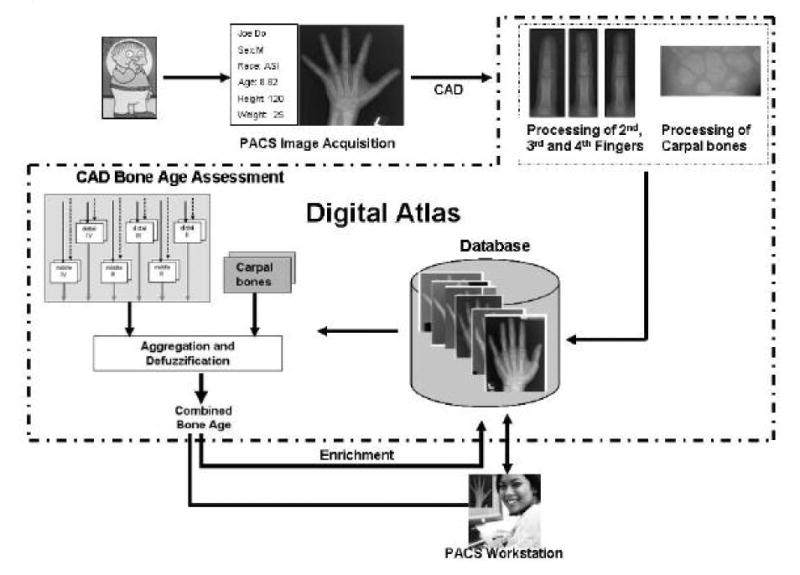
The workflow of CAD and PACS integration. Acquired hand image is processed by the CAD and compared with hand images in the database. The Digital Hand Atlas can be enriched by CAD bone age results.
Graphical User Interface
A graphical user interface (GUI) was designed to visualize CAD operation steps (Window 1) and results (Window 2). After the hand image is sent to the CAD and the processes are completed, the CAD workstation Window 1 displays the patient data (Fig. 4, Left), image with superimposed ROIs (Middle) and segmentation results (Right). In addition, detailed messages about currently CAD performed step can also be visualized in the Lower Right of Fig. 4 using a scroll bar at the bottom). The CAD results are shown in Window 2 as depicted in Fig. 5.
Figure 4.
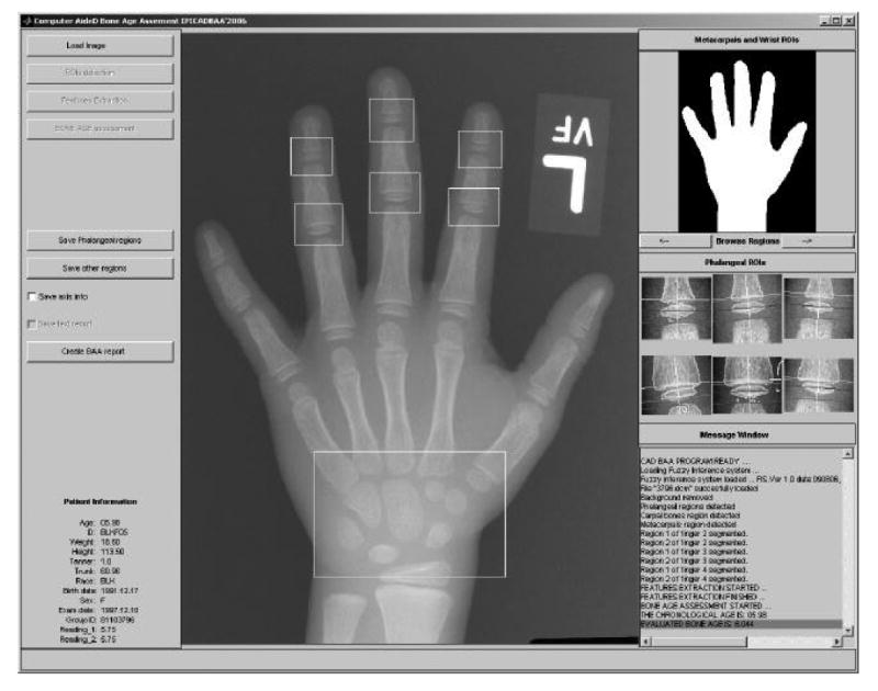
Graphical user interface of the CAD for BAA. The analyzed hand image with superimposed ROIs is in the GUI center. Patient data is on left side, right part of the GUI contains segmentation results and the message window.
Figure 5.
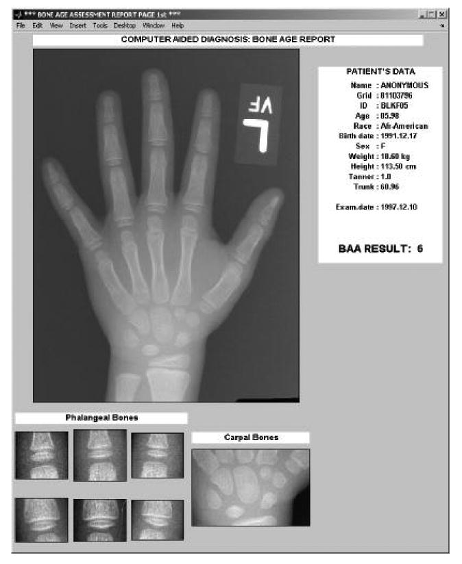
An example of a CAD report window with automatically extracted ROIs together with analyzed hand image. Patient data and CAD result are displayed in the upper right.
Integration of CAD BAA with PACS
The digital atlas has been integrated with a research PACS using a CAD-PACS integration toolkit for validation study [20]. The toolkit is based on the DICOM (Digital Imaging and Communications in Medicine) Standard and IHE (Integrating the Healthcare Enterprise) workflow profiles. The toolkit has two modules, the DICOM-based SC (secondary capture) module, and the DICOM SR – IHE module, both modules can output the CAD BAA results as shown in Fig. 5. The former module, a fast integration method, allows the CAD GUI results shown in Figs. 5 to be directly displayed on the PACS workstations using the DICOM SC. However, PACS workstations can only display these results but can not assess the image or the textual contents directly. The DICOM – IHE module is based on the DICOM Structured Report (SR) standard and the IHE Post-Processing Workflow Profile protocol. The integration is more elaborated but the PACS workstations, in addition to displaying CAD results as shown in Figs. 5, can also access the CAD result images and data for other research, teaching, and clinical services applications. Zhou [20] in this Special Issues provides detail description of the integration methods. Fig. 6 shows the workflow of the integration of CAD BAA with PACS. Within the dotted lines is the domain of the Digital Atlas, and PACS hardware components are the image acquisition and workstations. The CAD toolkit is integrated with the PACS software.
4. Results
With the large-scale data collection and elaborated design of the CAD module, many different types of meaningful clinical results could emerge. In this section, we provide results related to the evaluation of data collection methodology and its effect on the CAD bone age assessment. In particular, the effect of quality assurance protocol (QAP) to the performance of image analysis algorithm, the accuracy of image analysis algorithm in the phanlangeal ROIs (PROI), and the improvement of the CAD method in bone age assessment with the addition of second cycle data collection (See Table 2a,b) are given.
Effect of Quality Assurance Protocol (QAP) to the Performance of Segmentation Algorithm
Since data collection spanned about ten years, QAP is important to assure the consistence and quality of the data collected (See Appendix A). During the data collection process, by incorporating the QAP, about 5% of cases misspelled dates and other numeric data in the DICOM study description field were detected and corrected. Forty six images failed to pass the Step 1 of the QAP work flow (Appendix A) and were replaced by others. Main reasons were the hand placement was not aligned properly during the X-rays exam, and/or numerous artifacts on the image. Application of the Step 3 of the QAP also greatly improved the passage of all 1,400 images through the CAD in terms of smooth running of the CAD. All cases that caused errors were investigated and more robust error detection was implemented.
Evaluation of the image analysis algorithm
The evaluation of segmentation results involved 300 selected hand images from the second cycle data collection. The set covered distal and middle regions of interest with all possible stages of epiphyseal development, from those with epiphyses distinct in appearance, to epiphyses partially and completely fused with the metaphyses, otherwise. Other than these conditions, the selection process was random. The PROIs were automatically located in the hand image by the CAD software. All segmented regions with outlined cartilage and bony structure were presented to two radiologists of different clinical experience and training. Results of segmentation of each region were evaluated by these radiologists to one of three categories: good, acceptable, unacceptable. They were blind to the child's age, sex and race. Their subjective results are presented in Table 3. The average number of regions classified as good was 79.7%, and regions classified as good or acceptable was 93.7% which demonstrate that the automatic segmentation of the cartilage and bony structures is acceptable by the radiologists. For results in carpal bone ROI, see [17].
Table 3.
Experts' evaluation of PROIs outlined by the CAD. The average number of regions classified as good is 79.7%, and regions classified as good or acceptable is 93.7%
| Expert | good | acceptable | unacceptable |
|---|---|---|---|
| Expert 1 | 72.81% | 19,94% | 7.25% |
| Expert 2 | 86.5% | 8.08% | 5.24% |
Improvement of the CAD method with the addition of second cycle data collection
In Section 3 we described that the CAD consists of eleven fuzzy classifiers each of which is used to assess the bone age of the eleven categories: CAM, CAF, AAM, AAF, HIM, HIF, ASM, and ASF, female only, male only, and female and male (F & M) together, respectively. These eleven classifiers were trained in two times, with data from the first cycle collection, and with data from both cycles (first and second) collection. We discuss here the improvement of the CAD method for bone age assessment of the first eight categories with the addition of second cycle data collection. Comparison between races indicated that the addition of second cycle data collection does improve the performance of the CAD based on the comparison of the CAD bone age assessment (BAA) with the chronological age. Improvement is in the sense that less discrepancy between races was observed. Table 4 depicts an example of the girl BAA, where Table 4a) is the result using the first cycle data only, and Table 4b is the results using both first and second cycle data. The girl bone age development according to the female gender was divided into four stages as a gauge of comparison shown in the bottom of both Tables 4a) and 4b).
Table 4a.
Performance of the CAD Bone Age Assessment based on comparison with chronological age using first cycle data. The girl bone age development according to the female gender was divided into four stages as a gauge of comparison shown in the bottom. The values in these boxes represent the difference between CAD BAA and chronological age.
indicates that the difference has p-value < 0.05 which is significant.
Table 4b.
Performance of the CAD Bone Age Assessment based on comparison with chronological age using both first and second cycle data. Improvement is in the sense that less discrepancy between races was observed. The girl bone age development according to the female gender was divided into four stages as a gauge of comparison shown in the bottom. The values in these boxes represent the difference between CAD BAA and chronological age.
indicates that the difference has p-value < 0.05 which is significant.
5. Discussion and Summary
An overview of the computerized approach to bone age assessment utilizing Digital Hand atlas is presented. The Digital Hand Atlas serves as a reference data set reflecting current skeletal development of normal subjects of four descents, living in the US, in particular in the Los Angeles area. The Atlas is comprised of two components, a digital hand database with a collection of 1,400 digitized left hand radiographs and relevant data from evenly distributed normally children of Caucasian (CA), Asian (AS), African-American (AA) and Hispanic (HI) origin, male (M) and female (F), ranged from 1 to 18 year old; and a CAD module for bone age assessment. The automatic CAD approach utilizing quantitative knowledge of the Digital Hand Atlas can provide bone age value based on radiological findings sensitive to developmental changes.
We are in the beginning of validating and evaluating the performance of the digital hand atlas. Three types of results have been obtained: the effect of quality assurance protocol (QAP) to the performance of image analysis algorithm, the accuracy of image analysis algorithm in the phanlangeal ROIs (PROI), and the improvement of the CAD method in bone age assessment with the addition of second cycle data collection. Bone age assessment for children has been assessed based on the 1950 Greulich and Pyle Atlas (G & P) from a homogeneous population. With today's diverse ethnicities in the US, the G & P atlas may no longer be a good reference. We are in the process of analyzing the Digital Atlas results to address the following questions: 1. Does ethnicity and gender have different bone growth patterns? 2. Does the bone age of ethnic origins differ? 3. Is the bone growth for boys and girls different? And 4. Is the G & P atlas still a good reference for bone age assessment of today's children?
The atlas has been integrated with PACS which can directly access the hand image of a patient from the PACS and return the bone age assessment results to PACS workstations for on-line assisting clinicians to assess the bone age. The digital atlas can be expanded in bone age assessment of subjects of other ethnic origins by collecting digital hand images following the data collection protocol and the training of the fuzzy classifiers with methods discussed in this paper.
Acknowledgments
The authors would like to thank Alexis Wong, MD, and Bing Guo, MD, for their kind help in validating the segmentation results. This research work has been sponsored by NIH R01 EB 00298.
Biography
Arkadiusz Gertych received the master degree in 1995 and the PhD degree in 2003 in electrical engineering from the Silesian University of Technology, Poland (SUT). In 1996 he started working for SUT in Department of Biomedical Electronics and from 2003 he is also an Assistant Professor at this University. From 2004 he continues post-doctoral studies at University of Southern California Department of Radiology in Image Processing and Informatics Laboratory in Los Angeles. His research activities include signal and image processing, design and development of computer-aided (CAD) medical diagnosis support systems, medical informatics and radiation therapy systems.
Appendix A. Quality Assurance Protocol of the Images collected for the Digital Hand Atlas
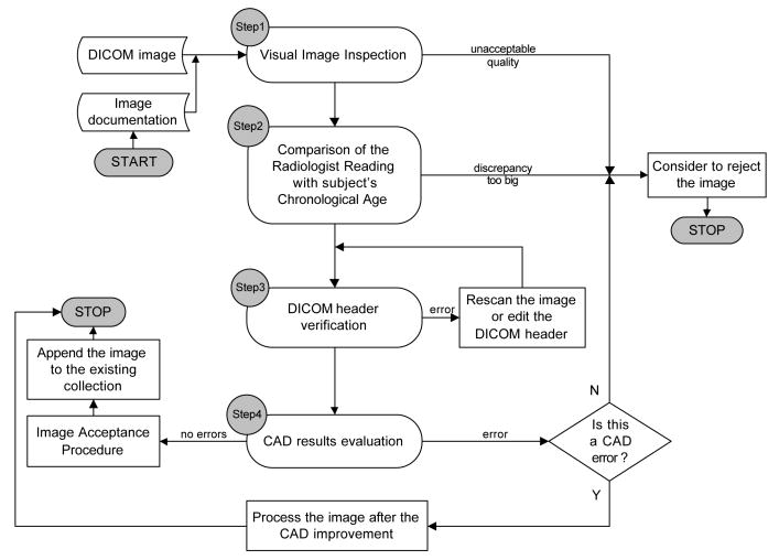
Testing and developing of a CAD requires the image data to be properly collected according to a standard protocol. Also, high quality of the data should be maintained if the images are intended to be used in a decision making process. A quality assurance protocol (QAP) has been developed and implemented in order to meet such needs. The QAP encompasses a manual and visual inspection of the image and the evaluation of its suitability for the automatic CAD process. The workflow of the QAP is presented in Fig. A1 which consists of four steps. Step 1 can be completed by the operator whereas Steps 2-4 are non-interactively performed by the CAD.
Figure A1.
The quality assurance protocol work flow with the CAD in the loop. Image inspection Step 1 and DICOM header verification: Step 2, 3 are performed first. In Step 4 errors and comparison of CAD result are evaluated. Acceptance of the CAD results allows appending the image to the existing collection.
In Step 1 (Fig. A1) the quality is first visually justified by the operator utilizing the graphical user interface of the CAD. If the hand image is not aligned properly with respect to the image plane, then it is immediately rejected. In Step 2, a comparison between the radiologist readings and the chronological age is performed. The image is rejected if the difference between the chronological age and the radiologist bone age reading is larger than a certain threshold trh. In this protocol, trh value has been set to three years. In Step 3, the content of the DICOM image header containing the subject's demographic data is compared with the data in the documentation used during the digitization procedure. Any inconsistencies between the documentation and patient record require corrections. In Step 4, image preprocessing procedures could reveal artifacts caused by nonuniform background, underexposed film borders, scratches and radiological markers that may cause difficulties in automatic image processing [12, 13]. Finally CAD results like: regions of interests and bone age value are evaluated. In Step 4 the CADBA result is also compared with subject's chronological age. If the discrepancy is smaller than the predefined threshold trh; the same as used in Step 2, then this image is subjected to the acceptance procedure and appended to the existing data collection. Otherwise it requires a CAD verification step to be taken. In such cases, the image will be subjected to bone age recalculation after a modification of the CAD.
In summary, the QAP applied to the Digital Hand Atlas encompasses checking the image features and patient data at various levels. Image quality is assessed in terms of correct hand placement, presence of image artifacts and capability of radiological findings extraction performed by the CAD. Verification of subject's demographic data can be performed in the QAP by the CAD operator or the CAD; however the latter option can be modified in a more automated fashion. Images that failed to comply with the QAP protocol have been replaced by other images. Using the QAP, the reliability of the data collected for the Digital Hand Atlas has been improved.
Footnotes
Publisher's Disclaimer: This is a PDF file of an unedited manuscript that has been accepted for publication. As a service to our customers we are providing this early version of the manuscript. The manuscript will undergo copyediting, typesetting, and review of the resulting proof before it is published in its final citable form. Please note that during the production process errors may be discovered which could affect the content, and all legal disclaimers that apply to the journal pertain.
References
- 1.Greulich WW, Pyle SI. Radiographic Atlas of Skeletal Development of Hand Wrist. 2. Stanford University Press; Stanford CA: 1971. [Google Scholar]
- 2.Roch AF, Rochman CG, Davila GH. Effect of training of replicaability of assessment of skeletal maturity (Greulich-Pyle) American Journal of Roentgenology. 1970;108:511–515. doi: 10.2214/ajr.108.3.511. [DOI] [PubMed] [Google Scholar]
- 3.Tanner JM. Growth at adolescence. 2. Oxford: Blackwell Scientific Publications; 1962. [Google Scholar]
- 4.Tanner JM, Whitehouse RH. Assessment of Skeletal Maturity and Prediction of Adult Height (TW2 Method) Academic Press; London: 1975. [Google Scholar]
- 5.Tanner JM, Healy MJR, Goldstein H, Cameron N. Assessment of Skeletal Maturity and Prediction of Adult Height (TW3 Method) Third. W. B. Saunders; London: 2001. [Google Scholar]
- 6.King DG, Steventon DM, O'Sullivan MP, Cook AM, Hornsby VP, Jefferson IG, King PR. Reproducibility of bone ages when performed by radiology registrars: an audit of Tanner and Whitehouse II versus Greulich and Pyle methods. Br J Radiol. 1994;67:848–851. doi: 10.1259/0007-1285-67-801-848. [DOI] [PubMed] [Google Scholar]
- 7.Mora S, Boechat MI, Pietka E, Huang HK, Gilsanz V. Skeletal Age Determinations in Children of European and African Descent: Applicability of the Greulich an Pyle Standards. Pediatric Research. 2001;50(5):624–628. doi: 10.1203/00006450-200111000-00015. [DOI] [PubMed] [Google Scholar]
- 8.Cao F, Huang HK, Pietka E, Gilsanz V. Digital hand atlas for Web-based bone age assessment: System design and implementation. Computerized Medical Imaging and Graphics. 2000;24:297–307. doi: 10.1016/s0895-6111(00)00026-4. [DOI] [PubMed] [Google Scholar]
- 9.Cao F, Huang HK, Pietka E, Gilsanz V, Dey PS, Gertych A, Pospiech-Kurkowska S. Image database for digital hand atlas; SPIE Medical Imaging. Proceedings of the SPIE; 2003. pp. 461–470. [Google Scholar]
- 10.Pietka E, McNitt-Gray MF, Huang HK. Computer-assisted phalangeal analysis in skeletal age assessment. IEEE Trans on Medical Imaging. 1991;10:616–620. doi: 10.1109/42.108597. [DOI] [PubMed] [Google Scholar]
- 11.Pietka E, Kaabi L, Kuo ML, Huang HK. Feature extraction in carpal-bone analysis. IEEE Trans on Medical Imaging. 1993;12:44–49. doi: 10.1109/42.222665. [DOI] [PubMed] [Google Scholar]
- 12.Pietka E, Gertych A, Pospiech S, Huang HK, Cao F. Computer assisted bone age assessment: Image pre-processing and ROI extraction. IEEE Trans on Medical Imaging. 2001;20:715–729. doi: 10.1109/42.938240. [DOI] [PubMed] [Google Scholar]
- 13.Pietka E, Pospiech S, Gertych A, Cao F, Huang HK, Gilsanz V. Computer Automated Approach to the extraction of epiphyseal regions in hand radiographs. Journal of Digital Imaging. 2001;14:165–172. doi: 10.1007/s10278-001-0101-1. [DOI] [PMC free article] [PubMed] [Google Scholar]
- 14. http://www.cdc.gov/growthcharts/
- 15.Pietka E, Gertych A, Pospiech-Kurkowska S, Cao F, Huang HK, Gilsanz V. Computer Assisted Bone Age Assessment: Graphical User Interface for Image Processing and Comparison. Journal of Digital Imaging. 2004;17(3):175–188. doi: 10.1007/s10278-004-1006-6. [DOI] [PMC free article] [PubMed] [Google Scholar]
- 16.Pietka E, Pospiech-Kurkowska S, Gertych A, Cao F. Integration of Computer assisted bone age assessment with clinical PACS. Comp Med Img Graph. 2003;27:217–228. doi: 10.1016/s0895-6111(02)00076-9. [DOI] [PubMed] [Google Scholar]
- 17.Zhang A, Gertych A, Liu BJ. Bone Age Assessment for Young Children from Newborn to 7-Year-Old Using Carpal Bones. Journal of Comp Med Img Graph. doi: 10.1016/j.compmedimag.2007.02.008. (in press) [DOI] [PMC free article] [PubMed] [Google Scholar]
- 18.Pietka E, Gertych A, Witko K. Informatics infrastructure of CAD system. Computerized Medical Imaging and Graphics. 2005;29:157–169. doi: 10.1016/j.compmedimag.2004.09.016. [DOI] [PubMed] [Google Scholar]
- 19.Gertych A, Pietka E, Liu BJ. Segmentation of Regions of Interest and Post-Segmentation Edge Location Improvement in Computer-Aided Bone Age Assessment. Pattern Analysis and Applications. (in press) [Google Scholar]
- 20.Zhou Z. CAD-PACS Integration Tool Kit based on DICOM Secondary Capture, Structured Report and IHE Workflow Profiles. Journal of Comp Med Img Graph. doi: 10.1016/j.compmedimag.2007.02.015. (in press) [DOI] [PubMed] [Google Scholar]
- 21.Mamdani EH. Application of fuzzy algorithms for control of simple dynamic plant. Proc IEEE. 1974;121:1585–8. [Google Scholar]


