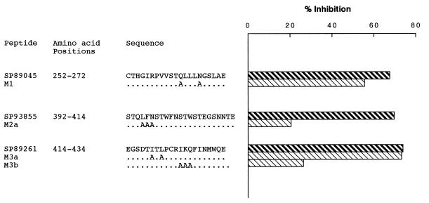
Figure 4.
Mutagenesis analysis of the sequence motifs involved in SAg binding. Dilutions of the wild-type and mutant peptides (M1, M2a, M3a, and M3b) were mixed with an equal volume of IgM LAN at a concentration that achieved 50% of maximal binding. After incubation, the mixtures were transferred to gp120MN-coated wells, and residual binding was revealed as indicated in the text. Represented is the inhibition achieved by the peptides used at a 5 μM concentration. The results are from one representative experiment.

