Abstract
The nonsegmented negative strand RNA viruses comprise hundreds of human, animal, insect, and plant pathogens. Gene expression of these viruses is controlled by the highly conserved order of genes relative to the single transcriptional promoter. We utilized this regulatory mechanism to alter gene expression levels of vesicular stomatitis virus by rearranging the gene order. This report documents that gene expression levels and the viral phenotype can be manipulated in a predictable manner. Translocation of the promoter–proximal nucleocapsid protein gene N, whose product is required stoichiometrically for genome replication, to successive positions down the genome reduced N mRNA and protein expression in a stepwise manner. The reduction in N gene expression resulted in a stepwise decrease in genomic RNA replication. Translocation of the N gene also attenuated the viruses to increasing extents for replication in cultured cells and for lethality in mice, without compromising their ability to elicit protective immunity. Because monopartite negative strand RNA viruses have not been reported to undergo homologous recombination, gene rearrangement should be irreversible and may provide a rational strategy for developing stably attenuated live vaccines against this type of virus.
The ability to alter the genotype of a virus in a manner that results in a predictable change in gene expression would be invaluable for studies of gene function and control. However, the genetic mobility of many viruses and RNA viruses in particular, engendered by homologous recombination, coupled with the innate polymerase error rate usually has a high potential to reverse both natural and engineered genetic changes. We show that it is possible to utilize the general mechanism for control of gene expression of a negative strand RNA virus to alter gene expression levels in a predictable way. In the present report, we describe how manipulation of the genome of a nonsegmented, negative strand RNA virus to translocate an essential gene for replication can be utilized to alter systematically the phenotype of the virus in a manner that should be irreversible.
The nonsegmented negative strand RNA viruses (order Mononegavirales) comprise many significant pathogens of humans, animals, plants, and insects. The four virus families in the order are: the Rhabdoviridae, such as rabies virus, vesicular stomatitis virus (VSV), and potato yellow dwarf virus; the Paramyxoviridae, such as measles, human, and bovine respiratory syncytial viruses; the Filoviridae, Ebola, and Marburg viruses, and the Bornaviridae. These viruses have an elegantly simple means of controlling gene expression by the highly conserved order of the genes relative to the single transcriptional promoter (1, 2). Genes proximal to the 3′ promoter site are transcribed at high levels whereas those at more distal positions are transcribed less abundantly as their distance from the promoter increases (3–6). Genes that encode proteins required in stoichiometric amounts such as the nucleocapsid protein N, which is necessary for genome encapsidation and replication, are located at or near the 3′ terminus, whereas genes that encode proteins needed in catalytic amounts such as the RNA dependent RNA polymerase L are more promoter distal. The importance of gene regulation is shown by the observation that the relative molar ratios of the nucleocapsid (N) protein, phosphoprotein (P), and polymerase (L) proteins are critical for optimal VSV viral RNA replication (7–9) and that RNA replication in vitro and in cultured cells is proportional to the amount of N protein synthesized (10, 11).
Gene regulation by progressive transcriptional attenuation provides a possible explanation for the strong conservation of gene order among the Mononegavirales, and it suggested a novel way to manipulate the viral phenotype—by rearranging the genes. Because both phylogenetic and experimental studies of these viruses indicate that homologous RNA recombination occurs rarely if at all (2, 12, 13), deliberate rearrangement of the viral genes should be irreversible by nature and have the potential to perturb the properties of the virus profoundly.
Infectious viruses have recently been recovered from cDNA clones of the rhabdoviruses rabies and VSV (14–16) as well as several other of the Mononegavirales (17), and this recovery makes possible the introduction of deliberate genetic changes. We examined whether the expression of a gene product critical for replication could be decreased and viral replication levels reduced in a systematic and predictable manner. We tested this hypothesis by moving the gene for the nucleocapsid protein of VSV from its wild-type promoter proximal position to successive positions down the viral genome. This paper describes how gene translocation reduced N gene expression in a stepwise manner thereby lowering the viral growth potential in cell culture and attenuating its lethality for mice. Importantly, these changes occurred without compromising the ability of the virus to elicit a protective host response, suggesting that this approach may provide a rational method to achieve a measured and stable degree of attenuation of this type of virus.
MATERIALS AND METHODS
Viruses and Cells.
The San Juan isolate of the Indiana serotype of VSV provided the original template for all cDNA clones used in this work except the G protein gene, which was derived from the Orsay isolate of VSV Indiana (15). Baby hamster kidney (BHK-21) cells were used to recover viruses from cDNAs, for single step growth experiments, and radioisotopic labeling of RNAs and proteins. BSC-40 cells were used for plaque assays.
Plasmid Construction and Recovery of Infectious Viruses.
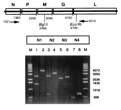
Each of the five genes of VSV contains the same sequence of the first five nucleotides: 3′-UUGUC-5′. We used this common sequence to construct molecular clones of individual genes from which DNA fragments precisely encompassing each gene could be released by restriction enzyme digestion and the genes could be reassembled without introducing other changes to the genome as described (L.A.B., C. Pringle, V.P.P., and G.W.W., unpublished work). The DNA segments that encompassed the individual genes were reassembled in any desired order to create a family of DNA plasmids whose nucleotide sequences corresponded precisely to that of wild-type VSV, except for the fact that their genes were rearranged. One deliberate mutation was introduced: the untranscribed, intergenic dinucleotide after the P gene is 3′-CA-5′ in the wild-type sequence, and this was made 3′-GA-5′ to conform with all the other gene junctions. These cDNA plasmids contained T7 promoters that expressed precise copies of the positive sense antigenomic VSV RNA (by use of a hepatitis δ virus ribozyme positioned to generate an exact 3′ terminus), and they were used for recovery of infectious viruses by transfection into VTF7–3-infected cells as described (15, 18, 19). The recovered viruses were amplified by low-multiplicity passage on BHK-21 cells in the presence of cytosine arabinoside to inhibit replication of VTF7–3, filtered through 0.2-μ filters to remove VTF7–3 and the process repeated until VTF7–3 was not detectable by plaque assay. The gene orders of the recovered viruses were verified by amplifying the rearranged portions of the viral genomes using reverse transcription and PCR followed by restriction enzyme analysis (Fig. 2).
Figure 2.
Gene order in recovered viral genomes. Genomic RNA was isolated from recovered viruses N1–N4 after five passages in cell culture and analyzed by reverse transcription and PCR followed by restriction enzyme analysis of the PCR products. PCR was carried out using primers that annealed to the N or L genes respectively (positions indicated by arrows) and the undigested products (lanes 2, 4, 6, and 8) or the products after digestion (lanes 1, 3, 5, and 7) with restriction endonucleases BglII (N1 and N2) or EcoRI (N3 and N4) were analyzed by electrophoresis on a 1% agarose gel in the presence of ethidium bromide. Lanes M = marker DNA fragments with sizes as indicated.
Single Cycle Virus Replication.
BHK-21 cells were infected at a multiplicity of three. After 1 hr of adsorption, the inoculum was removed, cultures were washed twice, and fresh medium was added and incubated at 31°C or 37°C. Samples were harvested at indicated intervals over a 36-hr period, and viruses were quantitated by plaque assay.
Analysis of Viral RNA and Protein Synthesis.
BHK-21 cells were infected with 5 plaque-forming units (pfu)/cell and viral RNA or protein synthesis analyzed by metabolic labeling with [3H]uridine (33 μCi/ml) for 2 hr in the presence of actinomycin D (5 μg/ml) or [35S]methionine (20 μCi/ml), for 30 min, respectively. After the labeling period, cells were harvested, cytoplasmic extracts were prepared, and RNA or protein was analyzed as described (8, 18, 19). RNAs were quantitated by densitometric analysis of autoradiographs, and proteins were quantitated by phosphorimaging and molar ratios were calculated.
Lethality in Mice.
The lethality of individual viruses was measured in male Swiss–Webster mice, 3 to 4 weeks old, obtained from Taconic Farms. Groups of five to seven animals were anesthetized lightly with ketamine/xylazine and inoculated with diluent PBS or with serial 10-fold dilutions of individual viruses. Mice inoculated intracerebrally (IC) received 30 μl inocula; those inoculated intranasally (IN) received 15 μl. Animals were observed daily, and the LD50 for each virus was calculated by the method of Reed and Muench (20).
Protection of Mice.
Groups of control mice inoculated IN with diluent or mice inoculated IN with nonlethal doses of individual viruses were monitored for neutralizing serum antibody production by tail bleeds. Fourteen days postinoculation, mice were challenged with 1.3 × 10⁶ pfu of N1 wild-type virus administered IN as described above. Challenged animals were monitored for 21 days.
RESULTS
Generation and Recovery of Rearranged Viruses.
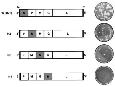
We rearranged the genes of VSV by manipulating an infectious cDNA clone made previously (15) to translocate the gene for the N protein from its normal promoter proximal position to second, third, or fourth in the gene order (Fig. 1). All other aspects of the viral nucleotide sequence were unaltered with the exception of the deliberate change of one nucleotide. The untranscribed, intergenic dinucleotide after the P gene that is 3′-CA-5′ in the wild-type virus was made 3′-GA-5′ to conform with all of the other intergenic junctions. Previous work indicates this change has little effect on transcription (21). The wild-type gene order also was reconstructed from the same components as a control for the validity of the strategy, and this virus was utilized as the wild-type virus control in all subsequent experiments. Viruses were recovered by transfection of the cDNAs into cells infected with recombinant vaccinia virus expressing the T7 polymerase (VTF7–3; ref. 22) and expressing the VSV N, P, and L proteins from plasmids with T7 promoters (15). The recovered viruses were designated according to the position of the N gene: N1 for the wild type and N2, N3, or N4 for the viruses with the N gene translocated to second, third, or fourth position (Fig. 1). Typical plaques of the recovered viruses are shown (Fig. 1) and their sizes decreased from that of the wild type (3 mm) to <0.5 mm for the N4 viruses.
Figure 1.
Gene order and plaque phenotype for wild-type (N1) VSV and rearranged viruses N2, N3, and N4.
The gene orders for each of the recovered viruses were verified after five passages by amplifying the rearranged portions of the viral genome using reverse transcription and PCR followed by restriction enzyme analysis of the PCR products. PCR was carried out using primers located in the N and L genes, and products prior to digestion decreased in size exactly as would be predicted (3998, 3175, 2335, and 661 nt for viruses N1-N4, respectively) as the N gene was moved to successive positions down the genome (Fig. 2, lanes 2, 4, 6, and 8). After digestion with restriction endonucleases BglII or EcoRI, which cleave uniquely in the M or L genes respectively, the observed sizes of the digestion products were exactly as predicted (Fig. 2, lanes 1, 3, 5, and 7). These data show that the gene order of the recovered viruses was as originally constructed and remained so after five passages in cell culture. The variant viruses were then examined for their levels of gene expression, growth potential in cell culture, and virulence in mice.
Effect of Gene Rearrangement on Expression of mRNAs and Proteins.
To ascertain how translocation of the N gene affected viral gene expression, the synthesis of viral RNAs and proteins was examined in infected BHK-21 cells by metabolic labeling with [3H]uridine or [35S]methionine, respectively. Gene rearrangement caused no change in the sizes of the 11.1-kb genomic RNA or the five encoded mRNAs, and no aberrant viral RNAs were observed (Fig. 3). However, the quantity of N mRNA decreased substantially from the wild-type level as its gene was moved successively away from the promoter in viruses N2, N3, and N4 (36, 6, and 3% of wild type, respectively; Fig. 3). Consistent with this decrease, an increase in the amount of G mRNA was observed with virus N4 in which the G gene was moved one position closer to the promoter as the N gene replaced it as the next to last in the gene order (Fig. 3). The amount of genomic RNA replication of N2, N3, and N4 declined relative to wild type (49, 26, and 4%, respectively) concomitant with the lowered expression of the N gene, as predicted if N protein synthesis was limiting for replication. The overall level of transcription was reduced also as the N gene was moved progressively promoter distal, presumably as a secondary effect because of the decrease in replication.
Figure 3.
RNA synthesis of viruses with N gene translocations. The pattern of RNA synthesis directed by viruses N2, N3, and N4 in BHK-21 cells is shown in comparison to wild-type (N1) VSV. VSV-specific RNA was labeled with [3H]uridine in the presence of actinomycin D from 3 to 5 hr postinfection and analyzed by gel electrophoresis. The positions of the genomic RNA and the five VSV mRNAs are indicated to the left of the figure. The P and M mRNAs comigrate. RNAs were quantitated by densitometric analysis of autoradiographs.
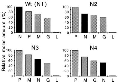
All five of the VSV proteins were expressed in cells infected with the rearranged viruses, and they all comigrated with those of the wild-type virus (data not shown). However, N protein synthesis declined as its gene was moved away from the 3′ position. The data presented in Fig. 4 show how the molar amounts of the proteins decrease as a function of their distance from the 3′ terminus in the wild-type virus N1. When the N gene was translocated, the data in Fig. 4 show that the molar ratios of the N protein relative to the P protein decreased progressively as the N gene was moved from first to second, third, or fourth in the gene order. These results confirm the predictions from previous analysis of gene expression in VSV and the sequential nature of transcription (3, 5, 6). Moreover, these data demonstrate directly that the position of a gene determines its level of expression.
Figure 4.
Molar ratios of proteins synthesized by rearranged viruses. The synthesis of virus-specific proteins by viruses N1 (WT), N2, N3, and N4 in BHK-21 cells was analyzed by metabolic labeling with [35S]methionine. Proteins were separated on a 10% polyacrylamide gel and quantitated by phosphorimager, and molar ratios of individual proteins were calculated relative to the most promoter proximal gene: N in the case of wild type or P in the rearranged viruses.
Examination of the levels of proteins in isolated, mature N1-N4 virions showed that the relative molar ratios of the proteins in mature virus particles remained essentially the same as that of the wild-type virus (data not shown). However, as shown below, less overall virus was produced from infections of N2-N4, correlating with the lowered level of genomic RNA replication.
Effect of Gene Rearrangement on Virus Replication.
Analysis of progeny virus production in cell culture showed that viruses replicated progressively less abundantly as the N gene was translocated downstream of its normal promoter proximal position. Single step growth curves showed that N2 was reduced in viral yield at 37°C by ≈15-fold, N3 was reduced by 50-fold, and N4 was reduced by 20,000-fold in replication ability as compared with the wild-type virus (Fig. 5). Comparison of virus growth at 31°C showed a similar progressive decline, but the effect was less pronounced, and overall, this temperature was more permissive for growth (Fig. 5, Inset). Replication of N4 virus was 300-fold better at 31°C compared with 37°C, and N2 and N3 virus replicated ≈10-fold better at 31°C. Wild-type N1 virus yields were only twofold different at the two temperatures. These data indicated that although the genes of N2, N3, and N4 were wild-type, rearrangement of the genes and the subsequent alterations of the protein molar ratios rendered some step of the viral replication process partially temperature sensitive. The plaque sizes of the viruses also varied as mentioned (Fig. 1), and this distinction was constant at both temperatures.
Figure 5.
Replication of viruses with N gene translocations. The rearranged viruses were assayed in comparison to N1 (WT) for replication potential by single step growth in BHK-21 cells infected at a multiplicity of 3 pfu. Titers of virus harvested at the indicated intervals were assayed in duplicate, and the results were averaged. Data shown are for replication at 37°C. Replication also was analyzed at 31°C, and viral yields per cell calculated at 36 hr postinfection for single step growth carried out at 37°C or 31°C are shown in the Inset.
Effect of N Gene Translocation on Lethality for Mice.
Mice are susceptible to fatal encephalitis after IC or IN inoculation of wild type VSV. In 1938, Sabin and Olitsky (23) described the neuropathology and comparative susceptibility of mice to encephalitis as a function of age and route of inoculation, and young mice have since served as a sensitive model for comparing the relative lethality of VSV and its mutants (24, 25). We compared the lethality of viruses N2, N3, and N4 for young mice in comparison with the N1 wild-type virus by both IC and IN inoculation. The amounts of virus required for a LD50 by each route are shown in Table 1. By IC inoculation, the LD50 dose for each of the viruses was 1–5 pfu although the average time to death was about twice as long with the N4 virus. These data show that when injected directly to the brain, thereby circumventing the majority of host defenses, the variant viruses eventually could cause fatal encephalitis.
Table 1.
Lethality of wild-type or rearranged VSV viruses for mice
| LD50 values* in pfu/mouse, days to death
| ||
|---|---|---|
| IC | IN | |
| N1 NPMGL(WT) | 1 (3–6) | 11 (5–10) |
| N2 PNMGL | 5 (3–7) | 250† (9–12) |
| N3 PMNGL | 5 (3–8) | 5,400† (7–9) |
| N4 PMGNL | 1 (4–11) | 30,000 (10–12) |
The LD50 was calculated from mortality among groups of five to seven mice inoculated IN or IC with five serial, 10-fold dilutions of virus. Data from a single, internally controlled experiment are shown; the duplicate experiments carried out for each route were similar.
Mortality data for these viruses yielded a bell-shaped death curve; the LD50 dose was calculated from the lower part of the curve.
Days to death are shown in parentheses.
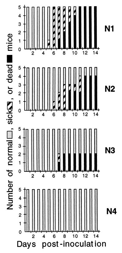
In contrast with these results, there were striking differences between the variant viruses in their LD50 values when administered IN (Table 1). Whereas the LD50 dose for the wild-type virus by IN administration was ≈10 pfu, the values for N2, N3, and N4 viruses were progressively greater. N2 required 20-fold more virus, N3 required 500-fold more virus, and N4 required 3,000-fold more virus than wild type, i.e., 30,000 pfu for the LD50. The time to onset of sickness (ruffled fur, lethargy, and hind limb paralysis) and extent of death increased progressively compared with wild type after infection with viruses N2, N3, and N4 (Fig. 6), and the extent of mortality was a function of dose (Table 1). These data show that when administered by a peripheral route, the progressive reduction in virus replication observed in cell culture correlated with a reduced lethality in mice.
Figure 6.
Relative lethality for mice. Pathogenesis of wild-type and rearranged viruses after IN inoculation into mice. Groups of mice inoculated with 300 pfu of each virus were monitored for time to onset of sickness and death.
Ability of Rearranged Viruses to Protect Against Wild-Type Challenge.
The observation that all of the viruses were lethal when inoculated IC indicated that even the most attenuated viruses were able to replicate in mice. This, coupled with the attenuation observed after IN administration, raised the possibility that the attenuated viruses might nevertheless be able to elicit a protective immune response. To test this possibility, mice were immunized by IN inoculation with serial 10-fold dilutions of the wild-type N1 or with variant viruses N2, N3, or N4. The surviving animals were challenged 14 days later by IN inoculation with 1.3 × 10⁶ pfu of wild-type virus. The percentage of animals surviving the challenge was a function of the immunizing dose in agreement with previous studies (24). For viruses N2, N3, and N4, 300 pfu/mouse was the lowest dose giving a 100% survival; 30 pfu yielded a 80–90% survival; 3–6 pfu gave a 45–85% survival; and doses below 3–6 pfu/mouse gave results that were not significantly different from those of age matched unimmunized controls (Fig. 7, dotted line in A). With the wild-type virus, the lethal dose and the protective dose were close, but in general, 80–85% of animals that survived administration of 3–6 pfu of virus were protected.
Figure 7.
Comparison of antibody production and ability to protect against lethal challenge. The ability to stimulate neutralizing antibody or to induce protection against a lethal wild-type challenge were compared for viruses N1-N4 as a function of dose of virus. Groups of mice were inoculated by IN instillation of serial dilutions of virus containing 3–6 pfu or 10-, 100-, or 1,000-fold more and analyzed for: (A) The ability of the IN inoculation to protect against a challenge with 1.6 × 10⁶ pfu of wild-type virus (an LD70 dose for 5- to 6-week old mice; dotted line in A shows lethality for unvaccinated controls) administered on day 14 postinoculation. Challenged animals were observed for 21 days for symptoms of sickness or death. The data from two independent experiments were averaged. (B) The presence of anti-VSV neutralizing antibody in serum at 14 days postinoculation prior to challenge. Neutralizing antibody titers are expressed as the reciprocal of the highest dilution, which gave 50% reduction in virus plaques.
Measurement of serum antibody prior to challenge on day 14 showed that despite attenuation for virulence in mice, the level of neutralizing antibody present in the serum of animals immunized with viruses N2, N3, and N4 was higher than that observed in the animals surviving inoculation of 3–6 pfu of wild-type virus and generally increased in a dose dependent manner (Fig. 7B). The lethality of the wild-type virus prevented direct comparison of antibody titers at higher doses, however, the neutralizing antibody titers in animals both vaccinated with viruses N1-N4 and then challenged with 1 × 10⁶ pfu of wild-type virus ranged from 1:625 to 1:3125. These data show that despite their attenuation for replication and lethality in animals, the N-rearranged viruses elicited a protective response that was undiminished compared with that of the wild-type virus.
DISCUSSION
The ability to introduce specific changes into the genome of a negative strand RNA virus allowed us to translocate the gene for the N protein to successive positions on the genome and demonstrate directly that the position of a gene relative to the promoter determined the level of expression. Previous work has shown that levels of N protein synthesis control the level of RNA replication (10, 11). Consistent with this work, we found that as the level of N mRNA synthesis and protein synthesis in cells infected with viruses N2, N3, and N4 was reduced, the level of genomic RNA replication also was reduced. Correspondingly, the production of infectious virus in cell culture was reduced in increments up to four orders of magnitude with virus N4 (Fig. 5). Finally, concomitant with reduced replication potential, the lethality of viruses N2, N3, and N4 for mice after IN inoculation was reduced by approximately one, two, or three orders of magnitude compared with the wild-type virus.
These data demonstrate that translocating a single gene essential for replication to successive positions located down the viral genome lowered the growth potential in cell culture and the lethality of the viruses for mice in a stepwise manner. However, the ability of the viruses to elicit a protective immune response in mice was not altered in correspondence with the reduction in virulence. We conclude that because the viruses all contained the wild-type complement of genes and all were competent to replicate, albeit at reduced levels, the level of replication was sufficient to induce a protective host response. For the wild-type virus, the protective dose and the lethal dose were similar, whereas they were separated by two to three orders of magnitude for viruses N3 and N4. Taken together, these data suggest a means of attenuating nonsegmented negative strand RNA viruses in a predictable, incremental manner that would allow one to determine an optimal level of attenuation to avoid disease production without loss of replication potential to induce a sufficient immune response. The work presented was carried out with VSV, which contains five genes. Other members of the Mononegavirales contain 6, 7, or 10 genes. Studies such as these will allow one to test for gene products required in precise amounts and exact molar ratios to support efficient replication and will examine whether limiting expression of a gene or genes essential for replication has similar effects on replication potential and virulence in cases where additional gene products may be involved in replication.
Because the Mononegavirales have not been observed to undergo homologous recombination (12), gene rearrangement is predicted to be irreversible, and for several reasons, it may provide a rational, alternative method for developing stably attenuated live vaccines against the nonsegmented negative strand RNA viruses. Live attenuated viruses have proven effective vaccines against many diseases, for example smallpox, yellow fever, measles, mumps, and poliomyelitis. However, the strategies for attenuation have been largely empirical. RNA virus genomes are notable for their high mutation rate, which is attributable to their lack of proof reading and error correction mechanisms. Most RNA viruses exist as complex quasispecies populations (reviewed in refs. 26 and 27), which has significant implications for their biology. The most obvious is that the viral population constitutes a huge reservoir of variants with potentially useful phenotypes in the face of environmental change. Advances in understanding the complex relationships that exist between the error rate of a viral polymerase, the viral population size, its passage history, and changes in the fitness landscape have revealed that empirical methods of attenuation may be intrinsically less reliable than previously thought (26, 28). Rearrangement of genes essential for replication utilizes the basic principle for control of gene expression, gene position, to effect changes in expression of genes requisite for replication and thus capitalizes on the lack of a natural means to reverse the rearrangement, i.e., lack of homologous recombination. This means of attenuation avoids the inherent potential of viruses attenuated by a limited number of single base mutations to revert to virulence through polymerase error and subsequent selection.
These experiments were carried out with VSV, considered the prototype negative strand RNA virus, to test the feasibility of the approach. However, an epidemic of VSV Indiana strain affecting not only cattle but also horses has occurred in the southwestern United States causing economic hardship, necessitating quarantines, and emphasizing the need for a vaccine. Studies to test the suitability and stability of this approach under suitable containment in the natural host are being organized. Based on the close similarity of the genome organization and mechanism for control of gene expression of the Mononegavirales, gene rearrangement provides a strategy to alter viral gene expression levels to investigate the role of gene products in replicative processes and, as shown by the work presented here, to systematically and incrementally attenuate virus replication and lethality. This approach to attenuation may prove applicable to some other members of this order and may provide a general approach for development of vaccines against numerous problematic viral pathogens.
Acknowledgments
We thank the members of our laboratories for helpful discussions and critical comments on the manuscript. This work was supported by Public Health Service grants from the National Institutes of Health, National Institute of Allergy and Infectious Diseases R37 AI 12464 (to G.W.W.) and R37 AI 18270 (to L.A.B.).
ABBREVIATIONS
- VSV
vesicular stomatitis virus
- pfu
plaque-forming unit
- IC
intracerebral
- IN
intranasal
- N
nucleocapsid protein
- P
phosphoprotein
- L
polymerase
References
- 1.Wagner R R. The Rhabdoviruses. New York: Plenum; 1987. [Google Scholar]
- 2.Pringle C R, Easton A J. Semin Virol. 1997;8:49–57. [Google Scholar]
- 3.Villareal L, Breindl M, Holland J. Biochemistry. 1976;15:1663–1667. doi: 10.1021/bi00653a012. [DOI] [PubMed] [Google Scholar]
- 4.Iverson L E, Rose J K. Cell. 1981;23:477–484. doi: 10.1016/0092-8674(81)90143-4. [DOI] [PubMed] [Google Scholar]
- 5.Ball L A, White C N. Proc Natl Acad Sci USA. 1976;73:442–446. doi: 10.1073/pnas.73.2.442. [DOI] [PMC free article] [PubMed] [Google Scholar]
- 6.Abraham G, Banerjee A K. Proc Natl Acad Sci USA. 1976;73:1504–1508. doi: 10.1073/pnas.73.5.1504. [DOI] [PMC free article] [PubMed] [Google Scholar]
- 7.Howard M, Wertz G W. J Gen Virol. 1989;70:2683–2694. doi: 10.1099/0022-1317-70-10-2683. [DOI] [PubMed] [Google Scholar]
- 8.Pattnaik A K, Wertz G W. J Virol. 1990;64:2948–2957. doi: 10.1128/jvi.64.6.2948-2957.1990. [DOI] [PMC free article] [PubMed] [Google Scholar]
- 9.Meier E, Harmison G G, Schubert M. J Virol. 1987;61:3133–3142. doi: 10.1128/jvi.61.10.3133-3142.1987. [DOI] [PMC free article] [PubMed] [Google Scholar]
- 10.Patton J T, Davis N L, Wertz G W. J Virol. 1984;49:303–309. doi: 10.1128/jvi.49.2.303-309.1984. [DOI] [PMC free article] [PubMed] [Google Scholar]
- 11.Arnheiter H, Davis N L, Wertz G, Schubert M, Lazzarini R A. Cell. 1985;41:259–267. doi: 10.1016/0092-8674(85)90079-0. [DOI] [PubMed] [Google Scholar]
- 12.Pringle C R. J Virol. 1981;39:377–389. doi: 10.1128/jvi.39.2.377-389.1981. [DOI] [PMC free article] [PubMed] [Google Scholar]
- 13.Pringle C R. In: The Genetics of Paramyxoviruses. Kingsbury D, editor. New York: Plenum; 1991. pp. 1–39. [Google Scholar]
- 14.Schnell M J, Mebatsion T, Conzelmann K K. EMBO J. 1994;13:4195–4203. doi: 10.1002/j.1460-2075.1994.tb06739.x. [DOI] [PMC free article] [PubMed] [Google Scholar]
- 15.Whelan S P J, Ball L A, Barr J N, Wertz G T W. Proc Natl Acad Sci USA. 1995;92:8388–8392. doi: 10.1073/pnas.92.18.8388. [DOI] [PMC free article] [PubMed] [Google Scholar]
- 16.Lawson N D, Stillman E A, Whitt M A, Rose J K. Proc Natl Acad Sci USA. 1995;92:4477–4481. doi: 10.1073/pnas.92.10.4477. [DOI] [PMC free article] [PubMed] [Google Scholar]
- 17.Conzelmann K-K. J Gen Virol. 1996;77:381–389. doi: 10.1099/0022-1317-77-3-381. [DOI] [PubMed] [Google Scholar]
- 18.Pattnaik A K, Ball L A, LeGrone A W, Wertz G W. Cell. 1992;69:1011–1020. doi: 10.1016/0092-8674(92)90619-n. [DOI] [PubMed] [Google Scholar]
- 19.Wertz G W, Whelan S, Legrone A, Ball L A. Proc Natl Acad Sci USA. 1994;91:8587–8591. doi: 10.1073/pnas.91.18.8587. [DOI] [PMC free article] [PubMed] [Google Scholar]
- 20.Reed E J, Muench H. Am J Hyg. 1938;27:493–497. [Google Scholar]
- 21.Barr J N, Whelan S P J, Wertz G W. J Virol. 1997;71:1794–1801. doi: 10.1128/jvi.71.3.1794-1801.1997. [DOI] [PMC free article] [PubMed] [Google Scholar]
- 22.Fuerst T R, Niles E G, Studier F W, Moss B. Proc Natl Acad Sci USA. 1986;83:8122–8126. doi: 10.1073/pnas.83.21.8122. [DOI] [PMC free article] [PubMed] [Google Scholar]
- 23.Sabin A, Olitsky P. J Exp Med. 1938;67:201–227. doi: 10.1084/jem.67.2.201. [DOI] [PMC free article] [PubMed] [Google Scholar]
- 24.Wagner R. Infection and Immunity. 1974;10:309–315. doi: 10.1128/iai.10.2.309-315.1974. [DOI] [PMC free article] [PubMed] [Google Scholar]
- 25.Youngner J S, Wertz G W. J Virol. 1968;2:1360–1361. doi: 10.1128/jvi.2.11.1360-1361.1968. [DOI] [PMC free article] [PubMed] [Google Scholar]
- 26.Holland J J, Torre J, Steinhauer D. Curr Top Microbiol Immunol. 1992;176:1–20. doi: 10.1007/978-3-642-77011-1_1. [DOI] [PubMed] [Google Scholar]
- 27.Domingo E, Escarmis C, Sevilla N, Moya A, Elena S, Quer J, Novella I, Holland J. FASEB J. 1996;10:859–864. doi: 10.1096/fasebj.10.8.8666162. [DOI] [PubMed] [Google Scholar]
- 28.Novella I, Clarke D, Quer J, Duarte E, Lee C, Weaver S, Elena S, Moya A, Domingo E, Holland J. J Virol. 1995;69:6805–6809. doi: 10.1128/jvi.69.11.6805-6809.1995. [DOI] [PMC free article] [PubMed] [Google Scholar]


