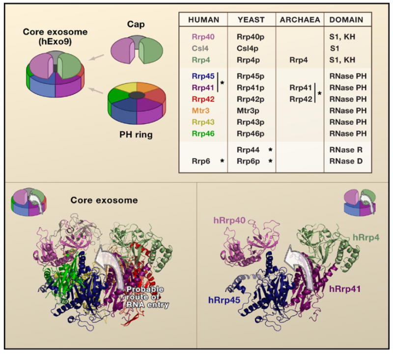
Figure 1. Structural Composition of the Exosome.

(Top) A schematic shows the nine subunit human exosome with the corresponding subunits in the table. The asterisk designates experimentally validated catalytic subunits. The human subunits are color coded. (Bottom) The structure of the exosome reveals the cleft between hRrp40 and hRrp4 within the context of the hExo9 complex crystal structure and schematic (left) and within the isolated subunits hRrp41, hRrp45, hRrp4, and hRrp40 (right). The arrow denotes the putative path of an RNA extending into the hRrp41/hRrp45 heterodimer through the cleft.
