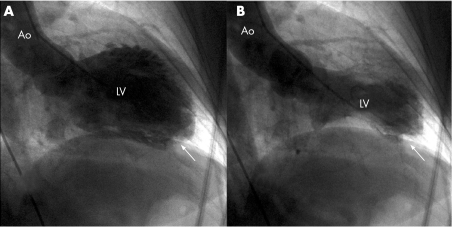
A 74‐year‐old woman who had been experiencing mental stress was admitted to our institution with a history of continuous atypical chest pain. The evolutive ECG showed T wave inversion in leads V2–V6, I AVL and III, with a prolonged QT interval. Mild enzymatic changes were found in blood chemistry examinations. Coronary angiography showed no significant stenosis, but left ventriculography demonstrated apical asynergy with basal hyperkinesia (apical ballooning). A striking filling defect highly suggestive of a thrombus was also viewed at the apex (panel A, diastole; panel B, systole; and video 1, white arrows; to see video footage visit the Heart website—http://heart.bmj.com/supplemental). Left ventricular ejection fraction was 40%. The apical ballooning and intraventricular thrombus were confirmed by transthoracic echocardiography. The patient was discharged under anticoagulant treatment.
After 2 months the ECG showed normal findings, and a transthoracic echocardiogram showed absolutely normal left ventricular (LV) wall motion and complete resolution of the apical thrombus.
Direct evidence of a LV thrombus associated with takotsubo‐like ventricular dysfunction has not been demonstrated, although there have been reports regarding the embolic complications of this disorder. Some articles reported that the akinetic LV wall in the setting of myocardial infarction is an important cause of LV thrombus. Given that the LV thrombus in the present case was caused by a wall motion abnormality, its clinical appearance seems rather late. We report a case of transient LV apical ballooning with LV thrombus demonstrated in the early angiographic procedure. With restoration of LV apical wall motion and warfarin treatment, the LV thrombus disappeared. LV thrombus should be considered an early and delayed complication of transient LV apical ballooning.
To view video footage visit the Heart website—http://heart.bmj.com/supplemental
Copyright © 2007 BMJ Publishing Group and British Cardiovascular Society.
End diastolic (panel A) and end systolic (panel B) ventriculograms of the patient, showing aquinesia of apical segments of the left ventricle (LV) and hypercontraction of basal and mid segments. A striking filling defect highly suggestive of a thrombus was also viewed at the apex (white arrow). Ao, aorta.
Supplementary Material
Associated Data
This section collects any data citations, data availability statements, or supplementary materials included in this article.


