Abstract
Botulinum neurotoxin (BoNT), the most poisonous substance known, causes naturally occurring human disease (botulism) and is one of the top six biothreat agents. Botulism is treated with polyclonal antibodies produced in horses which are associated with a high incidence of systemic reactions. Human monoclonal antibodies (mAbs) are under development as a safer therapy. Identifying neutralizing epitopes on BoNTs is an important step in generating neutralizing mAbs, and also has implications for vaccine development. Here we show that the three domains of BoNT serotype A (BoNT/A) can be displayed on the surface of yeast and used to epitope map six mAbs to the toxin domains they bind. The use of yeast obviates the need to express and purify each domain, and it should prove possible to display domains of other BoNT subtypes and serotypes for epitope mapping. Using a library of yeast displayed BoNT/A binding domain (HC) mutants and selecting for loss of binding, the fine epitopes of three neutralizing BoNT/A mAbs were identified. Two mAbs bind the C-terminal subdomain of HC, with one binding near the toxin sialoganglioside binding site. The most potently neutralizing mAb binds the N-terminal subdomain of HC, in an area not previously thought to be functionally important. Modeling the epitopes shows how all three mAbs could bind BoNT/A simultaneously and may partially explain the dramatic synergy observed on in vivo toxin neutralization when these antibodies are combined. The results demonstrate how yeast display can be used for domain-level and fine mapping of conformational BoNT antibody epitopes and the mapping results identify three neutralizing BoNT/A epitopes.
Keywords: epitope mapping, yeast surface display, botulinum neurotoxin, affinity, alanine scanning mutagenesis
Introduction
Botulinum neurotoxin (BoNT) is secreted by the spore-forming bacteria Clostridium botulinum and is the most poisonous substance known 1. The crystal structure of BoNT serotype A (BoNT/A) 2 shows three functional domains comprising a heavy and a light chain 2-4. The C-terminal portion of the heavy chain (HC) is the binding domain which docks the toxin to sialoganglioside receptors and a protein receptor on presynaptic neurons, resulting in toxin endocytosis 5-7. The translocation domain (HN), at the N-terminal portion of the heavy chain, mediates escape of the toxin light chain (LC) from the endosome. Depending on serotype, the LC cleaves one or more members of the SNARE complex of proteins, blocking acetylcholine release 8, 9.
Human botulism is caused by BoNT serotypes A, B, E, and F and is characterized by flaccid paralysis which, if not fatal, requires prolonged hospitalization in an intensive care unit and mechanical ventilation. Naturally occurring botulism results from ingestion of contaminated food, anaerobic wound infections, or gastrointestinal tract colonization by Clostridial bacteria 10. Botulinum neurotoxins are also classified by the Centers for Disease Control (CDC) as one of the six highest-risk threat agents for bioterrorism (the “Class A agents”) due to their extreme potency and lethality 11. Both Iraq and the former Soviet Union produced BoNT for use as weapons 12, 13 and the Japanese cult Aum Shinrikyo attempted to use BoNT for bioterrorism 11. Consequently, specific pharmaceutical agents are needed for treatment of intoxication.
Treatment of botulism in adults relies on the use of antitoxin 14, currently generated from immunized horses 15. This product is associated with a high incidence of side effects, including serum sickness and anaphylactic shock. As an alternative, monoclonal antibody (mAb) based antitoxins are under development 16, 17. Nowakowski and coworkers reported the generation of three mAbs, S25, C25, and 3D12, that neutralized BoNT/A both in vitro and in vivo 17. While in vivo neutralization for single mAbs was of relatively low potency, combining any two or all three mAbs led to highly potent neutralization of BoNT/A. Higher affinity derivatives of these three mAbs are now in cGMP production for anticipated toxicology studies and human clinical trials (http://www3.niaid.nih.gov/news/newsreleases/2005/27million_bioshield.htm).
We have been interested in mapping the epitopes of these and other BoNT/A mAbs. Such mapping can lead to an improved understanding of mechanism(s) of toxin neutralization, as well as shed light on the relationship between toxin structure and function. For example, putative sialoganglioside binding sites on toxin have been identified using X-ray crystallography 18, 19. Are these sites where neutralizing mAbs bind? Similarly, the BoNT/A docking site for the protein receptor remains unknown; identification of the epitopes for potently neutralizing BoNT/A mAbs might identify potential protein receptor binding sites on the BoNT/A HC. Finally, marked synergy in toxin neutralization has been observed when mAbs are combined 17. Identifying the sites and interactions between toxin and neutralizing mAbs could provide a structural model of the immune complexes and perhaps provide insights into mechanism of neutralization.
For this work, yeast display 20, 21 was utilized to successfully display all three BoNT/A domains, which could be used to map a panel of 6 mAbs to the domain level. This approach should prove useful for gross mapping of both polyclonal and monoclonal antibodies to BoNT/A and can probably be adapted for domains of other BoNT/A subtypes or other BoNT serotypes. This approach has an advantage of not requiring native protein expression and purification. Using yeast displayed libraries, the fine epitopes of neutralizing mAbs 3D12, HuC25, and S25 were identified down to the energetically important amino acid side chains. The results identify a number of neutralizing epitopes on BoNT/A and suggest mechanisms of toxin neutralization.
Results
Domain epitope mapping of BoNT/A neutralizing antibodies
To display the three functional domains of botulinum neurotoxin type A (BoNT/A HC, HN, and LC) on the surface of yeast, we used PCR to amplify the relevant gene from plasmids containing either a synthetic HC gene, or a synthetic LC-HN gene (Figure 1). PCR fragments encoding the HC, LC, HN, and LC-HN were then ligated into the yeast display vector pYD2 22 in frame with an SV5 epitope tag. This allowed for C-terminal fusion of the SV5 tag and a means to quantitatively measure the level of display on the yeast surface using a labeled SV5 mAb. Since Lacy and coworkers 2 previously described the presence of a ~50-residue belt (loop) region in the translocation domain that wraps around the LC domain of BoNT/A, we designed an HN variant which lacks the gene fragment that encodes for the belt region and also ligated this into pYD2. Ligation products were used to transform E. coli, clones containing the correct insert identified by DNA sequencing, and DNA prepared and used to transform S. cervesiae.
Figure 1. Yeast surface display of BoNT/A domains.
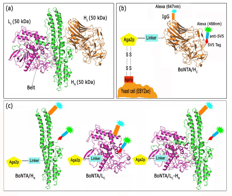
(a) Ribbon cartoon of the X-ray crystal structure of BoNT/A secondary structure (PDB accession name, 3BTA) showing catalytic light chain (LC, magenta), binding (HC, orange) and translocation (HN, green) domains 2. The belt region of HN, which wraps around the LC, is indicated. (b) Schematic representation of yeast displayed HC, fused to the yeast protein Aga2p via a flexible (Gly4Ser)3 linker. The level of yeast display is quantitated via direct or indirect (not shown) fluorescent labeling of an SV5 epitope-tag binding monoclonal antibody. A fluorescent labeled BoNT/A-specific IgG is used to independently verify display. (c) Yeast display of BoNT/A HN, BoNT/A LC, and BoNT/A LC-HN. SV5 epitope tag and domain specific mAbs are shown bound to their respective domains.
To demonstrate successful display of each of the BoNT/A domains, surface display was induced from each of the constructs and yeast co-stained with SV5 mAb labeled with Alexa-488 and BoNT/A specific mAbs labeled with Alexa-647. All five constructs (HC, HN with belt, HN without belt, LC, and HN-LC) were successfully displayed on the yeast surface as indicated by staining with SV5-488 (Figure 2a–e). To determine whether the displayed BoNT/A domains could be used to map monoclonal antibodies (mAbs), we studied a panel of BoNT/A mAbs previously shown to bind different toxin domains (Table 1). Both HC-specific mAbs 3D12 23 and HuC25 22 bound surface displayed HC, but not HN or LC (Figure 2a). In contrast, the HN specific mAb ING1 and the LC-specific mAb ING2 did not bind surface displayed HC (Figure 2a). Similarly, the LC-specific mAb ING2 bound surface displayed LC but did not bind HC or HN (Figure 2b). Surface displayed LC was not bound by HC or HN mAbs (Figure 2b). Yeast surface displayed HN domains (with or without the belt) were recognized by the HN specific murine 9D8 mAb but were not recognized by HC or LC mAbs (Figure 2c, d). Surface displayed LC-HN was bound by both LC (ING2) and HN (9D8) mAbs, but not by HC specific mAbs (Figure 2e). Finally, an interesting mAb (ING1) only bound surface displayed LC-HN, but did not bind displayed LC or HN. We had previously determined that this mAb bound recombinant LC-HN, but not LC, and had assumed that it recognized an HN epitope. The current results indicate that this mAb recognizes a complex epitope formed by the packing of the LC and HN, perhaps where the belt wraps around the LC.
Figure 2. Yeast display of BoNT/A domains and mapping of BoNT/A monoclonal antibodies by flow cytometry.
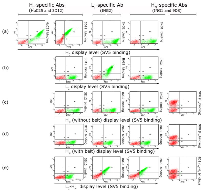
Specific binding to (a) yeast-displayed HC by human mAbs HuC25 and 3D12; (b) yeast-displayed LC by human mAb ING2; (c) and (d) yeast-displayed HN (with (c) or without (d) the belt region) by murine mAb 9D8, and (e) yeast-displayed LC-HN by mAbs ING2, 9D8 and ING1. All domains are well displayed as indicated by a greater than 2 log shift with anti-SV5. All domains are bound only by domain specific mAbs, with no binding of mAbs specific to other domains.
Table 1.
Monoclonal antibodies mapped using surface displayed BoNT/A domains.
| Antibody | BoNT/A KD (M) | Domain specificity | In vitro neutralization (fold over control) |
|---|---|---|---|
| HuC25 | 4.5 × 10⁻¹¹ | HC | 6.82* |
| 3D12 | 6.1 × 10⁻¹¹ | HC | 3.02 |
| S25 | 1.7 × 10⁻⁹ | HC | 1.85 |
| 9D8 | 1.2 × 10⁻⁹ | HN | ND |
| ING2 | 9.6 × 10⁻¹² | LC | ND |
| ING1 | 3.1 × 10⁻¹⁰ | LC-HN | ND |
Solution KD of HuC25, 3D12, ING1, and ING2 were measured in a flow fluorimeter (KinExA) (ref. 22 and unpublished data). KD of S25 and 9D8 were measured using surface plasmon resonance (BIAcore) 25. In vitro neutralization was determined as time to 50% neuroparalysis in the mouse hemidiaphragm assay and is expressed as fold over control 17.
In vitro neutralization data for C25, murine precursor of humanized C25, HuC25.
Fine epitope mapping of BoNT/A HC-specific neutralizing monoclonal antibodies
To identify neutralizing epitopes on BoNT/A, we epitope mapped three neutralizing BoNT/A HC mAbs (Table 1) down to the level of the energetically critical amino acid contacts between toxin and mAb. All three mAbs were initially single chain Fv (scFv) antibodies isolated from phage display libraries generated from immunized humans (3D12) or mice (S25 and C25) 16, 24. The C25 antibody was subsequently humanized (HuC25), resulting in an antibody with a 2-fold higher affinity for BoNT/A than C25. After conversion to IgG with human Fc, all three mAbs showed in vitro BoNT/A neutralization, as evidenced by a prolongation in the time to paralysis in the hemidiaphragm assay, and in vivo neutralization, as evidenced by a prolongation in time to death in mice challenged with toxin (Table 1, and ref. 17). For two of the mAbs, 3D12 and HuC25, fine epitope mapping was accomplished using an approach first described by Chao et al. 21 by: 1) generating a library of surface displayed BoNT/A HC mutants; 2) selecting for loss of binding of the mAb being epitope mapped; 3) analyzing the location of mutations leading to loss of mAb binding; and 4) constructing single yeast displayed BoNT/A HC alanine mutants in the putative mAb epitope and measuring the energetic contribution of the toxin side chain to mAb binding (ΔΔG). For the third mAb, S25, we took advantage of the fact that this mAb bound BoNT/A1 with high affinity but bound the BoNT/A2 subtype with much lower affinity 25. Combining this fact with the knowledge that S25 bound the C-terminal subdomain of HC, the HCC of BoNT/A, a number of BoNT/A HCC mutants were constructed substituting BoNT/A2 residues. The S25 epitope was identified based on loss of binding to a subset of the HCC mutants.
Construction of a randomly mutated BoNT/A HC library in yeast
To generate a library of BoNT/A HC mutants, we utilized error prone PCR and amplification conditions known to introduce a high rate of mutations. Briefly, a high rate of mutations (~1.1% error-rate) was introduced into the BoNT/A HC gene by amplification from the plasmid pYD2/HC, using error prone PCR and conditions previously described by Fromant and coworkers 26. The resulting gene repertoire was cloned into the yeast display vector pYD2 using gap repair homologous recombination to create a yeast library of ~ 3×10⁵ transformants with an insert of the appropriate size, as determined by PCR screening. The library was grown, BoNT/A HC epitope display induced and the library analyzed for display level by staining with SV5-Alexa-488, and mAb binding by costaining with 3D12-Alexa-647 and AR2-Alexa-647. AR2 is a higher affinity derivative of the HuC25 scFv 22. A relatively low frequency of display (approximately 0.5%) and antigen binding (approximately 0.4 %) was observed, probably as a result of the high mutation rate (Figure 3).
Figure 3. Selection of yeast-displayed BoNT/A HC with mutations that knocked out mAb binding.
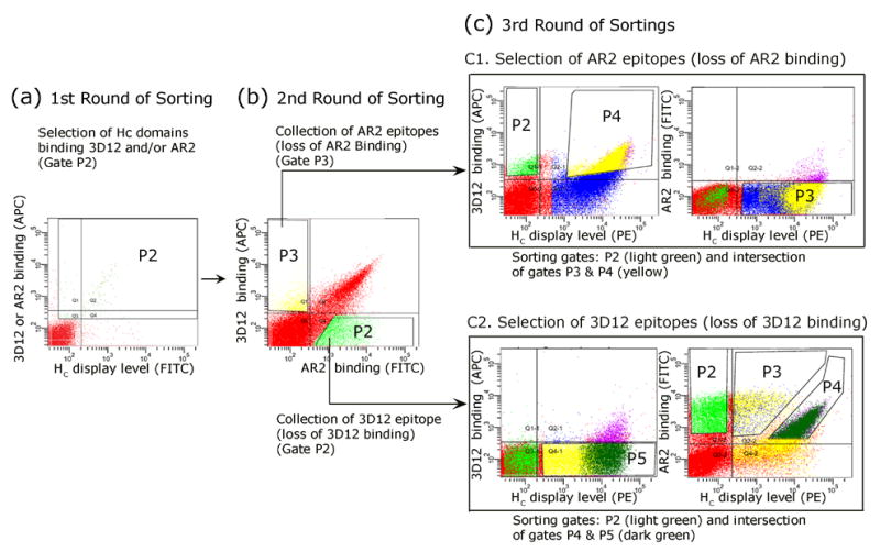
BoNT/A HC clones with mutations knocking out binding to mAb 3D12 or mAb AR2 were selected from a library of yeast displayed BoNT/A HC mutants using FACS. The first round of sorting (a) was designed to merely enrich for yeast displaying BoNT/A HC capable of binding 3D12 and/or AR2 in preparation for subsequent rounds of sorting. This was accomplished by simultaneously staining yeast with mAbs 3D12 and AR2 labeled with Alexa-647 and SV5-Alexa-488. Yeast displayed BoNT/A HC that were bound by mAbs 3D12 and/or AR2 (APC channel) were gated for selection (gate P2), regardless of display level (SV5-Alexa-488 (FITC) staining). The second round of sorting (b) was designed to isolate yeast displayed BoNT/A HC that contained mutations which resulted in loss of binding to one of the two mAbs but which retained binding to the other BoNT/A mAb. Requiring binding to the second BoNT/A mAb helped ensure that the selected domains were properly folded. This was accomplished by staining yeast with 3D12-Alexa-647 and AR2-Alexa-488. Yeast that lost binding to AR2 (gate P3), or that had lost binding to 3D12 (gate P2) were separately gated and sorted. The final round of sorting (c) was designed to complete the isolation of yeast displayed BoNT/A HC which had mutations that resulted in the loss of binding to one of the two BoNT/A mAbs but which retained binding to the other BoNT/A mAb. For the final round of sorting, yeast from second round gates P2 and P3 were stained in separate reactions with 3D12-Alexa-647 and AR2-Alexa-488 mAbs and anti-SV5-epitope tag mAb followed by anti-mouse PE. For sorting of yeast from the second round gate P3 selected for loss of binding to AR2, yeast without binding to AR2 (gate P3, blue dots) but with retained binding to 3D12 (gate P4, purple dots) were selected by intersecting the P3 and P4 gates (yellow dots) and collecting only those yeast in both gates (c, top panels). Yeast with the same phenotype, but without SV5 binding (truncation mutants, gate P2, green dots) were also sorted. For the final round of sorting of yeast from the second round gate P2 selected for loss of binding to 3D12, yeast without binding to 3D12 (gate P5, yellow dots) but with retained binding to AR2 (gate P4, purple dots) were selected by intersecting the P5 and P4 gates (dark green dots) and collecting only those yeast in both gates (c, bottom panels). Yeast with the same phenotype, but without SV5 binding (truncation mutants, gate P2, green dots) were also sorted.
Isolation of BoNT/A1 HC clones with selective loss of binding to 3D12 or HuC25
To identify the epitopes recognized by 3D12 and HuC25, the epitope library was subjected to serial rounds of flow sorting. For the first round of sorting, HC epitopes binding 3D12 and/or AR2 antibodies were recovered. This was accomplished by staining yeast with 3D12-Alexa-647, AR2-Alexa-647, and SV5-Alexa-488. Binding of 3D12 and AR2 mAbs to displayed HC was detected using the APC (647nm) channel and binding of SV5 was detected using the FITC (488nm) channel. At this round, all Alexa-647 positive yeast were gated and sorted (Figure 3a). Co-staining with both 3D12 and AR2 labeled yeast functionally displaying either, or both, epitopes. The gate was set to include yeast with low to absent levels of SV5 display (SV5 binding), as these might represent clones with stop codons that contained the mAb epitope. Such clones would prove useful for narrowing the potential epitope space (see below). The library sort output was then grown, induced and subjected to a second round of selection that resulted in the recovery of only 3D12 (Alexa-647, gate P3) or AR2 (Alexa-488, gate P2) binding yeast clones (Figure 3b). This was accomplished by co-labeling the first round output with 3D12-Alexa-647 and AR2-Alexa-488. Two separate sort gates were set, one for Alexa-647 positive yeast and one for Alexa-488 positive yeast, and yeast collected separately for the two gates. Presumably at this round, loss of binding of one of the mAbs resulted from the presence of BoNT/A HC mutations that resulted in minimal binding affinity for that mAb. Retained binding of the second mAb was utilized to ensure that the selected mutants were properly folded. Yeast from each sort gate were separately grown, induced and used for the next round of sorting.
For the final round of sorting, yeast from each of the previous two gates were stained in separate reactions with 3D12-Alexa-647, AR2-Alexa-488 and unlabeled SV5 mAb followed by anti-mouse-PE, to allow separate quantitation of the display level. For selection of well displayed epitopes bound only by mAb 3D12 (putative AR2/HuC25 epitopes), sort gates were set to include the 3D12-Alexa-647 positive population (APC positive) (sort gate P4, top panel, Figure 3c) and the AR2-Alexa-488 negative population (FITC negative) (sort gate P3, top panel, Figure 3c). These gates were set to include only the PE positive population, representing those yeast with positive SV5 binding (well displayed BoNT/A HC). The P3 and P4 gates were intersected and only yeast in both gates collected. A separate sort gate (P2, top panel, Figure 3c) was set to collect potential poorly expressed AR2/HuC25 epitopes bound by 3D12-Alexa-647 (APC positive and PE negative). Since this population was not bound by AR2-Alexa-488 (top left panel, Figure 3c), this gate was directly collected without the need for intersection. For selection of well displayed epitopes bound only by mAb AR2 (putative 3D12 epitopes), sort gates were set to include the AR2-Alexa-488 positive population (FITC positive) (sort gate P4, bottom panel, Figure 3c) and the 3D12-Alexa-647 negative population (APC negative) (sort gate P5, bottom left panel, Figure 3c). These gates were set to include only the PE positive population, representing those yeast with positive SV5 binding (well displayed BoNT/A HC). The P4 and P5 gates were intersected and only yeast in both gates collected. A separate sort gate (P2, bottom panel, Figure 3c) was set to collect potential poorly expressed 3D12 epitopes bound by AR2-Alexa-488 (APC positive and PE negative). Since this population was not bound by 3D12-Alexa-647 (bottom panel, Figure 3c), this gate was directly collected without the need for intersection.
Identification of the 3D12 and HuC25 epitopes
To identify the 3D12 and HuC25 epitopes, 50 individual yeast clones from the sort output of the last round of selections by flow cytometry were grown in liquid culture and BoNT/A HC display induced. Induced yeast cells were then incubated with HuC25 or 3D12 mAbs and binding quantitated by flow cytometry following incubation with APC-conjugated goat-anti-human F(AB)′2 and anti-SV5 (Alexa-488). The HC gene fragments from clones that were able to bind exclusively to 3D12 or HuC25 were amplified by PCR and the HC gene sequenced. DNA sequencing revealed multiple different HC mutants from each sorting, with each mutant containing between 3 and 15 different mutations. Molecular modeling of the locations of mutations on the X-ray crystal structure of BoNT/A enabled the selection of individual HC amino acid residues that could potentially form the 3D12 (mutations from clones that could only recognize HuC25) or the HuC25 (mutations from clones that could only recognize 3D12) functional epitopes. Of great assistance in identification of the HuC25 and 3D12 epitopes were clones sequenced from the sorts where there was no SV5 tag expression. A number of different mutants containing stop codons were identified which bound HuC25, but did not bind 3D12 or anti-SV5. The most C-terminal of these was at amino acid position 1101, demonstrating that the 3D12 epitope was distal to this amino acid in the HCC subdomain of BoNT/A HC. Conversely, since HuC25 bound the clone with the stop codon at this position, the HuC25 epitope must be proximal to this amino acid, in the HCN subdomain of the BoNT/A HC. To confirm this assumption, yeast displaying either the HCC (amino acid residues 1093 to 1296) or the HCN subdomain (amino acid residues 876 to 1092) were generated by PCR amplification from pYD2/HC. Induced yeast displaying HCN bound HuC25 but not 3D12 and yeast displaying HCC bound 3D12 but not HuC25, confirming the epitope location based on sequencing. Location of the epitopes to the subdomains allowed elimination of approximately half of the mutations from consideration.
Analysis of the location of mutations from selected clones resulted in the identification of a single loop of the HCC (residues 1127 to 1131, Table 2), which had substitutions in all clones sequenced that had lost binding to 3D12. To confirm this site as the functional epitope for 3D12, six single alanine mutants were made for each of these residues. Truncation of the amino acid side chain to alanine allows quantitation of the energetic contribution of that residue to binding 27, 28. Alanine substitutions were generated by a) PCR amplification of HC using primers that introduced the alanine mutations, and b) homologous recombination of the resulting PCR products by gap repair to yield the final pYD2/HC (alanine substituted) plasmids in yeast (Figure 4). 3D12 FAB was used to determine the impact of alanine mutations on antibody binding affinity and energetics to avoid bivalent binding of the two FAB heads of the IgG to the multiple copies of yeast displayed HC, which could lead to an avidity effect and an overestimation of the binding constant 29. The relative binding of the yeast displayed HC alanine mutants to KD concentrations of 3D12 or HuC25 FAB was analyzed by flow cytometry using APC-FAB-specific antisera and SV5-Alexa-488 labeled antibodies (Table 2 and Figure 5). Three mutations (G1129A, I1130A and R1131A) virtually eliminated binding of 3D12 FAB while binding of HuC25 FAB was unchanged (Table 2, Figure 5b). Confirming the relative binding results, the KD for binding of 3D12 FAB to G1129A, I1130A and R1131A was 7.3 nM to 18 μM, a 45 to more than 100,000 fold reduction in affinity compared to the wildtype affinity of 3D12 for BoNT/A HC (Table 3). Modeling of the location of the amino acids in the functional epitope of 3D12 on the BoNT/A crystal structure shows that 3D12 binds at the periphery of the BoNT/A HCC (Figure 6b). The 3D12 functional epitope spans a single loop with a compact and well defined lobe which protrudes outwards from the HC surface, the centermost residue of which is R1131. While this epitope may appear linear, binding of 3D12 FAB to yeast displayed BoNT/A HC could not be competed with a 19mer peptide epitope (data not shown).
Table 2.
Binding of BoNT/A HC alanine mutants to HuC25 or 3D12 FAB fragments.
| BoNT/A HC mutant | HuC25 FAB binding | BoNT/A HC mutant | 3D12 FAB binding |
|---|---|---|---|
| S896A | ++++ | N1126A | ++++ |
| K897A | ++++ | N1127A | ++++ |
| N899A | ++++ | V1128A | ++++ |
| I900A | ++++ | G1129A | − |
| G901A | +++ | I1130A | − |
| S902A | ++++ | R1131A | − |
| K903A | +++ | K1187A | ++++ |
| N905A | ++++ | ||
| Q915A | ++++ | ||
| F917A | ++++ | ||
| N918A | ++ | ||
| L919A | ++ | ||
| E920A | − | ||
| S921A | ++++ | ||
| E925A | ++++ | ||
| I927A | ++++ | ||
| K929A | ++++ | ||
| N930A | ++++ | ||
| F953A | − | ||
| N954A | ++++ | ||
| I956A | ++++ | ||
| C1060A | ++++ | ||
| R1061A | + | ||
| D1062A | + | ||
| T1063A | + | ||
| H1064A | − | ||
| Y1066A | ++ | ||
| *HCN (876-1092) | ++++ |
binding of Ala mutant similar to wild-type; −, complete loss of binding;
Deletion mutant of HC, lacking the C-terminus portion (amino acids 1093–1296) of the domain.
Figure 4. Generation of single alanine HC mutants by yeast gap repair.
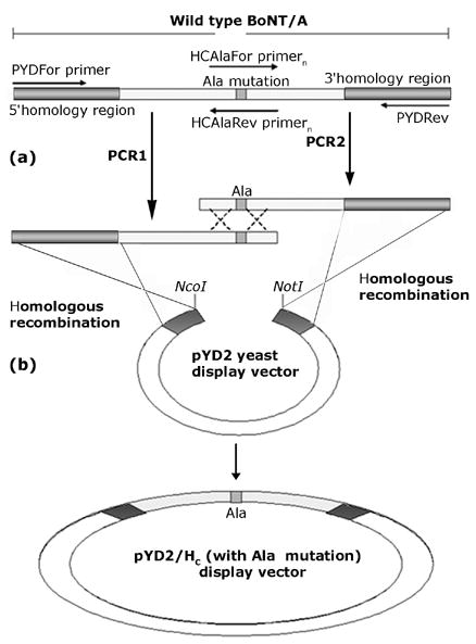
(a) The 5′ and 3′ ends of the HC gene were PCR amplified from wild type HC, replacing an HC amino acid residue with an alanine. Outside primers PYDFor, PYDRev and inside alanine-introducing sets of primers HCAlaForn and HCAlaRevn were used for PCR and introduction of the alanine mutation. (b) The PCR amplification products were then cotransformed with the NcoI-NotI digested display vector pYD2 into yeast to form the desired HC single alanine mutants in pYD2 by gap repair homologous recombination.
Figure 5. Binding of HuC25 and 3D12 FAB fragments to yeast displayed BoNT/A HC alanine mutants.
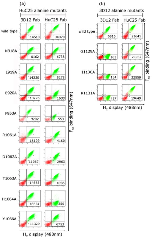
Antibody binding to yeast displayed HC was quantitated on a flow cytometer by co-staining with anti-SV5 (Alexa-488) and FABs HuC25 and 3D12 followed by FAB-specific-APC (647 nm) labeled antisera. Mean fluorescence intensity (MFI) values for FAB binding are shown. Comparison of the MFI for wildtype binding and mutant binding of HuC25 vs 3D12 FAB identified alanine mutations with reduced affinity.
Table 3.
Affinities and ΔΔG of alanine-substituted BoNT/A HC mutants.
| BoNT/A HC mutant | HuC25 FABKD (M) | ΔΔGmut-wt kcal/mol |
|---|---|---|
| wt | 1.07 ×10⁻¹⁰ | - |
| N918A | 6.70 ×10⁻¹⁰ | 0.9 |
| L919A | 7.44 ×10⁻¹⁰ | 1 |
| E920A | 1.31 ×10⁻⁹ | 1.3 |
| F953A | 2.15 ×10⁻⁸ | 3 |
| R1061A | 7.51 ×10⁻¹⁰ | 1 |
| D1062A | 6.06 ×10⁻¹⁰ | 0.9 |
| T1063A | 6.42 ×10⁻¹⁰ | 0.9 |
| H1064A | 1.35 ×10⁻⁵ | 6.8 |
| Y1066A | 4.27 ×10⁻¹⁰ | 0.7 |
|
| ||
| BoNT/A HC mutant | 3D12 FAB KD (M) | ΔΔGmut-wt kcal/mol |
|
| ||
| Wt | 1.75 ×10⁻¹⁰ | - |
| G1129A | 1.84 ×10⁻⁵ | 6.8 |
| I1130A | 7.35 ×10⁻⁹ | 2.2 |
| R1131A | 1.00 ×10⁻⁵ | 6.5 |
The dissociation equilibrium constant (KD) for HuC25 (or 3D12) FAB was calculated for each alanine mutant. The difference in binding free energy (ΔΔGala-wt) between the alanine-substituted and wild type (wt) HC was calculated according to the formula ΔΔG=RTln(KD,Ala/KD,wt) (Materials and Methods). Mutants in bold have ΔΔGmut-wt ≥1.
Figure 6. Model of the functional binding epitopes of neutralizing BoNT/A mAbs.
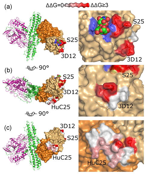
The epitopes of S25 (top panels), 3D12 (middle panels) and HuC25 (bottom panels) are shown, with the ΔΔG of important toxin side chain interactions colored red-white as indicated. The putative sialoganglioside contacting residues on HCC are colored blue, and the sialoganglioside is shown modeled as spheres, near the S25 epitope. HCC, space filling light orange; HCN, space filling dark orange, HN, green, LC, magenta. Molecular models were constructed using Pymol software (DeLano Scientific, LLC), using the coordinates of BoNT/A (3BTA) from the Protein Data Bank.
Identification of the HuC25 epitope was less straightforward. Analysis of the location of mutations reducing or eliminating binding to HuC25 suggested 4 different loops for evaluation (β25, β27, and β35, and β 36) 30. As a result, a much larger number of alanine mutants (27) were generated and analyzed compared to the 3D12 epitope (Table 2 and Figure 5a). Nine of the mutants (N918A, L919A, E920A, F953A, R1061A, D1062A, T1063A, H1064A, Y1066A) resulted in a significant reduction in binding of HC to HuC25 revealing that the epitope spanned four loops of the HC (β25, β27, β35, and β36 (Tables 2 and 3 and Figure 5a)). The most energetically important residues were F953 and H1064 (Table 3). Modeling of the location of the amino acids in the functional epitope of HuC25 on the BoNT/A crystal structure shows that HuC25 binds to the BoNT/A HCN, near the interface with HCC (Figure 6c). Unlike the 3D12 epitope, the HuC25 functional epitope is more discontinuous, spans a larger surface area on HC, and includes amino acid residues located in four distinct turns (N918, L919, E920 in one turn, F953 in a second turn, R1061, D1062, T1063, H1064 in a third turn and Y1066 in a fourth turn).
Identifying the epitope of S25
To identify the epitope of S25, we first measured binding to BoNT/A HCN and HCC and determined that S25 bound HCC. We then took advantage of the fact that S25 bound BoNT/A1 with high affinity but barely bound the BoNT/A2 subtype 25. Amino acid differences between BoNT/A1 and A2 HCC were identified and four yeast displayed BoNT/A mutants constructed with BoNT/A2 substitutions inserted as residues 1117, 1254-56, 1271-74, or 1294-95. Only the mutant 1254-56 showed loss of binding to S25, while retaining wildtype binding to 3D12. Modeling of the location of these amino acids on the BoNT/A crystal structure shows that S25 binds to the BoNT/A HCC very near the putative sialoganglioside binding site (Figure 6a) 31.
Modeling the interactions of C25, 3D12, and S25 with BoNT/A
We previously showed that a combination of C25, 3D12, and S25 IgG neutralized BoNT/A in vivo with markedly greater potency than either the single mAbs or mAb pairs 17. Molecular modeling based on the mAb epitopes indicates that all three mAbs can bind BoNT/A simultaneously confirming biophysical studies (Figure 7). The two mAbs which bind the BoNT/A HCC overlap the putative sialoganglioside binding site and cover a large portion of HCC, the domain thought to be more important for interaction with cellular receptors 31-33. C25 (HuC25) binds the HCN, near the interface with HCC. Recent data suggests that antibodies to regions around this interface are important for toxin neutralization 32. C25 is the most potently neutralizing mAb described 17, its epitope is near the HCN-HCC interface, and its epitope could represent a putative binding site for the recently identified BoNT/A protein receptors SV2 5, 7. The model of multiple mAb binding illustrates how a large portion of the toxin binding domain is occupied in regions known to be functionally important. This surface blockade, combined with a known synergistic effect on affinity of binding 17, may partially explain how these mAbs synergize to neutralize BoNT/A.
Figure 7. Model of the binding of three neutralizing mAbs to BoNT/A.
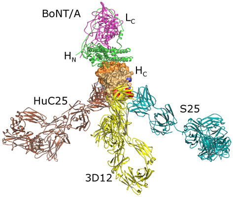
The binding of S25, 3D12, and HuC25 IgG to their epitopes on BoNT/A were modeled using the coordinates of BoNT/A (3BTA) and three copies of the coordinates of an arbitrary human IgG (1HZH) using Pymol software (DeLano Scientific, LLC). The model indicates that all three mAbs could bind simultaneously, creating a large tripod shaped immune complex.
Discussion
The three functional domains of BoNT/A were successfully displayed on the surface of yeast allowing the domain epitope mapping of a panel of six monoclonal antibodies. It was also possible to display a pair of domains (HN-LC, 100kDa), the largest protein displayed on the surface of yeast 21. At least three of these mAbs (HuC25, 3D12, and S25) are known to bind conformational epitopes on the HC 23. One of the BoNT/A mAbs studied (ING1) bound HN-LC, but did not bind HN or LC. A potential epitope for this mAb would be the belt as it wraps around the LC or a conformational epitope at the interface of the HN-LC. These results suggest that the domains are not only displayed, but properly folded. The affinities measured for mAb binding to yeast displayed domains were nearly identical to the affinities measured for pure neurotoxin. This is additional evidence that the domains are properly folded. Since the seven BoNT serotypes (A–G) share the same structural folds, it is likely that many, or all, domains of other BoNT serotypes could be successfully displayed on yeast. For example, we have been able to display at high level all three domains of BoNT serotype B (R. Levy and J.D. Marks, unpublished data). Display of BoNT domains of other serotypes would facilitate mAb domain epitope mapping and the measurement of binding affinity without the need to produce or purchase BoNT, which is a select agent and subject to the regulations governing its possession. In addition, at least 18 subtypes exist for the seven BoNT serotypes 25. These subtypes differ between each other by 2.6 to 31.6 % at the amino acid level, and these differences can result in differential mAb binding 25. It should also prove possible to generate yeast displayed subtype domains, which could be used to confirm mAb binding and measure the binding affinity, again without the need to express and purify each of the toxin subtypes.
It also proved possible to fine epitope map three HC mAbs down to the energetically important toxin amino acids. Many methods have been employed in the past to map antibody binding epitopes using either linear peptides on pins or spotted on cellulose membranes 34, peptide fragments expressed in E. coli 35, yeast 36, or bacteriophage display libraries23, 37, 38. Such approaches are limited to mAbs binding either linear epitopes, or epitopes not requiring fully folded and full length conformationally correct domains. For example, neither 3D12, C25, nor S25 bind linear 15mer peptide epitopes and do not recognize Western blotted BoNT/A 23. Selecting a library of BoNT/A HC fragments displayed on phage resulted in only the C-terminal subdomain of HC (HCC) being selected for the S25 and 3D12 epitopes and C25 only selected full length HC 23. Thus, while potentially narrowing the epitope space, such approaches are not useful for the fine mapping of mAbs recognizing conformational epitopes.
One approach for precise identification of conformational protein epitopes is alanine scanning, constructing individual alanine mutants of each residue, expressing and purifying protein, and measuring the binding affinity 27. Recently, shotgun scanning mutagenesis has been described, a high throughput method for mapping paratopes and protein-protein interactions using phage display 39, 40. In this approach, the relative functional impact of each amino acid is determined by the relative frequency of alanine mutants vs wildtype. Precise quantitation of ΔΔG requires expression and purification of each alanine mutant. More recently, it has proven possible to identify conformational antibody epitopes of complex proteins from yeast displayed libraries, including epitopes on EGF receptor and West Nile envelope protein21, 41. Like Oliphant et al, two mAbs binding non-overlapping conformational epitopes were utilized in this work for selection of loss of binding mutations. Retention of binding of the second mAb ensured that mutants were selected on the basis of mutations in the epitope, rather than selection of unfolded or poorly displayed mutants. The method was refined by also staining for SV5 epitope tag expression, as well as the binding of two mAbs. This allowed separation of well expressed mutants from mutants with C-terminal truncations due to the random insertion of stop codons. Analysis of such mutants helped identify the minimal binding fragment and allowed simplification of mutant analysis.
The results of mAb epitope mapping sheds further light on BoNT/A structure and function, the location of neutralizing epitopes, and mechanisms of mAb synergy in toxin neutralization. All three mAbs studied neutralize BoNT/A in vitro as evidenced by a prolongation in time to neuroparalysis in the hemidiaphragm assay and inhibition of cellular cleavage of the catalytic domain substrate SNAP-25 16, 17, 24, 42. As these mAbs all bind the toxin binding domain, neutralization is likely to occur via blockade of cellular binding and uptake. S25 blocks BoNT/A binding to gt1b sialoganglioside immobilized in a BIAcore (B. Yowler, unpublished data) and its epitope maps closely to the gt1b binding site on HCC. 3D12 also maps to HCC and along with S25 covers a large portion of HCC, the domain thought to be important for interaction with cellular receptors 31-33. C25 (HuC25) binds the HCN, near the interface with HCC in a neutralizing region 32 that could represent a putative binding site for the recently identified BoNT/A protein receptors SV2 5, 7. Our model of multiple mAb binding illustrates how a large portion of the toxin binding domain is occupied in regions known to be functionally important. This surface blockade, combined with a known synergistic effect on affinity of binding 17, may partially explain how these mAbs synergize to neutralize BoNT/A. The proximity of the three mAb Fc domains are also available for interaction with Fc receptors which could enhance toxin clearance from the circulation 43.
Materials and Methods
Strains, media and antibodies
The yeast strain EBY100Zeo was a gift from Dr. Feldhaus (Pacific Northwest National laboratory, Richland, WA). Briefly, EBY100Zeo was derived from EBY100 (GAL1-AGA1:URA3 ura3-52 trp1 leu2Δ1 his3Δ200 pep4::HIS2 prb1Δ1.6R can1 GAL) and carries the pTEF1 promoter Zeocin resistance gene (Sh ble gene). EBY100Zeo was maintained in YPD media (Current Protocols in Molecular Biology, John Wiley and Sons, Chapter 13.1.2). The bacterial strain E. coli DH5α, (K12,Δ(lac-pro), supE, thi, hsdD5/F′ traD36, proA+B+, lacIq, lacZΔM15) was used for cloning and preparation of plasmid DNA. The gene fragment encoding for botulinum neurotoxin type A (Hall) (BoNT/A) binding domain (HC) and the expression plasmid pET24a/(LC-HN) harboring the gene fragment that encodes for the fusion of light chain (LC) and translocation domains (HN) of BoNT/A were kindly provided by Dr. Leonard Smith (USAMRIID, MD)44-48. LiAc–treated EBY100Zeo cells were transformed, as previously described 49, with plasmid derivatives of the pYD2 yeast display vector (see below) and selected on SD-CAA medium (Current Protocols in Molecular Biology, John Wiley and Sons, Chapter 13.1.2). HC yeast surface display was induced by transferring yeast cultures from SD-CAA to SG-CAA medium (identical to SD-CAA medium except the glucose was replaced by galactose) supplemented with 12.5 μg ml−1 tetracycline, 50 μg ml−1 kanamycin and 25 μg ml−1 zeocin and growing at 18°C for 48 h, as described 50.
BoNT/A neutralizing monoclonal antibodies (mAbs) 3D12, HuC25, AR2, ING2, ING1, 9D8, and S25 were used 17, 22, 25. For IgG and FAB fragment detection by flow cytometry (FACS), Phycoerythrin (PE)-labeled goat-anti-human IgG, PE-labeled goat-anti-mouse IgG or FAB-specific Allophycocyanin (APC-647nm)-conjugated goat anti-human F(AB′)2 were employed (Jackson ImmunoResearch Laboratories, PA). Expression in yeast was monitored using an SV5 antibody that was purified from hybridoma supernatant with a Protein G column (GE Healthcare, NJ) and then directly labeled with Alexa-488 or Alexa-647 with a kit provided by Molecular Probes (Carlsbad, CA).
Oligonucleotide primers
The following oligonucleotides were purchased from Sigma-Proligo (St. Louis, MO):
HCFor: 5′-ATATAAACACCATGGCCACCTCCATCCTGAACCTGCGC-3′
HCRev: 5′-TAGTATATATGCGGCCGCCAGCGGACGTTCACCCCAACC-3′
LCHnFor: 5′-ATATATAATCCATGGTTCAGTTCG TTAACAAACAGTTCAACTAC-3′
LCRev: 5′-TGTATAATTGCGGCCGCTTTGTTGTAACCTTTGTCCAGAGATTTAG-3′
LCHnRev: 5′-TGTATAATTGCGGCCGCCTGGTTGTCAACGTATTTGGACAGC-3′
HNBeltFor: 5′-ATATATAATCCATGGCTCTGAACGACCTGTGCATCAAAGTTAAC-3′
HCFor: 5′-ATATATAATCCATGGCACTGGACAAATACACCATGTTCCACTACC-3′
LCFor: 5′-CGTGTTTTCAAAGTTAACACTAACG-3′
Gap5: 5′-TTAAGCTTCTGCAGGCTAGTG-3′
Gap3: 5′-GAGACCGAGGAGAGGGTTAGG-3′
PYDFor: 5′-AGTAACGTTTGTCAGTAATTGC-3′
GAP3Rev: 5′-GAGACCGAGGAGAGGGTTAGG-3′
GAP31Rev: 5′-GAGAGGGTTAGGGATAGG-3′
Construction and expression of plasmids for yeast display of BoNT/A domains
The yeast display vector pYD2 was derived from pYD1 (Invitrogen, Carlsbad, CA), as previously described 22. Primers HCFor and HCRev were designed to PCR amplify the HC gene fragment, adding the restriction sites NcoI and NotI. The resulting PCR product and pYD2 were then both digested with NcoI and NotI and their ligation resulted in the yeast display vector pYD2/HC. Primers LCHnFor and LCRev were designed to PCR amplify the LC gene fragment from the expression vector pET24a/(LC-HN), adding the restriction sites NcoI and NotI. Following digestion of both pYD2 and the resulting PCR amplification product with NcoI and NotI, LC was gel-purified and ligated into pYD2 to yield the yeast display vector pYD2/LC. Similarly, reverse primer LCHnRev and forward primers HNBeltFor or HCFor were used in order to PCR amplify the gene fragments encoding for the translocation domain HN with or without the belt region, respectively. Subsequently, ligation into pYD2 yielded the yeast display vectors pYD2/HN (including the belt) and pYD2/HN (without the belt). In order to construct the plasmid pYD2/LC-HN, primers LCFor and LCHnRev were first used to PCR amplify a C-terminal portion of LC-HN from plasmid pET24a/LC-HN adding the restriction sites BsrGI and NotI. Both the PCR amplification product and pYD2/LC were digested with BsrGI and NotI, purified and then ligated together to yield the final vector pYD2/LC-HN.
For domain epitope mapping of BoNT/A antibodies, the plasmids pYD2/HC, pYD2/LC, pYD2/HN (including the belt), pYD2/HN (without the belt) and pYD2/LC-HN were transformed to LiAc-treated EBY100Zeo cells. Yeast cultures were then grown and induced, as described above. To quantitate binding to botulinum neurotoxin domains, ING2 that recognizes BoNT/A LC, ING1 and 9D8 recognizing BoNT/A HN, HuC25, AR2 and 3D12 recognizing BoNT/A1 HC were used. Purified 3D12 and AR2 were directly labeled with Alexa-647 (Molecular Probes, CA). Purified HuC25, ING1, and ING2 were indirectly labeled with PE-goat-anti-human IgG and 9D8 with PE-goat-anti-mouse IgG.
To measure antibody binding within the yeast surface display context, only domain displaying yeast (binding to SV5 mAb) were included in the analysis by co-staining with SV5 (Alexa-488) or SV5 (Alexa-647).
Construction and expression of a mutant BoNT/A HC yeast displayed library
A library of HC mutants was generated by random mutagenesis using error prone PCR, as described by Fromant and coworkers 26. Briefly, primers Gap5 and Gap3 and Taq polymerase were used to PCR amplify HC from the plasmid pYD2/HC in a reaction mixture which included 0.5 mM MnCl2 and an excess concentration of one of the four substrate dNTPs. The amplified mutated HC PCR product was then gel purified using a gel extraction kit by Qiagen (Valencia, CA) and approximately 12 μg of HC and 12 μg of NcoI-NotI-digested-pYD2 vector (3:1 insert:vector molar ratio) were used to transform LiAc-treated EBY100Zeo cells by gap repair. Transformation mixtures were cultured and subcultured in SD-CAA media. The size of the library in yeast was calculated from the number of colonies appearing on SD-CAA plates that were plated with serial dilutions of the transformation mixture. The estimated error-rate of the mutagenic library was determined by colony-PCR amplification from single colonies with primers PYDFor and GAP3Rev, followed by sequencing (Elim Biopharmaceuticals, CA) with primer PYDFor and GAP31Rev. When the OD600 of the subcultured library reached 1, HC yeast surface display was induced by culturing in SG-CAA media for 48 hours at 18°C.
Selection and analysis of mutant HC libraries by FACS
In order to maintain the library or sort output diversity, an amount of yeast at least ten times larger than the library size or the sort output from the previous round were washed and resuspended in FACS buffer (phosphate-buffered saline (pH 7.4), 0.5% bovine serum albumin).
For the first round of selection, yeast were stained with AR2 (Alexa-647), 3D12 (Alexa-647) and SV5 (Alexa-488) at 1:200 dilution (in FACS buffer) for 2 h at 4°C. All yeast displaying HC binding AR2 and/or 3D12, regardless of display level (SV5 staining) were gated for selection on a FACSAria sorter (BD Biosciences, MD). For the second round of sorting, yeast were stained with 3D12 (Alexa-647) and AR2 (Alexa-488) and sort gates were set to collect yeast displaying HC binding 3D12 or AR2. For the final round of sorting, induced yeast cells were incubated with AR2 (Alexa-488), 3D12 (Alexa-647) and unlabeled SV5 at 1:200 dilution (in FACS buffer) for 2 h at 4°C. Cells were then washed and incubated with the secondary antibody PE-goat-anti-mouse IgG at 1:300 dilution for 1 h at 4°C. Yeast cells were then washed and resuspended in ice-cold FACS buffer. Gates on the FACSAria were set so that only yeast displaying HC that bound exclusively to either AR2 (Alexa-488) or to 3D12 (Alexa-647) would be selected (see Figure 3). Fluorescence compensation on FACSAria was necessary in order to subtract the inherent overlap of emission spectra that originate in the antibody PE and Alexa-488 fluorescent labels. Independent gates were set to collect separately yeast displaying HC that were poorly displayed or had no detectable display as measured by SV5 binding.
Following the third round of selection, individual clones from the sort output were inoculated in SD-CAA and then induced in SG-CAA media, as previously described. Induced cells were washed, resuspended in FACS buffer and incubated for 2 h at room temperature with 500 pM HuC25 IgG or 500 pM 3D12 IgG, followed by 1 h incubation with SV5 (Alexa-488) IgG and APC-goat anti-human FAB at 1:200 dilution at 4°C. Plasmids from yeast clones binding exclusively to HuC25 or 3D12 antibodies were recovered using a modified protocol of Qiagen (Valencia, CA) which uses acid-washed glass beads (Sigma, MO) for the efficient lysis of yeast cells. Recovered plasmids were then transformed into chemically competent E. coli DH5α cells. A high copy number of these plasmids could then be recovered using a miniprep kit (Qiagen, CA) and their sequences were confirmed by DNA sequencing (Elim Biopharmaceuticals, CA) with primers PYDFor and PYDRev.
Construction and yeast display of BoNT/A HC single alanine mutants
Genes encoding botulinum neurotoxin HC fragments with a single alanine substitution were constructed using a one-step polymerase chain reaction (PCR). Complementary sets of primers (HCAlaFor and HCAlaRev) were designed so that they could generate a single alanine substitution at the desired amino acid position of the HC gene fragment.
First, PYDFor and HCAlaRev (a different primer for each alanine mutation) were used to PCR amplify the 5′ end part of the HC gene from pYD2/HC, introducing an alanine mutation into the desired amino acid location. Similarly, PYDRev and HCAlaFor (a different primer for each alanine mutation) were used to PCR amplify the 3′ end portion of the HC gene (Figure 4). The 5′and 3′ end PCR products were then gel-purified using a gel extraction kit (Qiagen, CA) and were used to co-transform LiAc-treated EBY100Zeo cells with NcoI-NotI-digested pYD2 vector. The pYD2-HC (with alanine substitution) plasmids resulting from gap repair were then recovered from yeast cells, re-transformed into DH5α E. coli cells and their sequences were confirmed by DNA sequencing (Elim Biopharmaceuticals, CA) using PYDFor and PYDRev primers, as previously described.
Construction of BoNT/A1 HC mutants with BoNT/A2 amino acid substitutions
Similarly to the construction of the BoNT/A1 alanine mutants, four BoNT/A1 HC mutants were generated with BoNT/A2 substitutions at the specified amino acid locations: a) Y1117F, b) Q1254L, F1255Y, N1256D, c) I1271V, E1272G, R1273K, S1274A, d) R1294S, P1295S. The four pYD2-HC plasmids (with the BoNT/A2 substitutions) resulting from gap-repair were recovered from yeast cells and their sequences were confirmed with PYDFor and PYDRev primers, as previously described.
Generation and purification of FAB from IgG
FAB fragments were prepared from purified IgG using immobilized papain (Pierce Biotechnology, IL). Briefly, IgG was concentrated to ~12 mg ml−1 in 20 mM phosphate, 10 mM EDTA pH 7.0, then added to an equal volume of immobilized papain resin (washed with 20mM phosphate, 10 mM EDTA, 20 mM cysteine pH 7.0) and incubated at 37°C for 16 hours. The immobilized papain was removed by centrifugation, and the digest supernatant was dialyzed against 10 mM MES pH 5.6. The FAB fragment was separated from undigested IgG and FC fragments by cation exchange chromatography (HiTrap SP HP, GE Healthcare, NJ) using a salt gradient. The purified FAB was then dialyzed against PBS and stored at −80°C.
To ensure that the FAB retained the expected affinity, the KD of HuC25 and 3D12 IgG and FAB fragments for yeast-displayed HC were measured by flow cytometry. The measured KD values for 3D12 and HuC25 IgGs (90 pM and 20 pM respectively) were comparable to the solution KD previously measured (61 and 45 pM respectively) 22. As expected, the equilibrium KD of the monovalent 3D12 and HuC25 FAB fragments on yeast displayed HC, 175pM and 107 pM respectively, were also comparable to the solution KD but lower than the KD values of their bivalent IgG counterparts.
Affinity (KD) measurement of FAB fragments on wild type HC and alanine mutants
The dissociation equilibrium constants (KD) of 3D12 and HuC25 FAB fragments for the wild type or alanine-substituted mutants of yeast displayed HC were measured by flow cytometry on a LSRII flow cytometer (BD Biosciences, MD). First, EBY100Zeo yeast cultures harboring the pYD2/HC (wild type) or the pYD2/HC (alanine substituted) plasmids were grown and then induced as described above. Aliquots of approximately 1 × 10⁵ induced yeast cells (i.e., ~0.005 OD600 ml−1) were washed in FACS buffer and incubated with dilutions (ranging from 1μM-16pM) of 3D12 or HuC25 FAB fragments such that the KD would be spanned by at least 10-fold, where possible. Incubation volumes were chosen to ensure that a 10-fold molar excess of the antibody (ligand) over the displayed moiety (HC) would be maintained. For this purpose, we assumed that ~105 HC copies are displayed on the surface of a yeast cell. Incubation with 3D12 or HuC25 FAB was allowed to proceed for 2 hours at room temperature. At that point, cells were washed in FACS buffer and then resuspended in secondary APC-conjugated FAB-specific goat-anti-human F(AB)′2 at 1:200 dilution in FACS buffer. To measure the KD of 3D12 and HuC25 FAB fragments within the surface display context, only the HC displaying yeast were included in the analysis by co-staining with SV5 (Alexa-488) mAb.
Changes in free energy of binding (ΔΔGmut-wt)
For each HC alanine mutation that significantly decreased 3D12 or HuC25 FAB binding, the change of free energy (ΔΔGmut-wt) between the HC alanine (Ala) mutant relative to that of the wild type (wt) was calculated using the following standard formula and substituting with the previously measured KD constants:
These ΔΔGmut-wt calculations provided us with a measure of the energetic contribution of each one of the alanine-substituted amino acid residues on 3D12 or HuC25 binding, therefore indicating the position of their functional epitopes.
Competition of 3D12 FAB with a synthetic peptide epitope
The synthetic peptide N-KYVDVNNVGIRGYMYLKGP-C was purchased from Genemed Synthesis (South San Francisco, CA). Serial dilutions (ranging from 1 to 300μM) of the peptide in PBS buffer solution were allowed to interact for 1h at room temperature with KD concentration of 3D12 FAB. Following incubation with the 3D12 FAB, the 3D12 FAB-peptide mixtures were added to 106 HC-displaying EBY100Zeo (pYD2-HC) yeast cells for 45 min at room temperature. Cells were then washed, incubated for 30 min with secondary APC-conjugated FAB-specific goat-anti-human F(AB)′2 at 1:200 dilution in FACS buffer and SV5 (Alexa-488) and analyzed by FACS.
Acknowledgments
This work was partially supported by NIAID cooperative agreement U01 AI056493 and DoD contract DAMD17-03-C-0076.
Abbreviations used
- APC
allophycocyanin
- BoNT/A
botulinum neurotoxin serotype A
- BoNT/A HC
botulinum neurotoxin serotype A binding domain
- BoNT/A HN
botulinum neurotoxin serotype A translocation domain
- BoNT/A LC
botulinum neurotoxin serotype A light chain
- FAB
antibody binding fragment
- FACS
fluorescence activated cell sorting
- Fc
antibody crystalizable fragment
- IgG
immunoglobulin G
- KD
dissociation equilibrium constant
- MAb
monoclonal antibody
- MFI
mean fluorescence intensity
- PCR
polymerase chain reaction
- PE
phycoerythrin
- scFv
single-chain variable fragment
- SD-CAA
selective growth dextrose Casamino acids media
- SG-CAA
selective growth galactose Casamino acids media
Footnotes
Publisher's Disclaimer: This is a PDF file of an unedited manuscript that has been accepted for publication. As a service to our customers we are providing this early version of the manuscript. The manuscript will undergo copyediting, typesetting, and review of the resulting proof before it is published in its final citable form. Please note that during the production process errors may be discovered which could affect the content, and all legal disclaimers that apply to the journal pertain.
References
- 1.Gill DM. Bacterial toxins: a table of lethal amounts. Microbiol Rev. 1982;46:86–94. doi: 10.1128/mr.46.1.86-94.1982. [DOI] [PMC free article] [PubMed] [Google Scholar]
- 2.Lacy DB, Tepp W, Cohen AC, DasGupta BR, Stevens RC. Crystal structure of botulinum neurotoxin type A and implications for toxicity. Nat Struct Biol. 1998;5:898–902. doi: 10.1038/2338. [DOI] [PubMed] [Google Scholar]
- 3.Simpson LL. Kinetic studies on the interaction between botulinum toxin type A and the cholinergic neuromuscular junction. J Pharmacol Exp Ther. 1980;212:16–21. [PubMed] [Google Scholar]
- 4.Montecucco C, Schiavo G. Structure and function of tetanus and botulinum neurotoxins. Q Rev Biophys. 1995;28:423–472. doi: 10.1017/s0033583500003292. [DOI] [PubMed] [Google Scholar]
- 5.Dong M, Yeh F, Tepp WH, Dean C, Johnson EA, Janz R, Chapman ER. SV2 is the protein receptor for botulinum neurotoxin A. Science. 2006;312:592–596. doi: 10.1126/science.1123654. [DOI] [PubMed] [Google Scholar]
- 6.Dolly JO, Black J, Williams RS, Melling J. Acceptors for botulinum neurotoxin reside on motor nerve terminals and mediate its internalization. Nature. 1984;307:457–460. doi: 10.1038/307457a0. [DOI] [PubMed] [Google Scholar]
- 7.Mahrhold S, Rummel A, Bigalke H, Davletov B, Binz T. The synaptic vesicle protein 2C mediates the uptake of botulinum neurotoxin A into phrenic nerves. FEBS Lett. 2006;580:2011–2014. doi: 10.1016/j.febslet.2006.02.074. [DOI] [PubMed] [Google Scholar]
- 8.Schiavo G, Benfenati F, Poulain B, Rossetto O, Polverino de Laureto P, DasGupta BR, Montecucco C. Tetanus and botulinum-B neurotoxins block neurotransmitter release by proteolytic cleavage of synaptobrevin. Nature. 1992;359:832–835. doi: 10.1038/359832a0. [DOI] [PubMed] [Google Scholar]
- 9.Schiavo G, Rossetto O, Catsicas S, Polverino de Laureto P, DasGupta BR, Benfenati F, Montecucco C. Identification of the nerve terminal targets of botulinum neurotoxin serotypes A, D, and E. J Biol Chem. 1993;268:23784–23787. [PubMed] [Google Scholar]
- 10.Centers for Disease Control. Handbook for Epidemiologists, Clinicians,and Laboratory Workers. Atlanta: Georgia U.S. Department of Health and Human Services, Public Health Service; 1998. Botulism in the United States, 1899–1998. downloadable at http://www.bt.cdc.gov/agent/botulism/index,asp. [Google Scholar]
- 11.Arnon SS, Schechter R, Inglesby TV, Henderson DA, Bartlett JG, Ascher MS, Eitzen E, Fine AD, Hauer J, Layton M, Lillibridge S, Osterholm MT, et al. Botulinum toxin as a biological weapon: medical and public health management. JAMA. 2001;285:1059–1070. doi: 10.1001/jama.285.8.1059. [DOI] [PubMed] [Google Scholar]
- 12.United Nations Security Council. (1995). Tenth report of the executive committee of the special commision established by the secretary-general pursuant to paragraph 9 (b) (I) of security council resolution 687 (1991), and paragraph 3 of resolution 699 (1991) on the activities of the Special Commision Council, U.N.S.S/1995/1038.
- 13.Center for Nonproliferation Studies, Monterey Institute of International Studies. (1999). Former Soviet biological weapons facilities in Kazakhstan: past, present, and future. Bozheyeva, G., Kunakbayev, Y. & Yeleukenov, D. June 1999: 1–20. Occasional paper No.1.
- 14.Franz DR, Pitt LM, Clayton MA, Hanes MA, Rose KJ. In: Botulinum and Tetanus Neurotoxins: Neurotransmission and Biomedical Aspects. DasGupta BR, editor. Plenum Press; New York: 1993. pp. 473–476. [Google Scholar]
- 15.Black RE, Gunn RA. Hypersensitivity reactions associated with botulinal antitoxin. Am J Med. 1980;69:567–570. doi: 10.1016/0002-9343(80)90469-6. [DOI] [PubMed] [Google Scholar]
- 16.Amersdorfer P, Wong C, Smith T, Chen S, Deshpande S, Sheridan R, Marks JD. Genetic and immunological comparison of anti-botulinum type A antibodies from immune and non-immune human phage libraries. Vaccine. 2002;20:1640–1648. doi: 10.1016/s0264-410x(01)00482-0. [DOI] [PubMed] [Google Scholar]
- 17.Nowakowski A, Wang C, Powers DB, Amersdorfer P, Smith TJ, Montgomery VA, Sheridan R, Blake R, Smith LA, Marks JD. Potent neutralization of botulinum neurotoxin by recombinant oligoclonal antibody. Proc Natl Acad Sci U S A. 2002;99:11346–11350. doi: 10.1073/pnas.172229899. [DOI] [PMC free article] [PubMed] [Google Scholar]
- 18.Swaminathan S, Eswaramoorthy S. Structural analysis of the catalytic and binding sites of Clostridium botulinum neurotoxin B. Nat Struct Biol. 2000;7:693–699. doi: 10.1038/78005. [DOI] [PubMed] [Google Scholar]
- 19.Eswaramoorthy S, Kumaran D, Swaminathan S. Crystallographic evidence for doxorubicin binding to the receptor-binding site in Clostridium botulinum neurotoxin B. Acta Crystallogr D Biol Crystallogr. 2001;57:1743–1746. doi: 10.1107/s0907444901013531. [DOI] [PubMed] [Google Scholar]
- 20.Boder ET, Wittrup KD. Yeast surface display for screening combinatorial polypeptide libraries. Nat Biotechnol. 1997;15:553–557. doi: 10.1038/nbt0697-553. [DOI] [PubMed] [Google Scholar]
- 21.Chao G, Cochran JR, Wittrup KD. Fine epitope mapping of anti-epidermal growth factor receptor antibodies through random mutagenesis and yeast surface display. J Mol Biol. 2004;342:539–550. doi: 10.1016/j.jmb.2004.07.053. [DOI] [PubMed] [Google Scholar]
- 22.Razai A, Garcia-Rodriguez C, Lou J, Geren IN, Forsyth CM, Robles Y, Tsai R, Smith TJ, Smith LA, Siegel RW, Feldhaus M, Marks JD. Molecular evolution of antibody affinity for sensitive detection of botulinum neurotoxin type A. J Mol Biol. 2005;351:158–169. doi: 10.1016/j.jmb.2005.06.003. [DOI] [PubMed] [Google Scholar]
- 23.Mullaney BP, Pallavicini MG, Marks JD. Epitope mapping of neutralizing botulinum neurotoxin A antibodies by phage display. Infect Immun. 2001;69:6511–6514. doi: 10.1128/IAI.69.10.6511-6514.2001. [DOI] [PMC free article] [PubMed] [Google Scholar]
- 24.Amersdorfer P, Wong C, Chen S, Smith T, Desphande S, Sheridan R, Finnern R, Marks JD. Molecular characterization of murine humoral immune response to botulinum neurotoxin type A binding domain as assessed using phage antibody libraries. Infect Immun. 1997;65:3743–3752. doi: 10.1128/iai.65.9.3743-3752.1997. [DOI] [PMC free article] [PubMed] [Google Scholar]
- 25.Smith TJ, Lou J, Geren IN, Forsyth CM, Tsai R, Laporte SL, Tepp WH, Bradshaw M, Johnson EA, Smith LA, Marks JD. Sequence variation within botulinum neurotoxin serotypes impacts antibody binding and neutralization. Infect Immun. 2005;73:5450–5457. doi: 10.1128/IAI.73.9.5450-5457.2005. [DOI] [PMC free article] [PubMed] [Google Scholar]
- 26.Fromant M, Blanquet S, Plateau P. Direct random mutagenesis of gene-sized DNA fragments using polymerase chain reaction. Anal Biochem. 1995;224:347–353. doi: 10.1006/abio.1995.1050. [DOI] [PubMed] [Google Scholar]
- 27.Cunningham BC, Wells JA. High-resolution epitope mapping of hGH-receptor interactions by alanine-scanning mutagenesis. Science. 1989;244:1081–1085. doi: 10.1126/science.2471267. [DOI] [PubMed] [Google Scholar]
- 28.Wells JA. Systematic mutational analyses of protein-protein interfaces. Methods Enzymol. 1991;202:390–411. doi: 10.1016/0076-6879(91)02020-a. [DOI] [PubMed] [Google Scholar]
- 29.Nielsen UB, Adams GP, Weiner LM, Marks JD. Targeting of bivalent anti-ErbB2 diabody antibody fragments to tumor cells is independent of the intrinsic antibody affinity. Cancer Res. 2000;60:6434–6440. [PubMed] [Google Scholar]
- 30.Lacy DB, Stevens RC. Sequence homology and structural analysis of the Clostridial neurotxins. J Mol Biol. 1999;291:1091–1104. doi: 10.1006/jmbi.1999.2945. [DOI] [PubMed] [Google Scholar]
- 31.Rummel A, Mahrhold S, Bigalke H, Binz T. The HCC-domain of botulinum neurotoxins A and B exhibits a singular ganglioside binding site displaying serotype specific carbohydrate interaction. Mol Microbiol. 2004;51:631–643. doi: 10.1046/j.1365-2958.2003.03872.x. [DOI] [PubMed] [Google Scholar]
- 32.Tavallaie M, Chenal A, Gillet D, Pereira Y, Manich M, Gibert M, Raffestin S, Popoff MR, Marvaud JC. Interaction between the two subdomains of the C-terminal part of the botulinum neurotoxin A is essential for the generation of protective antibodies. FEBS Lett. 2004;572:299–306. doi: 10.1016/j.febslet.2004.06.094. [DOI] [PubMed] [Google Scholar]
- 33.Herreros J, Lalli G, Montecucco C, Schiavo G. Tetanus toxin fragment C binds to a protein present in neuronal cell lines and motoneurons. J Neurochem. 2000;74:1941–1950. doi: 10.1046/j.1471-4159.2000.0741941.x. [DOI] [PubMed] [Google Scholar]
- 34.Frank R, Overwin H. SPOT synthesis. Epitope analysis with arrays of synthetic peptides prepared on cellulose membranes. Methods Mol Biol. 1996;66:149–169. doi: 10.1385/0-89603-375-9:149. [DOI] [PubMed] [Google Scholar]
- 35.Christmann A, Wentzel A, Meyer C, Meyers G, Kolmar H. Epitope mapping and affinity purification of monospecific antibodies by Escherichia coli cell surface display of gene-derived random peptide libraries. J Immunol Methods. 2001;257:163–173. doi: 10.1016/s0022-1759(01)00461-6. [DOI] [PubMed] [Google Scholar]
- 36.Benichou S, Inchauspe G. Random fragment libraries using yeast expression plasmid. Methods Mol Biol. 1996;66:241–255. doi: 10.1385/0-89603-375-9:241. [DOI] [PubMed] [Google Scholar]
- 37.Wang LF, Yu M. Epitope identification and discovery using phage display libraries: applications in vaccine development and diagnostics. Curr Drug Targets. 2004;5:1–15. doi: 10.2174/1389450043490668. [DOI] [PubMed] [Google Scholar]
- 38.Mehra V, Sweetser D, Young RA. Efficient mapping of protein antigenic determinants. Proc Natl Acad Sci U S A. 1986;83:7013–7017. doi: 10.1073/pnas.83.18.7013. [DOI] [PMC free article] [PubMed] [Google Scholar]
- 39.Weiss GA, Watanabe CK, Zhong A, Goddard A, Sidhu SS. Rapid mapping of protein functional epitopes by combinatorial alanine scanning. Proc Natl Acad Sci U S A. 2000;97:8950–8954. doi: 10.1073/pnas.160252097. [DOI] [PMC free article] [PubMed] [Google Scholar]
- 40.Vajdos FF, Adams CW, Breece TN, Presta LG, de Vos AM, Sidhu SS. Comprehensive functional maps of the antigen-binding site of an anti-ErbB2 antibody obtained with shotgun scanning mutagenesis. J Mol Biol. 2002;320:415–428. doi: 10.1016/S0022-2836(02)00264-4. [DOI] [PubMed] [Google Scholar]
- 41.Oliphant T, Engle M, Nybakken GE, Doane C, Johnson S, Huang L, Gorlatov S, Mehlhop E, Marri A, Chung KM, Ebel GD, Kramer LD, et al. Development of a humanized monoclonal antibody with therapeutic potential against West Nile virus. Nat Med. 2005;11:522–530. doi: 10.1038/nm1240. [DOI] [PMC free article] [PubMed] [Google Scholar]
- 42.Hall YH, Chaddock JA, Moulsdale HJ, Kirby ER, Alexander FC, Marks JD, Foster KA. Novel application of an in vitro technique to the detection and quantification of botulinum neurotoxin antibodies. J Immunol Methods. 2004;288:55–60. doi: 10.1016/j.jim.2004.02.011. [DOI] [PubMed] [Google Scholar]
- 43.Montero-Julian FA, Klein B, Gautherot E, Brailly H. Pharmacokinetic study of anti-interleukin-6 (IL-6) therapy with monoclonal antibodies: enhancement of IL-6 clearance by cocktails of anti-IL-6 antibodies. Blood. 1995;85:917–924. [PubMed] [Google Scholar]
- 44.Clayton MA, Clayton JM, Brown DR, Middlebrook JL. Protective vaccination with a recombinant fragment of Clostridium botulinum neurotoxin serotype A expressed from a synthetic gene in Escherichia coli. Infect Immun. 1995;63:2738–2742. doi: 10.1128/iai.63.7.2738-2742.1995. [DOI] [PMC free article] [PubMed] [Google Scholar]
- 45.Potter KJ, Zhang W, Smith LA, Meagher MM. Production and purification of the heavy chain fragment C of botulinum neurotoxin, serotype A, expressed in the methylotrophic yeast Pichia pastoris. Protein Expr Purif. 2000;19:393–402. doi: 10.1006/prep.2000.1256. [DOI] [PubMed] [Google Scholar]
- 46.Smith LA, Jensen MJ, Montgomery VA, Brown DR, Ahmed SA, Smith TJ. Roads from vaccines to therapies. Mov Disord 19 Suppl. 2004;8:S48–52. doi: 10.1002/mds.20009. [DOI] [PubMed] [Google Scholar]
- 47.Ahmed SA, Smith LA. Light chain of botulinum A neurotoxin expressed as an inclusion body from a synthetic gene is catalytically and functionally active. J Protein Chem. 2000;19:475–487. doi: 10.1023/a:1026549431380. [DOI] [PubMed] [Google Scholar]
- 48.Jensen MJ, Smith TJ, Ahmed SA, Smith LA. Expression, purification, and efficacy of the type A botulinum neurotoxin catalytic domain fused to two translocation domain variants. Toxicon. 2003;41:691–701. doi: 10.1016/s0041-0101(03)00042-4. [DOI] [PubMed] [Google Scholar]
- 49.Gietz RD, Schiestl RH. Applications of high efficiency lithium acetate transformation of intact yeast cells using single-stranded nucleic acids as carrier. Yeast. 1991;7:253–263. doi: 10.1002/yea.320070307. [DOI] [PubMed] [Google Scholar]
- 50.Feldhaus MJ, Siegel RW, Opresko LK, Coleman JR, Feldhaus JM, Yeung YA, Cochran JR, Heinzelman P, Colby D, Swers J, Graff C, Wiley HS, et al. Flow-cytometric isolation of human antibodies from a nonimmune Saccharomyces cerevisiae surface display library. Nat Biotechnol. 2003;21:163–170. doi: 10.1038/nbt785. [DOI] [PubMed] [Google Scholar]


