Abstract
The Lepidium sativum plant and seeds are well known in the community of Saudi Arabia and some other Arabic countries as a good mediator for fracture healing in the human skeleton. However, there is no scientific proof for this phenomenon, except for the positive observation noted publicly by traditional medicine practitioners and people in the community as well as clinically by the author. Those healed fractures in human beings observed clinically due to the consumption of L sativum seeds propagated the attention of the author to carry out this study, with the goal of proving it in the laboratory by inducing fractures in the midshaft of the left femur of 6 adult New Zealand White rabbits divided into 2 groups (control, n = 3 and test, n = 3). The test rabbits were fed soon after surgery with L sativum seeds mixed with their normal diet, whereas no seeds were given to the control group. X-rays of the induced fractures were taken at 6 and 12 weeks postoperatively to assess the healing of the fractures and documenting the healing by direct measurements of callus formation in millimeters at the longitudinal medial (LM) and longitudinal lateral (LL) and circumferential (CM) areas. The test group had a statistically significant increase in the healing of fractures compared with the control group (P < .001 for CM/6 weeks and P < .004 for CM and P < .043 for LM/12 weeks). We concluded that L sativum seeds had a marked influence on fracture healing in rabbits, clearly supporting their effects on human beings as a well-known natural element to promote fracture healing in traditional medicine. This, of course, has a marked clinical implication that needs to be investigated further.
Introduction
Fracture healing and its pathophysiologic process have been the axis of enormous studies and observations. Factors accelerating or hindering healing were diverse and unpredictable.[1] Examples were the utilization of recombinant osteogenic protein-1, which accelerates fracture healing,[2,3] mechanical vibration along the axis of the fracture,[4] ion resonance electromagnetic field stimulation,[5] and static magnetic force with samarian cobalt magnets.[6] However, the use of nutritional elements to treat some ailments and fractures is as ancient as the history of human beings.[7,8] One of those plants that was used in traditional medicine was Lepidium sativum.[7,8] It was given the name of Le Cresson (the cress) and known as a division of crucifers.[8] The plant was well recognized in European communities as Herba Lepidii Sativi, and its consumption had increased in the former Soviet Union and Western European countries as a source of vitamins, diuresis effect, a stimulant of bile function, and a cough reliever.[9] In addition, this plant was used in the community of Saudi Arabia as an important element in Saudi folk medicine for multiple applications, but mainly in fracture healing.[7,10] Different Arabic names, such as Rashad/Hurf/Thuffa, were given to L sativum in Arabic countries, including Saudi Arabia, which has the plant grown in Hijaz, AlQaseem, and the Eastern province.[7] The roots of the plant, leaves, and their seeds were used traditionally, but the effect of the seeds on fracture healing was noticed publicly in folk medicine and has been reported in rats.[10]
The cost of the seeds in the local market is 20-30 Saudi riyals (5-6 Euros) per 1 kg. The average human adult weight (between 50-70 kg) requires 2-3 kg of seeds for 1 month consumption on the basis of an average daily oral intake of 75-105 g. This, of course, is very cheap when compared with the other fracture healing adjuvants, which are quite expensive.
The personal clinical observation of the author in the traditional treatment of fractures, which clearly showed that L sativum seeds have marked effects on the acceleration of fracture healing in the human skeleton, has propagated interest to study this in the laboratory to document this phenomenon radiologically from callus formation and compare it with the control group for the presence or absence of any statistically significant relationships.
The specific objective of this study is to prove that L sativum seeds have a positive effect on accelerating fracture healing in vivo in rabbits, which by itself supports the observation noticed in traditional medicine. This, in fact, carries a great impact for the treatment of fractures to be applied clinically in the future.
Method
The setting of the laboratory was sufficient to conduct this study under the direct operation and supervision of the author. Six adult New Zealand White rabbits (Oryctolagus cuniculus) of 6 months of age and weighing 4-5 kg were used and divided into 2 groups – the control (C/no. = 3) and the test (T/no. = 3). Surgery was carried out under intramuscular general anesthesia with ketamine HCl 10-20 mg/kg body weight and xylazine 1-3 mg/10 kg body weight, exposing the midshaft of the left femur and inducing subperiosteal transverse fractures, which were then reduced and immobilized by intramedullary K-wires. All rabbits had uneventful recovery, were housed in cages, and were allowed to move freely without external support. Consequently, they were fed with a normal diet, but the test animals had, in addition, 6 g of L sativum seeds in their food daily. These seeds were obtained from the local market of the type grown in the Al-Qaseem area in Saudi Arabia from the L sativum plant (Figure 1).
Figure 1.
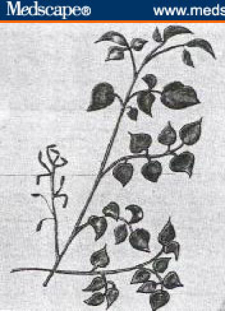
Lepidium sativum plant.
Follow-up of the diet, wound care, and behavior were carried out daily, and it was observed early the next morning that the rabbits were eating all of their food before adding 6 g of the seeds in their new food.
After 6 weeks postoperatively, all 6 rabbits (control and test) had their left femurs x-rayed in a nearby hospital. The study was continued as before and repeat x-rays in the same areas of the left femur were done at 12 weeks.
The study was ended and the animals were sacrificed followed by collection of data, tabulation, and statistical analysis with the SPSS package.
These data showed that the P values of the control and test groups at 6 weeks were P = NS, P = NS, and P < .001 in the longitudinal lateral (LL), longitudinal medial (LM), and circumferential (CM) callus measurements, respectively.
The P values at 12 weeks had more significant areas of P < .043 and P < .004 in the LM and CM callus, respectively. The LL callus remained nonsignificant (P = NS).
Results
This study, which lasted 12 weeks from the time of surgery, divided rabbits into control (no. = 3/C1, C2, C3) and test groups (no. = 3/T1, T2, T3). Soon after their recovery from general anesthesia, the control group was fed with a normal diet, but the test animals had a normal diet plus 6 g of L sativum seeds for the whole period of the study. The results of daily follow-up showed uneventful recovery, good healing of wounds, and weight gain in all rabbits. Documentation of fracture healing in the left femur of all groups was carried out and clinically based on the presence or absence of crepitus plus abnormal movements at the fracture sites, and radiologically from the site of callus formation. X-rays of the left femur at the fracture sites in all 6 rabbits were taken in a nearby hospital on 2 occasions – the first at 6 weeks and the second at 12 weeks postoperatively. Both groups showed good healing of fractures and intact intramedullary K-wires (Figures 2 and 3).
The healing of fractures continued at 12 weeks and was almost complete in all groups (Figures 4 and 5).
Further documentation of the healing of fractures was carried out by a direct measurement, in millimeters, of the callus at the fracture sites LM, LL, and CM on the corresponding x-rays.
Data collected at 6 and 12 weeks postoperatively had callus formation measured at those sites and tabulated for further statistical analysis.
The SPSS statistical package was used to compare the test and control groups for any significant relationships or differences with regard to the healing of fractures in rabbits due to the consumption of L sativum seeds.
A statistically significant difference was found in one area (CM; P < .001) at 6 weeks of measurements of callus formation, as shown in Table 1 .
Table 1.
Callus Formation in Millimeters at 6 and 12 Weeks Postoperatively
| Callus Formation at 6 Weeks Postoperatively (mm) | Callus Formation at 12 Weeks Postoperatively (mm) | |||||
|---|---|---|---|---|---|---|
| Number of Cases | Longitudinal Lateral (mm) | Longitudinal Medial (mm) | Circumferential (mm) | Longitudinal Lateral (mm) | Longitudinal Medial (mm) | Circumferential (mm) |
| C1 | 30 | 18 | 7 | 28 | 17 | 9 |
| C2 | 23 | 30 | 2 | 32 | 15 | 2 |
| C3 | 25 | 21 | 4 | 22 | 11 | 4 |
| T1 | 23 | 26 | 22 | 25 | 23 | 17 |
| T2 | 40 | 42 | 18 | 43 | 49 | 18 |
| T3 | 33 | 33 | 21 | 34 | 39 | 19 |
| t | −1.121 | −1.817 | −8.485 | −1.12 | −2.915 | −6.018 |
| P < | NS | NS | .001 | NS | .043 | .004 |
However, more areas revealed statistically significant differences at 12 weeks of measurements of callus formation in the LM and CM sites (P < .043 and P < .004, respectively; Table 1).
It was obvious that callus formation at 12 weeks continued to predominate in all areas of the test group and became more prominent in the LM and CM areas. This difference in callus formation between the test and control groups persisted at 6 and 12 weeks and was more evident in the test group.
Nevertheless, the weights of all rabbits, when documented weekly for 4 weeks, showed mild gradual increases in both groups with a slight tendency to be more evident in the test group, but this was not statistically significant (Table 2).
Table 2.
Weight of Rabbits in Kilograms
| Weight (kg) | |||||
|---|---|---|---|---|---|
| Number of Cases | First Week | Second Week | Third Week | Fourth Week | Total (kg) |
| C1 | 3.000 | 4.605 | 4.700 | 4.400 | 16.705 |
| C2 | 2.500 | 4.305 | 5.045 | 4.445 | 16.295 |
| C3 | 4.200 | 4.500 | 4.300 | 4.750 | 17.750 |
| T1 | 4.000 | 5.502 | 5.250 | 5.495 | 20.247 |
| T2 | 3.000 | 4.411 | 4.350 | 4.395 | 16.156 |
| T3 | 3.800 | 4.170 | 4.920 | 5.250 | 18.140 |
| t | −0.622 | −0.535 | −0.466 | −1.467 | |
| P < | NS | NS | NS | NS | |
Discussion
Healing of fractures was a major task that was studied immensely clinically, experimentally, and traditionally aiming at facilitating this phenomenon positively and documenting it by different methods, such as ultrasound,[11] biomechanical measurements,[12] and dual-energy x-ray absorptiometry.[13] The influences of many factors and medications on the healing of fractures were noted as well.[2,3]
However, alternative medicines, such as traditional folk medicine, have used natural elements from ancient times to now.[8] This was practiced for the treatment of many ailments in different societies.[7,9] L sativum and its seeds, in particular, were publicly used in Saudi Arabia as a traditional medicine, mostly for the treatment of recent traumatic fractures and less commonly in delayed or nonunited fractures. Good results of healing of fractures were observed over decades in the hands of traditional folk medicine practitioners. This was also noted by the author, who was encouraged to conduct laboratorial studies to investigate the effect of L sativum seeds on fracture healing in vivo. Recently, rats fed with L sativum seeds had their induced fractures tested for healing, and an increase of collagen deposition and tensile strength was found at the fracture sites.[10]
Our study aimed at confirming this phenomenon of accelerated healing of induced fractures in New Zealand White rabbits under the effects of L sativum seeds, which supports the observations that have been noted in clinical and traditional folk medicine practices.
The surgical procedure of open induction and reduction of fractures was unique, more accurate, and informative when compared with the other previous studies – especially its application in clinical practice. The documentation of callus formation with 2 new methods, radiography and measurements in millimeters, was also new.
Rabbits, of average weight 4-5 kg, were fed 6 g of seeds daily. In comparison, the average human adult weight of 50-70 kg will require an intake of 75-105 g per day; this means 25-35 g (teaspoonful) if divided into 3 times a day for better tolerance and efficacy. Hence, the total consumption of seeds by a human adult will be 2-3 kg per month, and this is, of course, much cheaper than other adjuvant therapy and expensive medicine.
The callus formations in induced fractures of the test rabbits fed with L sativum seeds were statistically significant when compared with the control group, and these clearly indicated that they played a major role in promoting and accelerating callus formation in those fractures.
Looking at the current concepts of fracture healing on the basis of blood supply and stability, it was considered that the molecular activity of the fracture exudates was the most decisive factor for bone healing.[14] These fracture exudates have a high concentration of osseous morphogens, growth factors, osteoprogenitor cells, and bone-specific vessels,[15] which through them different factors can influence fracture healing by accelerating or inhibiting their activities.
Hence, the effects of L sativum seeds were possibly on 1 or more of these constituents, such as fatty acids,[16] protein,[17,18] or through their activities[19] as well as possible incorporation in the biological activities similar to the stable isotope tracers.[20]
Furthermore, the emulsifying properties of mucilages of the seeds, as stated in some studies,[21,22] were also possible contributory factors in the acceleration of the fracture healing phenomenon through their effects on fracture exudates.
Consequently, the tensile strength, stiffness of fractures, and other inhibitory factors – which are also important elements[23–25] influencing fracture healing together with all previously mentioned factors – will open a major field of further studies to be carried out under the influence of L sativum seeds.
Of note, L sativum seeds had no significant effects on the weight of rabbits, which was similarly noted by others.[10]
Conclusions
We conclude that L sativum seeds showed a significant effect on fracture-induced healing in rabbits in vivo, which supports the observation noted in the community and in traditional folk medicine. These clearly reveal the importance of further potential studies to be continued by the author and others who are interested in this new field. They will also have important clinical implications for future applications and studies.
Figure 2.
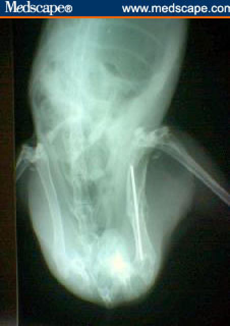
X-ray of control group at 6 weeks postoperatively.
Figure 3.
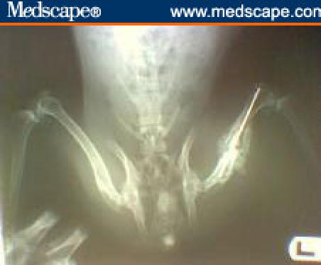
X-ray of test group at 6 weeks postoperatively.
Figure 4.
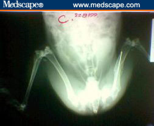
X-ray of control group at 12 weeks postoperatively.
Figure 5.
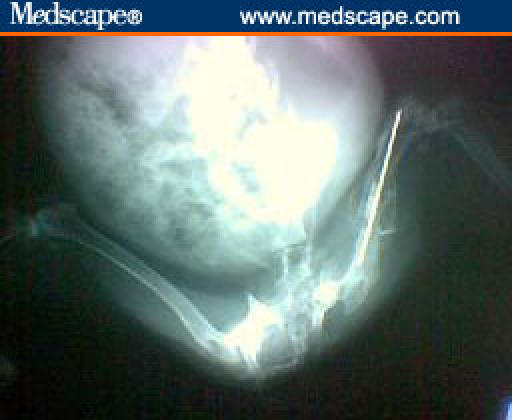
X-ray of test group at 12 weeks postoperatively.
Footnotes
Readers are encouraged to respond to the author at ahaj73@yahoo.com or to Paul Blumenthal, MD, Deputy Editor of MedGenMed, for the editor's eyes only or for possible publication via email: pblumen@stanford.edu
References
- 1.Ketchen EE, Porter WE, Bolton NE. The biological effects of magnetic fields on man. Am Ind Hyg Assoc J. 1978;39:1. doi: 10.1080/0002889778507706. [DOI] [PubMed] [Google Scholar]
- 2.Cook SD, Barrack RL, Santman M, Patron LP, Salkeld SL, Whitecloud TS. The Otto Aufranc Award. Strut allograft healing to the femur with recombinant human osteogenic protein-1. Clin Orthop Relat Res. 2000;381:47–57. doi: 10.1097/00003086-200012000-00006. [DOI] [PubMed] [Google Scholar]
- 3.Ripamonti U. Bone induction by recombinant human osteogenic protein-1 (hOP-1, BMP-7) in the primate Papio ursinus with expression of mRNA of gene products of the TGF-beta superfamily. J Cell Mol Med. 2005;9:911–928. doi: 10.1111/j.1582-4934.2005.tb00388.x. [DOI] [PMC free article] [PubMed] [Google Scholar]
- 4.Han ZB, Chen LP, Yang XZ. Experimental study of fracture healing promotion with mechanical vibration in rabbits [in Chinese] Chung Hua Wai Ko Tsa Chih. 1994;32:215–216. [PubMed] [Google Scholar]
- 5.Diebert MC, McLeod BR, Smith SD, Liboff AR. Ion resonance electromagnetic field stimulation of fracture healing in rabbits with a fibular osteotomy. J Orthop Res. 1994;12:878–885. doi: 10.1002/jor.1100120616. [DOI] [PubMed] [Google Scholar]
- 6.Bruce GK, Howlett CR, Huckstep RL. Effect of a static magnetic field on fracture healing in a rabbit radius preliminary result. Clin Orthop Related Res. 1987;222:300–305. [PubMed] [Google Scholar]
- 7.Ageel AM, Tariq M, Mossa JS, Al-Yahya MA, Al-Said MS. Plants Used in Saudi Folk Medicine. Riyadh, Saudi Arabia: KACST, King Saud University Press; 1987. pp. 245–415. [Google Scholar]
- 8.Qudamah A. Dictionary of Food and Treatment by Plants. Beirut: Dar Alnafaes; 1995. pp. 241–244. [Google Scholar]
- 9.Czimber G, Szabo LG. Therapeutical effect and production of garden cress (Lepidium Sativum L) Gyogyszereszet. 1988;32:79–81. [Google Scholar]
- 10.Ahsan SK, Tariq M, Ageel M, Al-Yahya MA, Shah AH. Studies on some herbal drugs used in fracture healing. Int J Crude Drug Res. 1989;27:235–239. [Google Scholar]
- 11.Ricciardi L, Perissinotto A, Dabala M. Mechanical monitoring of fracture healing using ultrasound imaging. Clin Orthop. 1993;293:71–76. [PubMed] [Google Scholar]
- 12.Cunningham JL, Kenwright J, Kershaw CJ. Biomechanical measurement of fracture healing. J Med Eng Technol. 1990;14:92–101. doi: 10.3109/03091909009015420. [DOI] [PubMed] [Google Scholar]
- 13.Muir P, Markel MD, Bogdanske JJ, Johnson KA. Dual energy X-ray absorptiometry and force-plate analysis of gait in dogs with healed femora after leg lengthening plate fixation. Vet Surg. 1995;24:15–24. doi: 10.1111/j.1532-950x.1995.tb01288.x. [DOI] [PubMed] [Google Scholar]
- 14.Hulth A. Current concepts of fracture healing. Clin Orthop Related Res. 1989;249:265–284. [PubMed] [Google Scholar]
- 15.Pennig D. The biology of bones and of bone fracture healing. Unfallchirurg. 1990;93:488–491. [PubMed] [Google Scholar]
- 16.Kolodziejski J, Mruk-Luczkiewicz A, Mionskowski H. Physiochemical investigations on the oil from seeds of genus Lepidium L. Cruciferae. Diss Pharm Pharmacol. 1969;21:235–239. [Google Scholar]
- 17.Burghardt H, Brunner H, Oelmuller R, Lottspeich F, Oster U, Rudiger W. Natural inhibitors of germination and growth, VII synthesis of Ribulosebisphosphate carboxylase in darkness and its inhibition by Coumarin. Z Naturforsch [C] 1994;49:321–326. doi: 10.1515/znc-1994-5-607. [DOI] [PubMed] [Google Scholar]
- 18.Koropp K, Volkmann D. Monoclonal antibody CRA against a fraction of action from Cress roots recognized its antigen in different plant species. Eur J Cell Biol. 1994;64:153–162. [PubMed] [Google Scholar]
- 19.Iori R, Rollin P, Streicher H, Thiem J, Palmieri S. The myrosinase-glucosinolate interaction mechanism studied using some synthetic competitive inhibitors. FEBS Lett. 1996;385:87–90. doi: 10.1016/0014-5793(96)00335-3. [DOI] [PubMed] [Google Scholar]
- 20.Giussani A, Heinrichs U, Roth P, Werner E, Schramel P, Wendler I. Biokinetic studies in humans with stable isotopes as tracers. Part 1: a methodology for incorporation of trace metals into vegetables. Isotopes Environ Health Stud. 1998;34:291–296. doi: 10.1080/10256019808234062. [DOI] [PubMed] [Google Scholar]
- 21.Patel MM, Chauhan GM, Patel LD. Mucilages of Lepidium Sativum, Linn (Asario) and Ocimum Canum Sims (Bavchi) as emulgents. Indian J Hosp Pharm. 1987;24:200–202. [Google Scholar]
- 22.Wendt L, Meler J. Rheological study of pharmaceutical emulsions containing mucilage from the seeds of garden Cress instead of gum arabic. Farm Pol (Farmacja-Polska) 1988;44:87–91. [Google Scholar]
- 23.Pollak D, Floman Y, Simkin A, Avinezer A, Freund HR. The effect of protein malnutrition and nutritional support on the mechanical properties of fracture healing in the injuried rat. J Parenter Enteral Nutr. 1986;10:564–567. doi: 10.1177/0148607186010006564. [DOI] [PubMed] [Google Scholar]
- 24.Delgado Martinez AD, Martinez ME, Carrascal MT, Rodriguez Avial M, Munuera L. Effect of 25-OH vitamin D on fracture healing in elderly rats. J Orthop Res. 1998;16:650–653. doi: 10.1002/jor.1100160604. [DOI] [PubMed] [Google Scholar]
- 25.Lindgren JU, DeLuca HF, Mazess RB. Effects of 1, 25 (OH) 2D3 on bone tissue in the rabbit: studies on fracture healing, disuse osteoporosis and prednisolone osteoporosis. Calcif Tissue Int. 1984;36:591–595. doi: 10.1007/BF02405372. [DOI] [PubMed] [Google Scholar]


