Abstract
The occurrence rate of transient cortical blindness after contrast media exposure has been reported to be as high as 1% to 4% after cerebral or vertebral angiography, but such blindness has been described in only a few cases of coronary angiography with modern, non-ionic, low-osmolality radio-contrast agents. In this study, we present a case of abrupt cortical blindness after exposure to contrast media during diagnostic coronary angiography; to our knowledge, this is the 1st report in the medical literature that describes transient cortical blindness after iobitridol use.
Key words: Blindness/etiology/radiography, bloodbrain barrier/drug effects, cerebral cortex/drug effects, cognition disorders/etiology, contrast media/administration & dosage/drug effects, coronary angiography iobitridol, iohexol/adverse effects/diagnostic use, visual cortex/drug effects
Transient cortical blindness is an infrequent complication of cerebral and vertebral angiography, with occurrence rates of 0.3% to 1.0%,1 but the rate can be as high as 4% when hyperosmolar iodinated contrast agents are used.2 Cortical blindness is rarely encountered after angiography of native coronary arteries or of bypass grafts.3–5 Our review of the English-language literature revealed only 18 such cases: 7 after angiography of the native coronary vessels and 11 after angiography of coronary artery bypass grafts.3,5–7 Herein, we present the case of a 70-year-old woman who developed cortical blindness after exposure to contrast media during diagnostic coronary angiography performed with iobitridol. This, to our knowledge, is the 1st such report in the literature.
Case Report
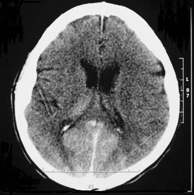
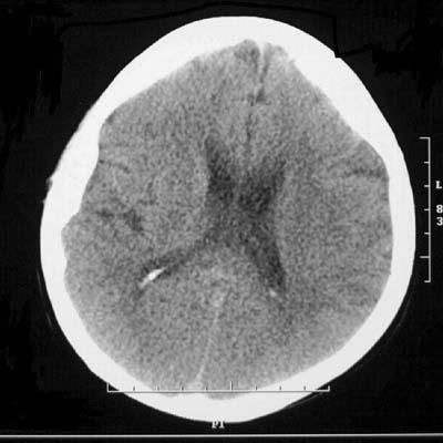
In January 2005, a 70-year-old woman who had angina on exertion was admitted to our clinic for coronary angiography. She had a history of hypertension of 20 years' duration, of diabetes mellitus, and of hypercholesterolemia. Physical examination revealed normal findings, and her blood pressure was 140/85 mmHg. Selective angiography of her native coronary arteries and a left ventriculogram were completed without difficulty by use of the Judkins technique from the right femoral artery. A total of 75 mL of non-ionic, low-osmolar contrast agent (Xenetix® 350 [350 mg iodine/ mL], Guerbet; Aulnay-sous-Bois, France) containing iobitridol was used. The procedure was performed without complications. Two hours later, the patient experienced visual loss, headache, and confusion, with retrograde amnesia and disorientation. Her blood pressure rose to 190/100 mmHg. Neurologic findings were normal: extraocular movements were preserved and pupillary reflex was normal. Ophthalmic examination of the fundi also yielded normal results; however, bilateral cortical blindness was diagnosed. A cranial computed tomographic (CT) scan was performed 3 hours after coronary angiography. No additional contrast material was used during the procedure. The CT scan showed marked bilateral contrast enhancement in the occipital lobes and no evidence of cerebral hemorrhage (Fig. 1). Low-molecular-weight heparin (enoxaparin sodium given subcutaneously, 1 mg/kg, twice daily) and amlodipine (10 mg/day) were started. The patient remained confused and amnesic for 24 hours after visual loss. Then her vision gradually improved, progressing to complete recovery within 72 hours. Cranial CT scanning—repeated at 36 hours after visual loss—showed clearing of the contrast media (Fig. 2). She was discharged 4 days after admission with no residual blindness or neurologic deficit. Subsequently, the patient was well at her 6-month and 1-year visits.

Fig. 1 Cranial computed tomographic scan, 3 hours after coronary angiography, shows marked bilateral contrast enhancement of the medial aspect of both occipital lobes in a symmetric distribution. The accumulation of contrast media shows no relationship to avascularity, which argues against an embolic event as the pathogenesis.

Fig. 2 Computed tomographic scan, 36 hours after coronary angiography, when the patient recovered complete sight. There is no contrast enhancement in the cranium.
Discussion
The mechanism of cerebral injury that causes cortical blindness remains speculative. Some previous studies3 have suggested that cortical blindness may be related to contrast-induced hypotension during the angiographic procedure, to the presence of hypertensive vascular disease, or to the osmolality and total amount of contrast material injected.
Hinchey and colleagues8 have suggested that there might be a relationship between cortical blindness and hypertensive encephalopathy—a clinical syndrome that can include visual disturbances. Our patient had a long history of hypertension and had a documented acute rise in blood pressure after the procedure. Most of the patients in previous reports also had chronic hypertension, although the details of pressure changes during angiography have not usually been reported. Hypertensive encephalopathy is thought to result from sudden elevation of systemic blood pressure that exceeds the autoregulatory capacity of the cerebral vessels, thereby producing regions of vasodilation and vasoconstriction, with a breakdown of the blood–brain barrier and focal transudation of fluid.9,10 Imaging in hypertensive encephalopathy—like the CT findings in our patient— has shown bilateral abnormalities in the occipital lobes, involving the subcortical white matter and often extending to the cortical surface. Reversible edema, localized mainly in the occipital lobes, is a prominent feature. Edema secondary to disturbance of autoregulation of the posterior cerebral vessels is another possible mechanism for transient cortical blindness.9 Blood–brain barrier injury during the use of contrast media has been reported before, especially in patients with uncontrolled hypertension.11 Other reported risk factors for blood– brain barrier injury include renal insufficiency, eclampsia, and immunosuppressive drug abuse.8
Experimentally, the blood–brain barrier can be disrupted with the introduction of hyperosmolar solutions into the spinal cord or blood. Whether disruption is causally related to the hypertonicity of the contrast agent or to the volume of the dose is not yet clear. Various investigators1–7 have reported using from 80 to 400 mL of contrast agent (we used only 75 mL). Although non-ionic agents are less toxic than their ionic counterparts, several cases of transient cortical blindness have now been reported with use of these low-osmolality agents.3,10 Zwicker and Sila12 have confirmed, by magnetic resonance imaging (MRI), the mechanism of a transient vasculopathy with disruption of the blood– brain barrier as the cause of transient cortical blindness after angiography.12
Most of the patients in whom transient cortical blindness has been reported have undergone angiography of bypass grafts. It is likely, in these patients, that direct injection into the vertebral artery occurs during angiography of the internal mammary artery conduit. The patients' prolonged supine posture might also play a role in the intracranial enhancement of the contrast solution in the occipital region.
It is known that certain patients are more prone than others to idiosyncratic reactions after contrast injection. Direct idiosyncratic neurotoxicity still appears to be the most reasonable explanation to account for transi ent blindness after coronary angiography, although the exact mechanism is not known.3 This line of reasoning finds support herein, because the volume of contrast agent that we used was very small (a total of 75 mL).13 To our knowledge, ours is the 1st report of transient ischemic blindness in association with iobitridol.
There is no specific measure to be taken for protection against this unusual and alarming complication. Prompt neurologic consultation and MRI or CT scanning of the brain are indicated to confirm the diagnosis. Forced diuresis to remove the offending agent has not been shown to be beneficial, but the blood pressure must be controlled. The cerebral reaction is not patient specific. When the contrast medium has been excreted— which takes an average of 3 days (range, 15 minutes to 3 weeks)—normal vision returns as the normal protective function of the blood–brain barrier returns. In most cases, the return of vision is gradual, proceeding from light and motion perception to a final return of color vision.3
Footnotes
Address for reprints: Hakan Ozhan, MD, Department of Cardiology, Duzce University School of Medicine, 81620 Duzce, Turkey. E-mail: ozhanhakan@yahoo.com
References
- 1.Horwitz NH, Wener L. Temporary cortical blindness following angiography. J Neurosurg 1974;40:583–6. [DOI] [PubMed]
- 2.Studdard WE, Davis DO, Young SW. Cortical blindness after cerebral angiography. Case report. J Neurosurg 1981;54:240–4. [DOI] [PubMed]
- 3.Kinn RM, Breisblatt WM. Cortical blindness after coronary angiography: a rare but reversible complication. Cathet Cardiovasc Diagn 1991;22:177–9. [DOI] [PubMed]
- 4.Fischer-Williams M, Gottschalk PG, Browell JN. Transient cortical blindness. An unusual complication of coronary angiography. Neurology 1970;20:353–5. [DOI] [PubMed]
- 5.Henzlova MJ, Coghlan HC, Dean LS, Taylor JL. Cortical blindness after left internal mammary artery to left anterior descending coronary artery graft angiography. Cathet Cardiovasc Diagn 1988;15:37–9. [DOI] [PubMed]
- 6.Rama BN, Pagano TV, DelCore M, Knobel KR, Lee J. Cortical blindness after cardiac catheterization: effect of rechallenge with dye. Cathet Cardiovasc Diagn 1993;28:149–51. [DOI] [PubMed]
- 7.Sticherling C, Berkefeld J, Auch-Schwelk W, Lanfermann H. Transient bilateral cortical blindness after coronary angiography. Lancet 1998;351:570. [DOI] [PubMed]
- 8.Hinchey J, Chaves C, Appignani B, Breen J, Pao L, Wang A, et al. A reversible posterior leukoencephalopathy syndrome. N Engl J Med 1996;334:494–500. [DOI] [PubMed]
- 9.Schwartz RB, Jones KM, Kalina P, Bajakian RL, Mantello MT, Garada B, Holman BL. Hypertensive encephalopathy: findings on CT, MR imaging, and SPECT imaging in 14 cases. AJR Am J Roentgenol 1992;159:379–83. [DOI] [PubMed]
- 10.Lim KK, Radford DJ. Transient cortical blindness related to coronary angiography and graft study. Med J Aust 2002;177: 43–4. [DOI] [PubMed]
- 11.Johansson BB. Hypertension and the blood-brain barrier. In: Neuwelt EA, editor. Implications of the blood-brain barrier and its manipulation. Vol 2. New York: Plenum Press; 1989. p. 389–410.
- 12.Zwicker JC, Sila CA. MRI findings in a case of transient cortical blindness after cardiac catheterization. Catheter Cardiovasc Interv 2002;57:47–9. [DOI] [PubMed]
- 13.Petersein J, Peters CR, Wolf M, Hamm B. Results of the safety and efficacy of iobitridol in more than 61,000 patients. Eur Radiol 2003;13:2006–11. [DOI] [PubMed]


