Abstract
Chronic post-hypoxic myoclonus, also known as Lance-Adams syndrome (LAS), is a rare complication of successful cardiopulmanry resuscitation often accompanied by action myoclonus and cerebellar ataxia. It is seen in patients who have undergone a cardiorespiratory arrest, regained consciousness afterwards, and then developed myoclonus days or weeks after the event. Worldwide, 122 cases have been reported in the literature so far, including 1 case of Chinese. Here we report 2 Chinese LAS patients with detailed neuroimagings. Cranial single photon emission computed tomography (SPECT) of patient 1, a 52-year-old woman, showed a mild hypoperfusion in her left temporal lobe, whereas patient 2, a 54-year-old woman, manifested a mild bilateral decrease of glucose metabolism in the frontal lobes and a mild to moderate decrease of the N-acetyl aspartate (NAA) peak in the bilateral hippocampi by cranial [18F]-fluorodeoxyglucose positron emission tomographic (PET) scan and cranial magnetic resonance spectroscopy (MRS), respectively. We also review the literature on the neuroimaging, pathogenesis, and treatment of LAS.
Keywords: Lance-Adams syndrome, Chronic post-hypoxic myoclonus, Action myoclonus, Cerebellar ataxia, Single photon emission computed tomography, Positron emission tomography, Magnetic resonance spectroscopy
INTRODUCTION
With the development of emergency medicine and improvements in emergency medical services, the number of patients who survived a cardiorespiratory arrest has increased. The survivors may develop neurological complications, such as post-hypoxic myoclonus (PHM). Two types of PHM can occur in patients with hypoxic injury of the brain: the acute and the chronic. The acute PHM, some termed post-hypoxic myoclonic status epilepticus (MSE), occurs soon after a hypoxic insult and is characterized by generalized myoclonic jerks in patients who are deeply comatose (Hallett, 2000), which implies a poor prognosis. The chronic PHM, also known as Lance-Adams syndrome (LAS), is predominantly characterized by action myoclonus that starts days to weeks after cardiorespiratory resuscitation (CPR) in patients who regained consciousness (Lance and Adams, 1963). PHM, both the acute and the chronic, is not yet well understood. Occurrence of LAS is rare, and only 122 cases worldwide, including one Chinese case (Guo et al., 2002), have been described so far (Kowalczyk et al., 2006; Polesin and Stern, 2006). Here we present two Chinese LAS cases with detailed studies of neuroimagings. A review of literature on the neuroimaging, pathogenesis, and treatment of LAS is also reported.
CASE REPORTS
Case 1
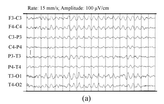
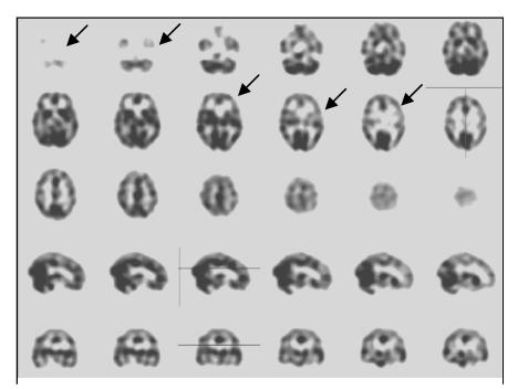
A 52-year-old woman had a cardiorespiratory arrest after a cholecystectomy in a local hospital on Feb. 27, 2006. Although her cardiac output was restored within 4 min after the operation, the patient remained in coma and needed a respiratory support. Five hours after CPR, she developed a severe generalized myoclonus, which temporarily responded to midazolam, but was primarily controlled by intramuscular phenobarbital and intravenous sodium valproate. Three days later, the patient regained her consciousness and had no hemiparesis or aphasia. However, she continued to have a moderate generalized myoclonus in the face, trunk, and limbs, accompanied by dysmetria, dysarthria, and ataxia. These made her unable to sit from a supine position, stand after sitting in a chair awhile, walk without aid, or perform simple, coordinated, manual tasks. Stimuli, such as touches, sounds, and startles, triggered and aggravated the myoclonic jerks, which would disappear with relaxation of her body and limbs or during a sleep. Three weeks after CPR, cranial magnetic resonance (MR) imaging and diffusion weighted (DW) imaging showed no abnormalities. Her Mini-Mental State Examination (MMSE) score was 18, suggesting a moderate cognitive impairment. She had only received three years’ primary school education, and according to the study by Crum et al.(1993), the median MMSE score of a population reference group was 22 for those with 0 to 4 years of schooling. She was disoriented to time and had a mild short-term memory impairment and difficulties with calculation and attention. The blood concentration of neuron specific enolase (NSE), an indication of a poor prognosis for ischemic brain damage, was normal. The therapeutic regime of intramuscular phenobarbital and intravenous sodium valproate was maintained, and 4 weeks later, the patient’s generalized myoclonus was improved. However, after she stopped taking phenobarbital, the severe generalized myoclonus returned, during which she developed two secondary generalized tonic-clonic seizures. An electroencephalogram (EEG) showed low-amplitude alpha waves, diffuse delta activity, and a moderate amount of theta waves (predominant in the left temporal lobe) in daytime, and some sharp and slow waves at night; however, there were no epileptiform activities during myoclonus (Fig.1). After the administration of phenobarbital was resumed, the patient had no further generalized tonic-clonic seizures, and the generalized myoclonus became well controlled. Two months later, cranial single photon emission computed tomography (SPECT) demonstrated a mild hypoperfusion on her left temporal lobe (Fig.2). She was diagnosed with LAS. During the consequent treatment, the dose of intramuscular phenobarbital was gradually reduced, and her generalized myoclonus was well controlled with clonazepam tablets (1 mg po tid) and sodium valproate tablets (0.4 g po bid). However, when the dose of clonazepam or sodium valproate was reduced, the generalized myoclonus recurred. Three months after CPR, she could sit up from the supine position and walk 100 m with only a little help, and the dose of clonazepam was reduced to 2 mg per day, with the dose of sodium valproate not changed (0.4 g po bid).
Fig. 1.
Four weeks after cardiorespiratory resuscitation (CPR), electroencephalogram (EEG) of case 1 showed low-amplitude alpha waves, diffuse delta activity, and a moderate amount of theta waves in daytime (a), and some sharp and slow waves detected at night (b)
Fig. 2.
Cranial single photon emission computed tomography scan of case 1 shows mild hypoperfusion in the left temporal lobe (arrows) two months after cardiorespiratory resuscitation (CPR)
Case 2
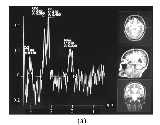
The second case is a 54-year-old woman with a history of depression, who had a cardiorespiratory arrest due to an attempted suicide by hanging. She hanged herself during a 15-min absence of her husband. When taken down by her husband, she was unconscious, with her face being purple. The husband performed a chest pressing on her, and 2 min later her face turned from purple to red and her respiration resumed. Ten minutes later, a rescue team arrived to find her heart beat, blood pressure, and respiration were within normal ranges, except her consciousness. She also developed a generalized seizure before she was transferred to the intensive care unit of a local hospital 40 min later. In the subsequent 3 weeks, she remained unconscious, after which she waked up and developed generalized myoclonic jerks that were aggravated by voluntary movements, sounds, and touches, but disappeared with a relaxation of the body and limbs or during a sleep. Meanwhile, cerebellar ataxia also emerged. She was unable to feed herself, sit up from a chair, get up, or walk by herself. She scored 27 in HAMD (Hamilton Depression Scale) and 5 in HAMA (Hamilton Anxiety Scale), which indicated a severe depression. Nine months after CPR, a video-EEG showed a few low-amplitude theta waves intermingled with a few faster waves against a background of low-amplitude alpha waves, without epileptiform activities during myoclonus. Also, cranial computed tomography (CT) scans, T1 and T2 weighted MR imaging, and DW imaging showed no abnormality, either. Ten months post the resuscitation, cranial magnetic resonance spectroscopy (MRS) showed a moderate decrease in the N-acetyl aspartate (NAA) peak in her left hippocampus and a mild decrease in the right hippocampus, suggesting a neuronal loss due to ischemia. The NAA/(Cho+Cr) ratio in the left hippocampus was 0.479, while the one in the right hippocampus was 0.54 (Fig.3). Cranial MRS of other brain areas did not show any abnormalities. Cranial [18F]-fluorodeoxyglucose positron emission tomographic (PET) scan displayed a mild decrease of glucose metabolism in both frontal lobes (Fig.4). Thus, the diagnosis of LAS was established. The patient was treated with clonazepam tablets (1 mg po tid), sodium valproate tablets (0.4 g po bid), sertraline tablets (an antidepressant of the selective serotonin reuptake inhibitor type) (75 mg po qd), and topiramate (75 mg po bid). Under such regimen, her generalized myoclonus was well controlled; the situation worsened only when the dose of sodium valproate was reduced. On discharge, clonazepam and topiramate were discontinued, the dosage of sodium valproate was increased to 1.0 g per day, and the same dosage of sertraline remained. At the 1-year follow-up, the patient could sit in a chair for 2 h and walk 300 m with the assistance of relatives, but she still could not sit from the supine position or walk by herself. Her MMSE score was only 14, implying a severe cognitive impairment. The neuropsychological assessments disclosed prominent impairments of the immediate memory and short-term memory, disorientation to time, dyscalculia, slow coordination, and poor concentration. The patient could obey simple commands such as folding a paper, but could not draw or write because of the triggered myoclonic jerks. Her dysarthria had persisted to some extent.
Fig. 3.
Ten months after cardiorespiratory resuscitation (CPR) cranial magnetic resonance spectroscopy of case 2 shows a moderate decrease of the N-acetyl aspartate (NAA) peak in the left hippocampus (a) and a mild decrease in the right hippocampus (b). The NAA/(Cho+Cr) ratio in the left and right hippocampus was 0.479 and 0.54, respectively
Fig. 4.
Ten months after cardiorespiratory resuscitation (CPR) [18F]-fluorodeoxyglucose positron emission tomographic (PET) scan of case 2 shows a mild decrease of glucose metabolism in bilateral frontal lobes
DISCUSSION
Myoclonus refers to sudden, shock-like, involuntary body movements that can occur in various patterns, i.e., focal, multifocal, or generalized. The chronic PHM, also known as LAS (Lance and Adams, 1963), is predominantly characterized by action myoclonus that has no consistent correlation with EEG abnormalities. LAS occurs in patients after they have regained consciousness days to weeks after CPR. The myoclonic jerks are specifically triggered by action, startle, and tactile stimulation, and they usually disappear with relaxation of the body and limbs or with a sleep. The severity of the myoclonus is proportional to the precision of the task that is required. Both of our patients presented with the generalized myoclonus accompanied by cerebellar ataxia 3 d to 3 weeks after CPR, respectively. In both patients, the EEG had no characteristic abnormalities. These clinical features fulfilled the criteria for the diagnosis of Lance-Adams-type myoclonus.
LAS has diverse clinical, electrophysiological, and neurochemical abnormalities, and the loss of neurotransmitter serotonin (5-hydroxytryptophan, 5-HT) within the inferior olive has been thought to be an important causal factor (Welsh et al., 2002a). It is postulated that estrogen, through its regulation of serotonergic activity, may influence the clinical course of PHM (Kompoliti et al., 2001). Other neurotransmitter systems, such as gamma-aminobutyric acid (GABA), may also be involved and interact with the 5-HT system to suppress PHM (Jaw et al., 1996; Matsumoto et al., 2000). Clonazepam was found to have dramatic effects on PHM. Welsh et al.(2002b) found that Purkinje cells were not uniformly sensitive to transient ischemia, but those located in the paravermal and vermal areas died most frequently. These cells project mainly to the ventrolateral thalamic nucleus but are deficient in aldolase C and EAAT4 (excitatory amino acid transporter 4), which could allow them to survive the pathologically intense synaptic input from the inferior olive after the restoration of blood flow. The authors considered that certain brainstem structures, the paravermal and vermal areas of the cerebellum, and the diencephalons may be implicated in human PHM. The loss of GABAnergic inhibition in cerebellar afferent neurons after ischemia leads to diaschisis of the motor thalamus and reticular formation, which, in turn, is responsible for enhanced motor excitability and myoclonus.
Recently, neuroimaging has been used to detail the anatomical and pathophysiological basis of PHM. Dubinsky et al.(1991) demonstrated a significant increase in the metabolism of the medulla in patients with palatal myoclonus, presumably due to hypertrophy of the inferior olivary nucleus, which is said to possess intrinsic pacemaker properties. Using PET scan, Frucht et al.(2004) reported that, compared with control subjects, 7 patients with LAS had significantly increased glucose metabolism in the pontine tegmentum, spreading to the mesencephalon and the ventrolateral thalamus. In our first patient, cranial SPECT (Fig.2) showed mild hypoperfusion in the left temporal lobe, whereas in our second patient, cranial PET scan showed a mild bilateral decrease of glucose metabolism in the frontal lobes (Fig.4), and cranial MRS showed a mild to moderate decrease of the NAA peak in bilateral hippocampui (Fig.3). These findings are in agreement with a previous PET study in post-anoxic patients with permanent amnesia, which showed hypometabolism in the thalamus, frontal cortex, and temporal cortex (Reed et al., 1999). While there have been few reports on LAS studied by MRS, it appears a promising approach. The cranial MRS of our second case showed abnormal NAA peak in bilateral hippocampui (Fig.3), suggesting a cause of memory impairment; however, it did not show any abnormalities in the frontal lobes where cranial PET scan showed a mild bilateral decrease of glucose metabolism (Fig.4). Some PET studies disclosed a reduced blood flow and metabolism in the prefrontal cortex of the depressed patients (Videbech, 2000; Kimbrell et al., 2002). We postulated that the abnormality on PET scan of our second patient might be related with the severe depression she had suffered before the event.
While the treatment for LAS has been limited, due mainly to a lack of clear understanding of the pathophysiological mechanisms involved, LAS has an excellent prognosis if treated early (Werhahn et al., 1997). A combination of medications based on the neurotransmitter hypotheses is often needed to obtain adequate control of symptoms. Frucht and Fahn (2000) reviewed more than 100 cases of LAS and found that clonazepam, sodium valproate, and piracetam were significantly effective in approximately 50% of patients. Recently, several groups have confirmed the efficacy of levetiracetam in PHM (Frucht et al., 2001; Krauss et al., 2001; Lim and Ahmed, 2005). Clonazepam, sodium valproate, piracetam, and levetiracetam may be recommended as first-line agents to treat patients with LAS. A combination of medications including clonazepam and sodium valproate well controlled the myoclonus of our patients, whereas a previously reported Chinese LAS case (Guo et al., 2002) was effectively treated with 5-hydroxytryptophan, fluoxetine hydrochloride and carbidopa tablets. Apart from these drugs, based on the observation of hypermetabolism in the ventrolateral thalamus on PET scan, stereotactic targeting of the ventrolateral thalamus using deep brain stimulation has been suggested for selected patients with severe, medication-refractory PHM (Frucht et al., 2004).
Up to now, there have been no reports dealing with successful treatment of cerebellar vermis ataxia. In our cases, cerebellar vermis ataxia was the predominant symptom and sign and was difficult to be controlled. Patients even could not get up or stand up by themselves although their muscle strength was quite less affected and myoclonus had been effectively controlled. Cerebellar ataxia was the most threatening cause of poor life quality of these patients. Further studies to clarify the pathogenesis of LAS are important to find novel therapeutic methods for LAS.
Footnotes
Project supported by the National Natural Science Foundation of China (No. 30600193), the Youth Talent Special Fund of the Health Bureau of Zhejiang Province, China (No. 2004QN012), and the Health Bureau of Zhejiang Province, China (Nos. 2000A114 and 2007A100)
References
- 1.Crum RM, Anthony JC, Bassett SS, Folstein MF. Population-based norms for the Mini-Mental State Examination by age and educational level. JAMA. 1993;269(18):2386–2391. doi: 10.1001/jama.269.18.2386. [DOI] [PubMed] [Google Scholar]
- 2.Dubinsky RM, Hallett M, Di Chiro G, Fulham M, Schwankhaus J. Increased glucose metabolism in the medulla of patients with palatal myoclonus. Neurology. 1991;41(4):557–562. doi: 10.1212/wnl.41.4.557. [DOI] [PubMed] [Google Scholar]
- 3.Frucht S, Fahn S. The clinical spectrum of posthypoxic myoclonus. Mov Disord. 2000;15(Suppl. 1):2–7. doi: 10.1002/mds.870150702. [DOI] [PubMed] [Google Scholar]
- 4.Frucht SJ, Louis ED, Chuang C, Fahn S. A pilot tolerability and efficacy study of levetiracetam in patients with chronic myoclonus. Neurology. 2001;57(6):1112–1114. doi: 10.1212/wnl.57.6.1112. [DOI] [PubMed] [Google Scholar]
- 5.Frucht SJ, Trost M, Ma Y, Eidelberg D. The metabolic topography of posthypoxic myoclonus. Neurology. 2004;62(10):1879–1881. doi: 10.1212/01.wnl.0000125336.05001.23. [DOI] [PubMed] [Google Scholar]
- 6.Guo XH, Yu SY, Liu J, Wu WP, Pu CQ, Zhu K. Posthypoxic myoclonus treated with 5-hydroxytryptophan: a case report. J Clin Neurol. 2002;15(5):313. (in Chinese) [Google Scholar]
- 7.Hallett M. Physiology of human posthypoxic myoclonus. Mov Disord. 2000;15(Suppl. 1):8–13. doi: 10.1002/mds.870150703. [DOI] [PubMed] [Google Scholar]
- 8.Jaw SP, Nguyen B, Vuong QT, Trinh TA, Nguyen M, Truong DD. Effects of GABA uptake inhibitors on posthypoxic myoclonus in rats. Brain Res Bull. 1996;39(3):189–192. doi: 10.1016/0361-9230(95)02103-5. [DOI] [PubMed] [Google Scholar]
- 9.Kimbrell TA, Ketter TA, George MS, Little JT, Benson BE, Willis MW, Herscovitch P, Post RM. Regional cerebral glucose utilization in patients with a range of severities of unipolar depression. Biol Psychiatry. 2002;51(3):237–252. doi: 10.1016/S0006-3223(01)01216-1. [DOI] [PubMed] [Google Scholar]
- 10.Kompoliti K, Goetz CG, Vu TQ, Carvey PM, Leurgans S, Raman R. Estrogen supplementation in the posthypoxic myoclonus rat model. Clin Neuropharmacol. 2001;24(1):58–61. doi: 10.1097/00002826-200101000-00010. [DOI] [PubMed] [Google Scholar]
- 11.Kowalczyk EE, Koszewicz MA, Budrewicz SP, Podemski R, Slotwinski K. Lance-Adams syndrome in patient with anoxic encephalopathy in the course of bronchial asthma. Wiad Lek. 2006;59(7-8):560–562. (in Polish) [PubMed] [Google Scholar]
- 12.Krauss GL, Bergin A, Kramer RE, Cho YW, Reich SG. Suppression of post-hypoxic and post-encephalitic myoclonus with levetiracetam. Neurology. 2001;56(3):411–412. doi: 10.1212/wnl.56.3.411. [DOI] [PubMed] [Google Scholar]
- 13.Lance JW, Adams RD. The syndrome of intention or action myoclonus as a sequel to hypoxic encephalopathy. Brain. 1963;86(1):111–136. doi: 10.1093/brain/86.1.111. [DOI] [PubMed] [Google Scholar]
- 14.Lim LL, Ahmed A. Limited efficacy of levetiracetam on myoclonus of different etiologies. Parkinsonism Relat Disord. 2005;11(2):135–137. doi: 10.1016/j.parkreldis.2004.07.010. [DOI] [PubMed] [Google Scholar]
- 15.Matsumoto RR, Truong DD, Nguyen KD, Dang AT, Hoang TT, Vo PQ, Sandroni P. Involvement of GABA (A) receptors in myoclonus. Mov Disord. 2000;15(Suppl. 1):47–52. doi: 10.1002/mds.870150709. [DOI] [PubMed] [Google Scholar]
- 16.Polesin A, Stern M. Post-anoxic myoclonus: a case presentation and review of management in the rehabilitation setting. Brain Inj. 2006;20(2):213–217. doi: 10.1080/02699050500442972. [DOI] [PubMed] [Google Scholar]
- 17.Reed LJ, Marsden P, Lasserson D, Sheldon N, Lewis P, Stanhope N, Guinan E, Kopelman MD. FDG-PET analysis and findings in amnesia resulting from hypoxia. Memory. 1999;7(5-6):599–612. doi: 10.1080/096582199387779. [DOI] [PubMed] [Google Scholar]
- 18.Videbech P. PET measurements of brain glucose metabolism and blood flow in major depressive disorder: a critical review. Acta Psychiatr Scand. 2000;101(1):11–20. doi: 10.1034/j.1600-0447.2000.101001011.x. [DOI] [PubMed] [Google Scholar]
- 19.Welsh JP, Placantonakis DG, Warsetsky SI, Marquez RG, Bernstein L, Aicher SA. The serotonin hypothesis of myoclonus from the perspective of neuronal rhythmicity. Adv Neurol. 2002;89:307–329. [PubMed] [Google Scholar]
- 20.Welsh JP, Yuen G, Placantonakis DG, Vu TQ, Haiss F, O'Hearn E, Molliver ME, Aicher SA. Why do Purkinje cells die so easily after global brain ischemia? Aldolase C, EAAT4, and the cerebellar contribution to posthypoxic myoclonus. Adv Neurol. 2002;89:331–359. [PubMed] [Google Scholar]
- 21.Werhahn KJ, Brown P, Thompson PD, Marsden CD. The clinical features and prognosis of chronic posthypoxic myoclonus. Mov Disord. 1997;12(2):216–220. doi: 10.1002/mds.870120212. [DOI] [PubMed] [Google Scholar]


