

Abstract
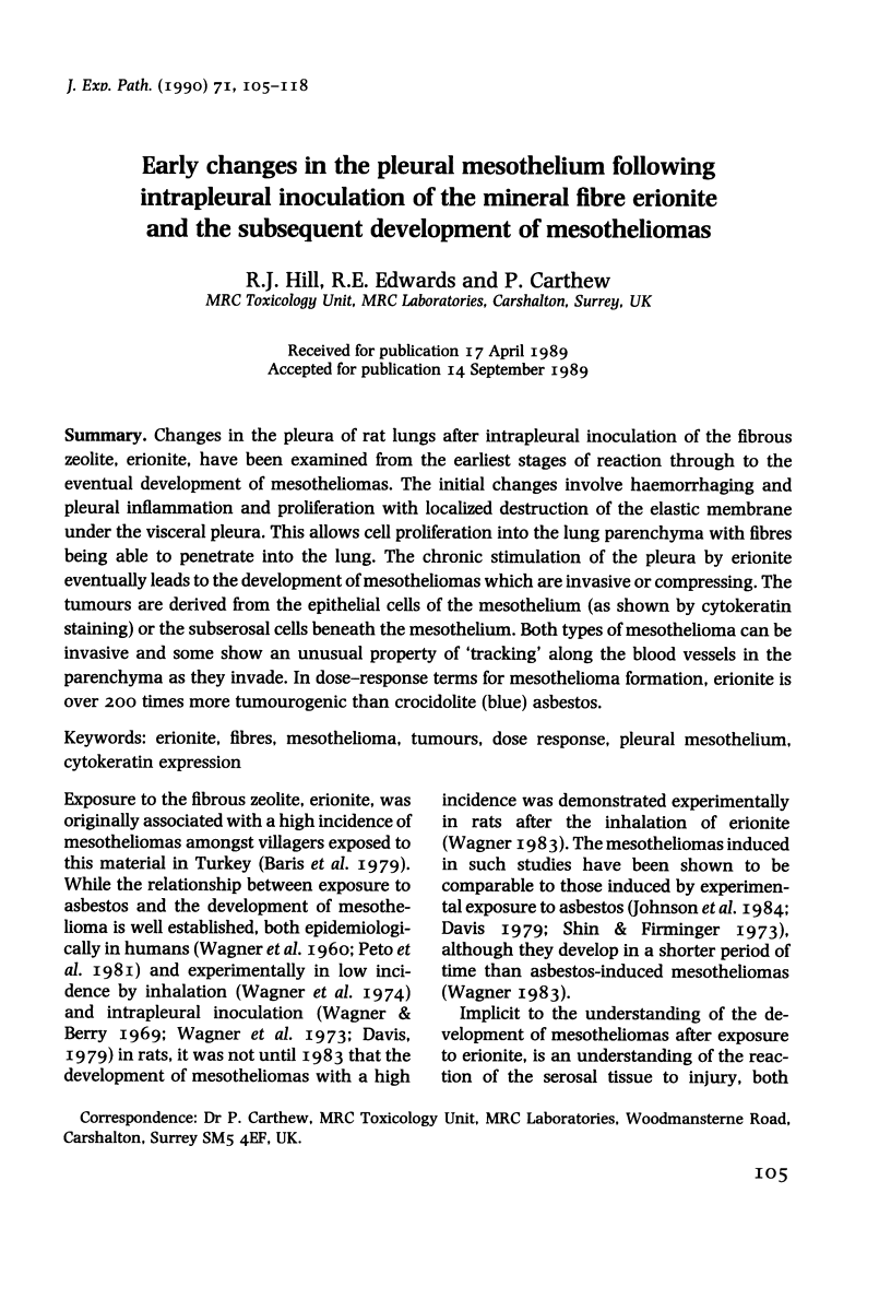
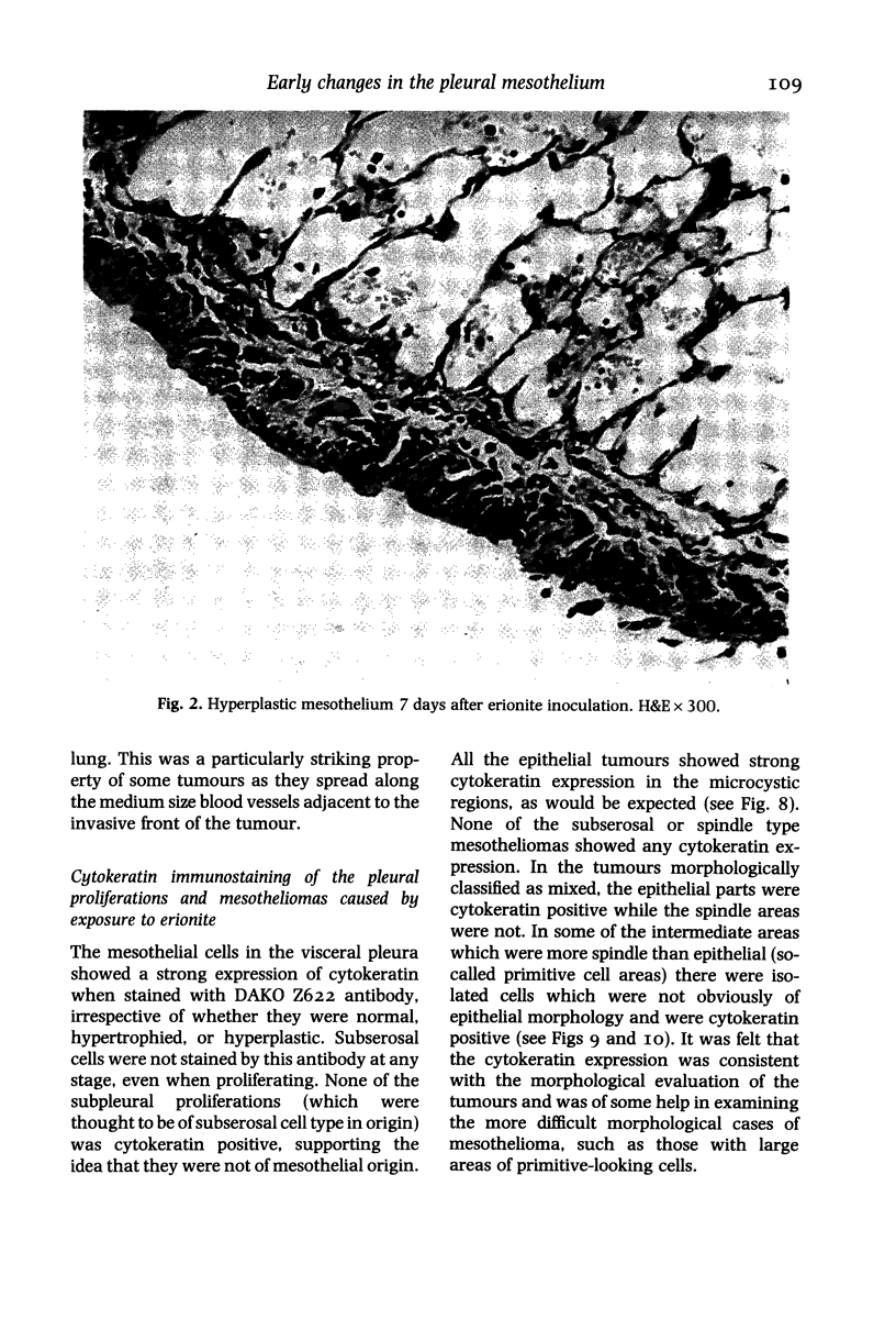
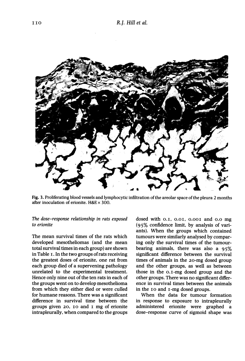
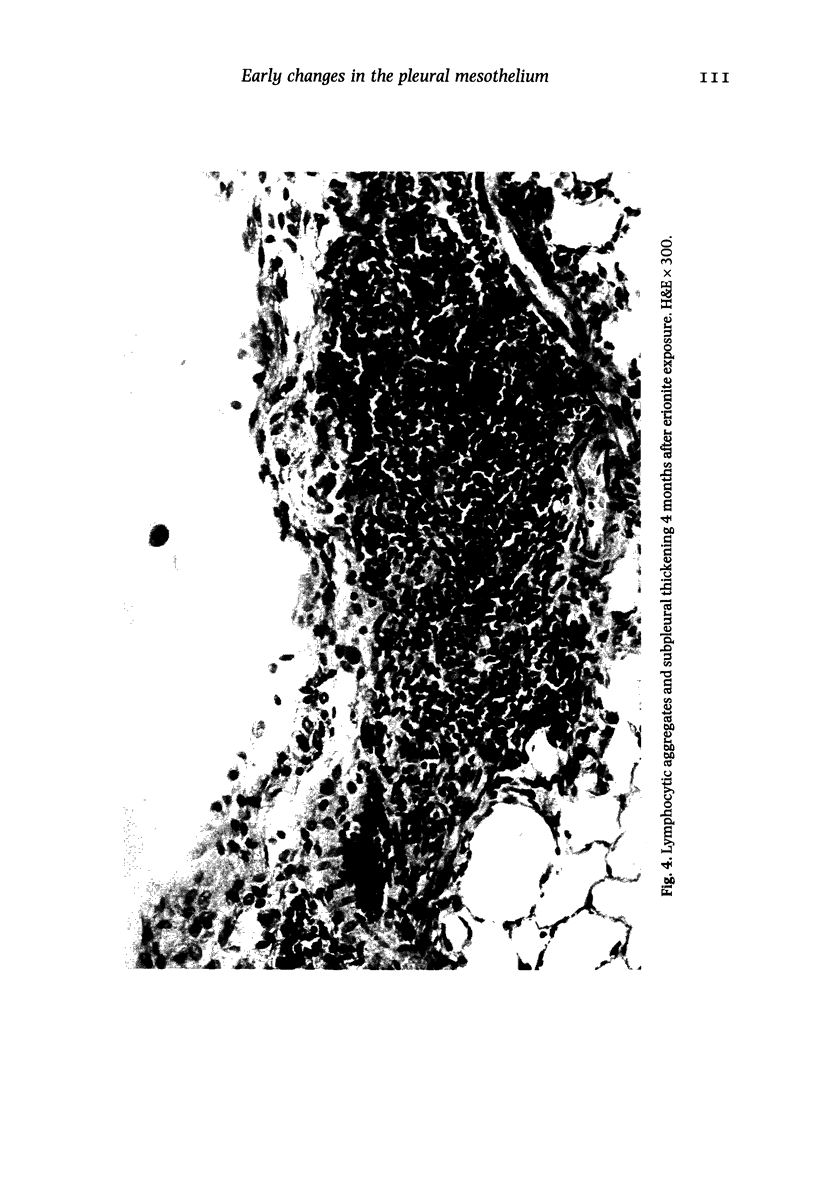
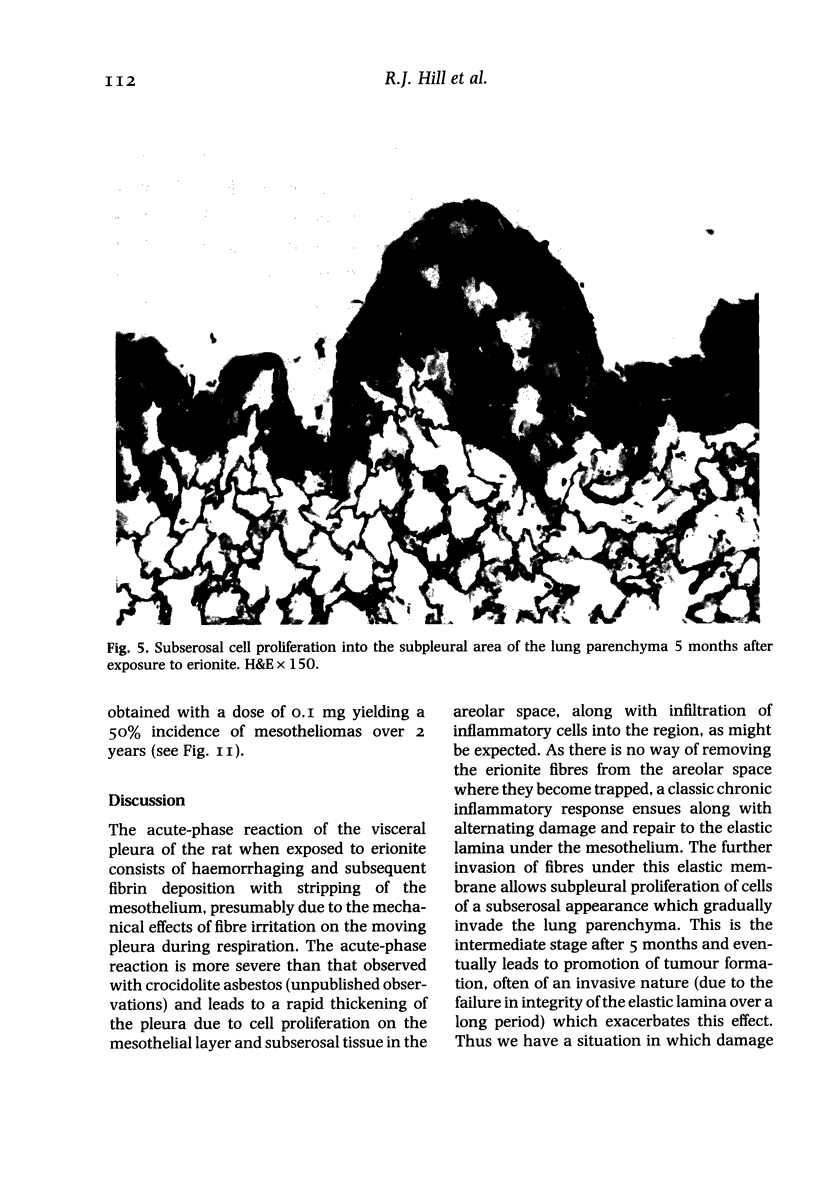
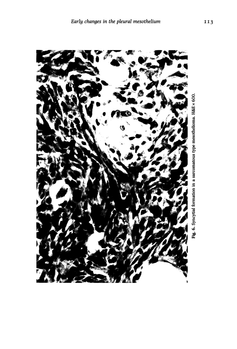
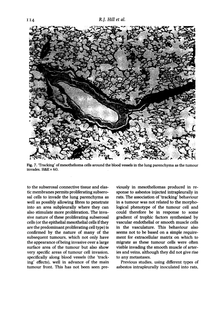
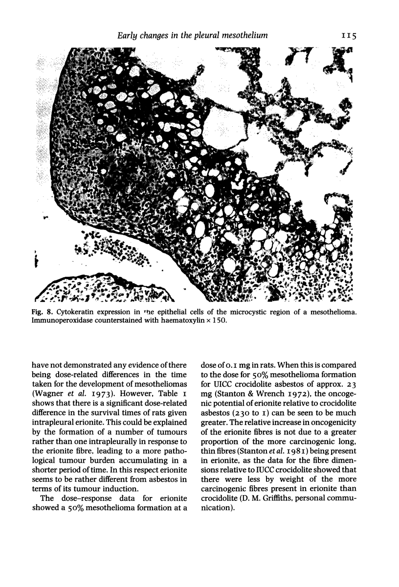
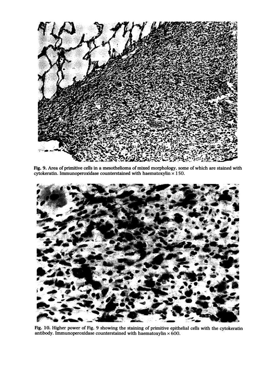
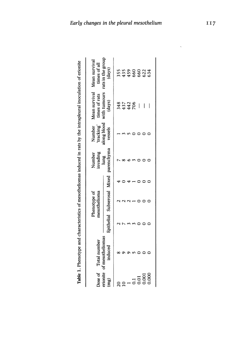
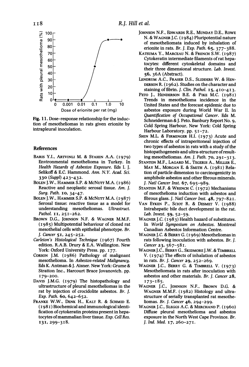
Changes in the pleura of rat lungs after intrapleural inoculation of the fibrous zeolite, erionite, have been examined from the earliest stages of reaction through to the eventual development of mesotheliomas. The initial changes involve haemorrhaging and pleural inflammation and proliferation with localized destruction of the elastic membrane under the visceral pleura. This allows cell proliferation into the lung parenchyma with fibres being able to penetrate into the lung. The chronic stimulation of the pleura by erionite eventually leads to the development of mesotheliomas which are invasive or compressing. The tumours are derived from the epithelial cells of the mesothelium (as shown by cytokeratin staining) or the subserosal cells beneath the mesothelium. Both types of mesothelioma can be invasive and some show an unusual property of 'tracking' along the blood vessels in the parenchyma as they invade. In dose-response terms for mesothelioma formation, erionite is over 200 times more tumourogenic than crocidolite (blue) asbestos.
Full text
PDF
Images in this article


Selected References
These references are in PubMed. This may not be the complete list of references from this article.
- Bariş Y. I., Artvinli M., Sahin A. A. Environmental mesothelioma in Turkey. Ann N Y Acad Sci. 1979;330:423–432. doi: 10.1111/j.1749-6632.1979.tb18744.x. [DOI] [PubMed] [Google Scholar]
- Bolen J. W., Hammar S. P., McNutt M. A. Reactive and neoplastic serosal tissue. A light-microscopic, ultrastructural, and immunocytochemical study. Am J Surg Pathol. 1986 Jan;10(1):34–47. doi: 10.1097/00000478-198601000-00005. [DOI] [PubMed] [Google Scholar]
- Bolen J. W., Hammar S. P., McNutt M. A. Serosal tissue: reactive tissue as a model for understanding mesotheliomas. Ultrastruct Pathol. 1987;11(2-3):251–262. doi: 10.3109/01913128709048326. [DOI] [PubMed] [Google Scholar]
- Brown D. G., Johnson N. F., Wagner M. M. Multipotential behaviour of cloned rat mesothelioma cells with epithelial phenotype. Br J Cancer. 1985 Feb;51(2):245–252. doi: 10.1038/bjc.1985.35. [DOI] [PMC free article] [PubMed] [Google Scholar]
- Shin M. L., Firminger H. I. Acute and chronic effects of intraperitoneal injection of two types of asbestos in rats with a study of the histopathogenesis and ultrastructure of resulting mesotheliomas. Am J Pathol. 1973 Mar;70(3):291–313. [PMC free article] [PubMed] [Google Scholar]
- Stanton M. F., Wrench C. Mechanisms of mesothelioma induction with asbestos and fibrous glass. J Natl Cancer Inst. 1972 Mar;48(3):797–821. [PubMed] [Google Scholar]
- Wagner J. C., Berry G. Mesotheliomas in rats following inoculation with asbestos. Br J Cancer. 1969 Sep;23(3):567–581. doi: 10.1038/bjc.1969.70. [DOI] [PMC free article] [PubMed] [Google Scholar]
- Wagner J. C., Berry G., Skidmore J. W., Timbrell V. The effects of the inhalation of asbestos in rats. Br J Cancer. 1974 Mar;29(3):252–269. doi: 10.1038/bjc.1974.65. [DOI] [PMC free article] [PubMed] [Google Scholar]
- Wagner J. C., Berry G., Timbrell V. Mesotheliomata in rats after inoculation with asbestos and other materials. Br J Cancer. 1973 Aug;28(2):173–185. doi: 10.1038/bjc.1973.134. [DOI] [PMC free article] [PubMed] [Google Scholar]
- Wagner J. C., Johnson N. F., Brown D. G., Wagner M. M. Histology and ultrastructure of serially transplanted rat mesotheliomas. Br J Cancer. 1982 Aug;46(2):294–299. doi: 10.1038/bjc.1982.197. [DOI] [PMC free article] [PubMed] [Google Scholar]