Abstract
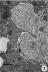
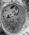
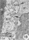
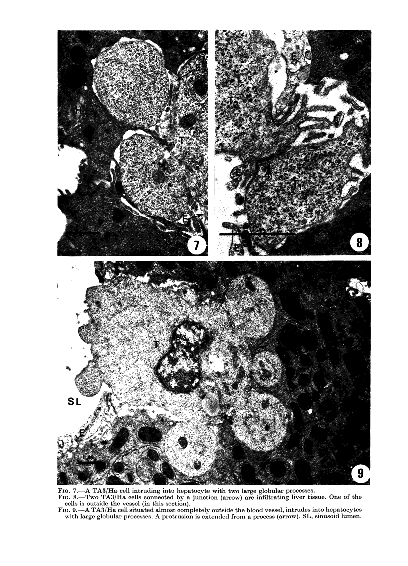
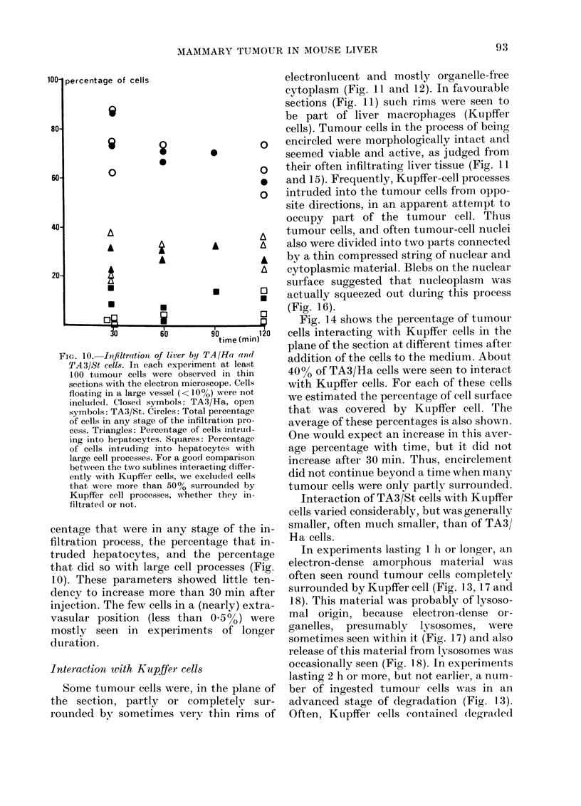
Interactions between TA3 mammary-carcinoma cells and liver cells were studied with the electron microscope in mouse livers that had been perfused with a defined medium containing the tumour cells. Infiltration of liver tissue by the TA3 cells proceeded in the following steps. First, numerous small protrusions were extended through endothelial cells and into hepatocytes. Next, some cells had larger processes deeply indenting hepatocytes. Finally a few tumour cells became located outside the blood vessels. Two variant cell lines, TA3/Ha and TA3/St, differing in cell coat and surface charge, did not differ in the extent of infiltration. TA3/Ha cells were often encircled by thin processes of liver macrophages (Kupffer cells). Encircled cells were initially intact, but later some of them degenerated. These observations suggest that TA3/Ha cells were phagocytized by the Kupffer cells. Encirclement appeared to be inhibited after only 30 min, when many cells were still partly surrounded. Encirclement of TA3/St was much less frequent. After injection of tumour cells intra-portally in vivo, similar results were obtained, which demonstrated the validity of the perfused liver model. TA3/Ha cells formed much fewer tumour nodules in the liver than TA3/St cells.
Full text
PDF




Images in this article
Selected References
These references are in PubMed. This may not be the complete list of references from this article.
- Alexander P., Evans R. Endotoxin and double stranded RNA render macrophages cytotoxic. Nat New Biol. 1971 Jul 21;232(29):76–78. doi: 10.1038/newbio232076a0. [DOI] [PubMed] [Google Scholar]
- Babai F., Tremblay G. Ultrastructural study of liver invasion by Novikoff hepatoma. Cancer Res. 1972 Dec;32(12):2765–2770. [PubMed] [Google Scholar]
- Bucana C., Hoyer L. C., Hobbs B., Breesman S., McDaniel M., Hanna M. G., Jr Morphological evidence for the translocation of lysosomal organelles from cytotoxic macrophages into the cytoplasm of tumor target cells. Cancer Res. 1976 Dec;36(12):4444–4458. [PubMed] [Google Scholar]
- Carr I., McGinty F., Norris P. The fine structure of neoplastic invasion: invasion of liver, skeletal muscle and lymphatic vessels by the Rd/3 tumour. J Pathol. 1976 Feb;118(2):91–99. doi: 10.1002/path.1711180205. [DOI] [PubMed] [Google Scholar]
- Carr I., Underwood J. C., McGinty F., Wood P. The ultrastructure of the local lymphoreticular response to an experimental neoplasm. J Pathol. 1974 Jul;113(3):175–182. doi: 10.1002/path.1711130307. [DOI] [PubMed] [Google Scholar]
- Chambers V. C., Weiser R. S. Brief communication: An electron microscope study of the cytophagocytosis of sarcoma I cells by alloimmune macrophages in vitro. J Natl Cancer Inst. 1973 Oct;51(4):1369–1375. doi: 10.1093/jnci/51.4.1369. [DOI] [PubMed] [Google Scholar]
- Chambers V. C., Weiser R. S. The ultrastructure of sarcoma I cells and immune macrophages during their interaction in the peritoneal cavities of immune C57BL-6 mice. Cancer Res. 1972 Feb;32(2):413–419. [PubMed] [Google Scholar]
- Di Luzio N. R., McNamee R., Olcay I., Kitahama A., Miller R. H. Inhibition of tumor growth by recognition factors. Proc Soc Exp Biol Med. 1974 Jan;145(1):311–315. doi: 10.3181/00379727-145-37800. [DOI] [PubMed] [Google Scholar]
- Dingemans K. P. Behavior of intravenously injected malignant lymphoma cells. A morphologic study. J Natl Cancer Inst. 1973 Dec;51(6):1883–1895. doi: 10.1093/jnci/51.6.1883. [DOI] [PubMed] [Google Scholar]
- Dingemans K. P. Invasion of liver tissue by blood-borne mammary carcinoma cells. J Natl Cancer Inst. 1974 Dec;53(6):1813–1824. doi: 10.1093/jnci/53.6.1813. [DOI] [PubMed] [Google Scholar]
- Dingemans K. P., Roos E., van den Bergh Weerman M. A., van de Pavert I. V. Invasion of liver tissue by tumor cells and leukocytes: comparative ultrastructure. J Natl Cancer Inst. 1978 Mar;60(3):583–598. doi: 10.1093/jnci/60.3.583. [DOI] [PubMed] [Google Scholar]
- FISHER E. R., FISHER B. Electron microscopic, histologic, and histochemical features of the Walker carcinoma. Cancer Res. 1961 May;21:527–531. [PubMed] [Google Scholar]
- Friberg S., Jr Comparison of an immunoresistant and an immunosusceptible ascites subline from murine tumor TA3. I. Transplantability, morphology, and some physicochemical characteristics. J Natl Cancer Inst. 1972 May;48(5):1463–1476. [PubMed] [Google Scholar]
- Friberg S., Jr, Lilliehök B. Evidence for non-exposed H-2 antigens in immunoresistant murine tumour. Nat New Biol. 1973 Jan 24;241(108):112–114. doi: 10.1038/newbio241112a0. [DOI] [PubMed] [Google Scholar]
- Hauschka T. S., Weiss L., Holdridge B. A., Cudney T. L., Zumpft M., Planinsek J. A. Karyotypic and surface features of murine TA3 carcinoma cells during immunoselection in mice and rats. J Natl Cancer Inst. 1971 Aug;47(2):343–359. [PubMed] [Google Scholar]
- Hibbs J. B., Jr Heterocytolysis by macrophages activated by bacillus Calmette-Guérin: lysosome exocytosis into tumor cells. Science. 1974 Apr 26;184(4135):468–471. doi: 10.1126/science.184.4135.468. [DOI] [PubMed] [Google Scholar]
- Miller S. C., Hay E. D., Codington J. F. Ultrastructural and histochemical differences in cell surface properties of strain-specific and nonstrain-specific TA3 adenocarcinoma cells. J Cell Biol. 1977 Mar;72(3):511–529. doi: 10.1083/jcb.72.3.511. [DOI] [PMC free article] [PubMed] [Google Scholar]
- Roos E., Dingemans K. P., van de Pavert I. V., van den Bergh-Weerman M. Invasion of lymphosarcoma cells into the perfused mouse liver. J Natl Cancer Inst. 1977 Feb;58(2):399–407. doi: 10.1093/jnci/58.2.399. [DOI] [PubMed] [Google Scholar]
- Sanford B. H., Codington J. F., Jeanloz R. W., Palmer P. D. Transplantability and antigenicity of two sublines of the TA3 tumor. J Immunol. 1973 May;110(5):1233–1237. [PubMed] [Google Scholar]
- Snodgrass M. J., Hanna M. G., Jr Ultrastructural studies of histiocyte-tumor cell interactions during tumor regression after intralesional injection of Mycobacterium bovis. Cancer Res. 1973 Apr;33(4):701–716. [PubMed] [Google Scholar]


