Abstract
Background
Canal wall down and canal wall up mastoidectomy represent two surgical approaches to middle ear cleft pathology. Very few studies have examined the effects of these procedures both on the patients' well being and on the resources needed to maintain that state. In this study the authors report the outpatient attendance pattern of canal wall down mastoidectomy patients
Methods
This is a retrospective case-note review of 101 patients who underwent a CWD mastoidectomy at Derriford Hospital, Plymouth, UK. All surgery was performed by the senior author (PCW-T) between 1985 and 1997. The main outcome measures were the frequency of outpatients' visits, clinical problems at visit and the percentage of discharged patients.
Results
The studied patients made a total of 1341 outpatient visits between November 1985 and December 1998 with an average of 13.3 visits per patient (median of 11 visits). Almost two thirds of the group still attend for regular follow up. The greatest number of visits occurred in the first 24 months after surgery. The commonest reasons for outpatient visits were the removal of the clinical features of chronic cavity inflammation. Residual/recurrent cholesteatoma, residual perforations and structural cavity problems were infrequent.
Conclusion
CWD mastoidectomy carries an intrinsic morbidity resulting in a long term attendance in the outpatients.
Background
Canal wall down (CWD) and canal wall up (CWU) mastoidectomy represent two surgical approaches to middle ear cleft pathology. A significant amount of literature is available comparing the advantages and disadvantages of both techniques. Hulka and McElveen (1998), in a randomised, blinded, temporal bone study, suggested that canal wall down mastoidectomy was significantly superior to the intact canal wall technique in visualising middle ear pathology. [1]
Numerous modifications have been introduced to CWD mastoidectomy to avoid some of its drawbacks whilst maintaining the good exposure it provides. On the other hand, the use of endoscopes has improved visualisation in CWU techniques. [2,3]
Merchant et al (1997) studied the efficacy of tympanomastoid surgery for control of infection in chronic otitis media. They found that the outcome was not influenced by variables such as CWU versus CWD, primary versus revision surgery, and the extent of disease. [4]
Murphy and Wallis (1998, came to the conclusion that CWU and CWD mastoid surgeries in paediatric patients had similar results. They suggested that variables other than hearing should be used to make treatment decisions regarding the canal wall in paediatric candidates for mastoid surgery. [5]
No study has addressed the outpatient attendance and workload of CWD mastoidectomy patients. The authors believe that it behoves clinicians not only to consider the particular procedures from a technical point of view, but also to examine the effects of those procedures both on the patients' well being and on the resources needed to maintain that state.
Methods
The hospital records of a hundred and one patients who underwent a CWD mastoidectomy by the senior author (PCW-T) during the period 1985–1997 were reviewed and a database of the studied patients was set up using Microsoft Access 97. The end limit was chosen to allow a significant follow-up interval for the group.
Results
Of the 101 patients included in the study, 64 patients were males and 37 were females. The mean age of the studied patients was 43.16 years (± 19.61) with an age range between 4 and 87 years.
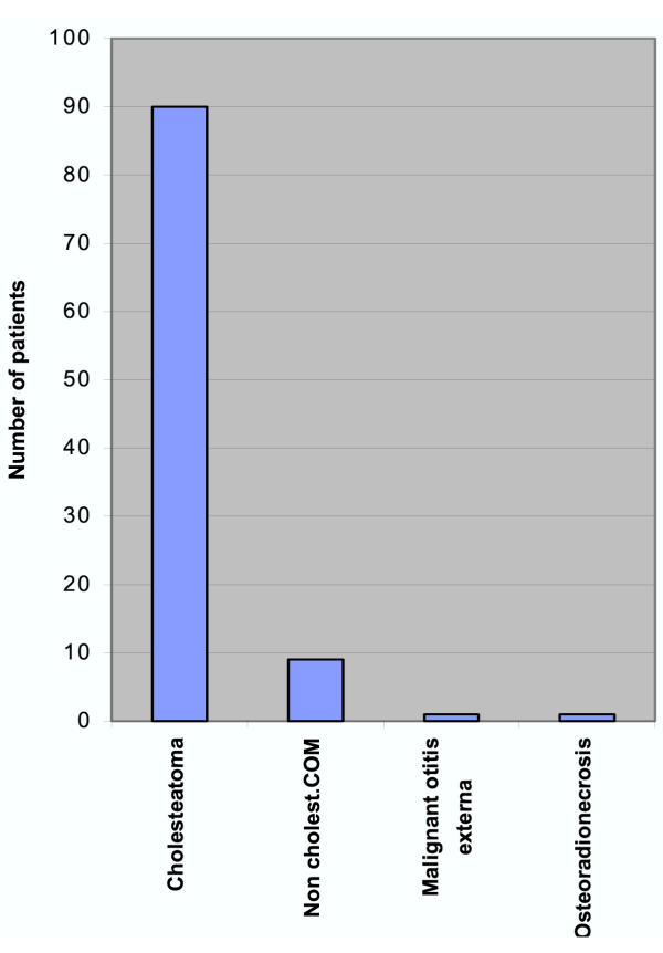
Figure 1 show the clinical diagnosis of the patients. Almost all procedures were carried out for cholesteatoma. In 9 cases the diagnosis was non-cholesteatomatous chronic suppurative otitis media unresponsive to intensive and protracted medical treatment. Two patients with other diagnoses required an open cavity: for osteoradionecrosis following parotid radiotherapy, and for malignant otitis externa as an adjunct to intensive medical therapy.
Figure 1.
Clinical diagnosis
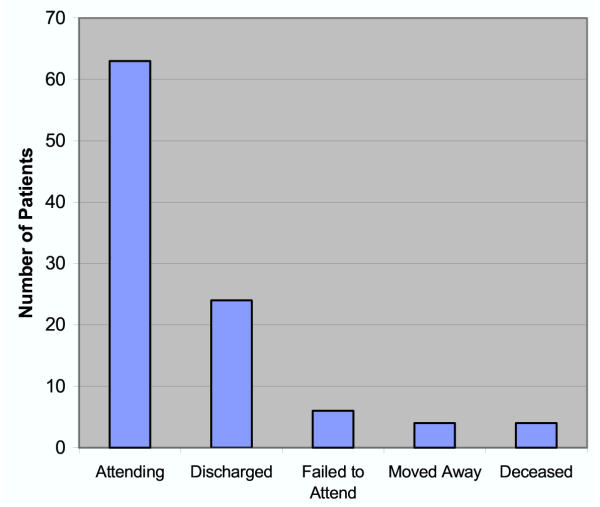
Figure 2 details their current clinical status. Almost two thirds of the group still attend for regular follow-up visits.
Figure 2.
Current Status (n = 101)
There were only two significant postoperative complications: one patient developed a postauricular wound infection with partial dehiscence, and was treated with antibiotics. The second patient developed a mild facial paresis that recovered totally in a few weeks.
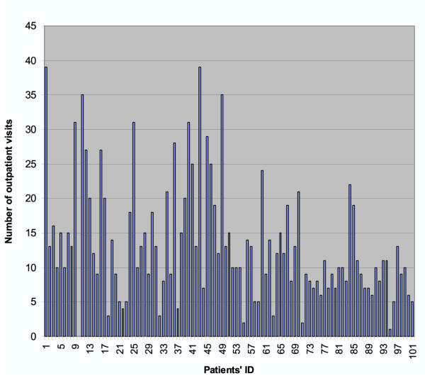
The studied patients made a total of 1341 outpatient visits between November 1985 and December 1998 with an average of 13.3 visits per patient (median of 11 visits). Figure 3 shows the total number of outpatient visits per patient. It is structured chronologically, the first patient being the earliest in the operative series. One patient in the series (case 10) was lost to follow up immediately after the procedure, having moved away. Apart from the obvious decrease in visit numbers that would be expected as the time between the date of operation and the group cut-off limit approaches, there is no discernible trend in visit frequency.
Figure 3.
Total number of outpatient visits for each patient (1985–1998)
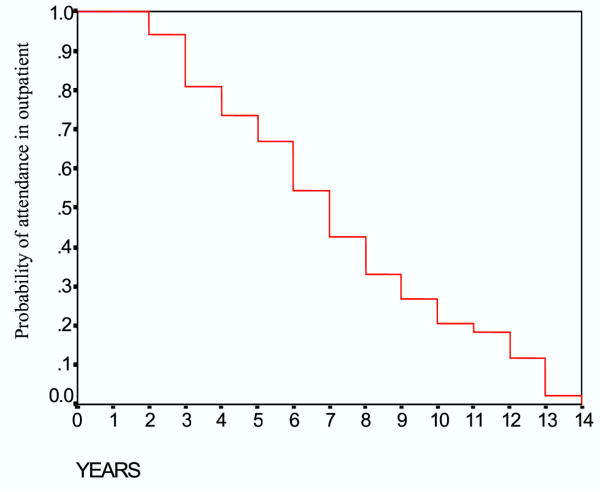
Figure 4 is a life table analysis of outpatient's attendance over the studied period of 158 months. The greatest number of visits occurred in the first 24 months after surgery. Most of these patients were reviewed by the senior author (PCW-T) (Table 1).
Figure 4.
Survival 'attendance' of postoperative visits
Table 1.
Examining clinician in postoperative visits.
| CLINICIAN | NUMBER OF VISITS |
| Consultant | 1076 (80.24%) |
| Registrar | 250 (18.64%) |
| Senior House Officer | 15 (1.12%) |
| Total | 1341 (100%) |
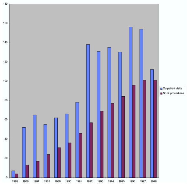
Figure 5 shows number of outpatient visits per year compared with the cumulative number of CWD procedures performed by the senior author during the same period. Only in the last two years of the series has the number of procedures annually shown any decrease. The abrupt change in numbers of visits occurring after 1991 is as yet unexplained.
Figure 5.
Outpatient visits and cumulative number of CWD procedures per year
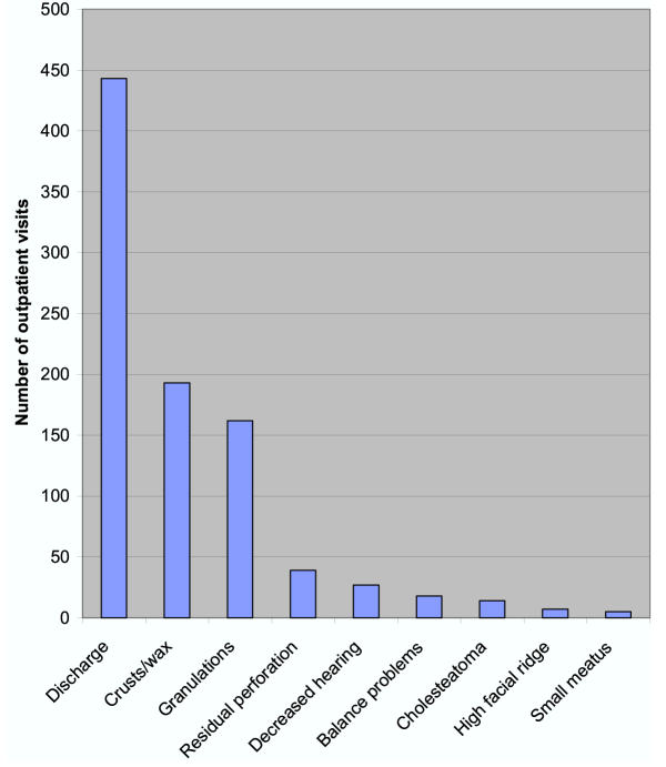
The clinical findings at each visit are shown in figure 6. The commonest reasons for visits were the removal of the clinical features of chronic cavity inflammation (wax, keratin accumulations, discharge, debris and granulation tissue). The other findings, with the exception of balance problems are fixed anatomical features of the cavity, and were usually recorded only once. Nineteen patients (18.81%) had their surgery revised by the senior author (PCW-T). Of these, two patients had obliteration of their mastoid cavities, three had split thickness skin grafts applied and one patient had an obliteration of the eustachian tube orifice. The findings at revision surgery are detailed in Table 2. The remainder (13 patients) had a re-exploration of the cavity with attention to the main areas known to give rise to problems in cavities: high facial ridges, inadequate meati and tympanic segment perforation. Three of these patients were eventually discharged from clinic, and the rest (10 patients) are attending for annual reviews; an indication of an easily manageable cavity.
Figure 6.
Findings noted at the postoperative outpatient visits. (Number of visits = 1341)
Table 2.
Pathological findings at revision surgery (n = 19)
| DIAGNOSIS | OCCURRENCE |
| Discharging cavity | 7 |
| Residual/recurrent cholesteatoma | 5 |
| Granulations | 5 |
| Residual perforation | 4 |
| Small meatus | 3 |
| High facial ridge | 2 |
| Sensorineural Hearing loss | 1 |
Discussion
The choice of the surgical technique for chronic ear disease depends on a number of factors including both the philosophy and preference of the surgeon, the nature of the pathology, and the general health of the patient. The various arguments between the opposing schools of CWD and CWU mastoidectomy have been rehearsed so many times in print that it is unnecessary to repeat them.
CWD mastoidectomy is a safe procedure when performed by experienced otologists or properly supervised trainees. A recent advance, which is now our current practice, is routine facial nerve monitoring. The majority of CWD procedures were performed for chronic suppurative otitis media with cholesteatoma. In our series, there were no major complications. In most cases, the decision to create a cavity is made pre-operatively, the decision being based both on clinical features of the ear disease and the patient's medical status.
The technique is via a postauricular incision, the canal wall being progressively enlarged until the cavity is created (a technique variously described as epi-tympanomastoidectomy, trans-canal mastoidectomy or an "inside-out" mastoidectomy). The philosophy behind the technique as practised by the senior author (PCW-T) is to obtain, by a thorough and meticulous surgical technique, a smooth cavity of the appropriate size relative to the degree of mastoid pneumatisation, no significant facial ridge (the word "ridge" is an historic misnomer, since the course of the nerve is sub-vertical through its mastoid course) and an adequate meatoplasty both to ensure good aeration of the cavity and ease of post-operative toilet. The meatoplasty is formed from an inferiorly based flap, with a wide removal of scaphoid fossa cartilage. At the end of the procedure a series of packs of ribbon gauze impregnated with BIPP (Bismuth Iodoform Paraffin Paste) are inserted. These are removed sequentially over the initial post-operative period, the last pack remaining for up to two months.
The results of a national comparative audit of 611 mastoidectomies by 55 consultants were published by the Royal College of Surgeons of England in 1995. The audit showed that there were a statistically significant greater number of "wet" ears with canal wall down than with canal wall up mastoidectomies. [6] This unsurprising result from an admittedly very small sample of otologists underlines the most significant problem with the open cavity
Variation in the quality of healing in mastoid cavities has never been clearly understood. Rambo, in 1979, suggested that buried mucosa leading to cystic formation is the principal factor responsible for the wide variation in healing, even though all chronic disease has been removed. [7] Youngs studied the histopathological features of material removed from 159 mastoid cavities at revision surgery. The findings included squamous epithelium with acute and chronic inflammation, foreign body granuloma and aural polyps. Of particular note was the very infrequent finding of discharging cavities lined with respiratory epithelium, implying that retained mucosa in mastoid air cells is not a common cause of persistent otorrhoea. [8]
Youngs also studied epithelial migration in 20 mastoid cavities. His findings cast doubt on the assumption that clean trouble-free cavities are maintained by a satisfactorily functioning epithelial migration. [9]
The most frequent findings in our outpatient reviews were discharging cavities, crusts, wax, and granulations. We believe that these problems are inherent to mastoid cavities, and seem to occur at one time or another in most patients with mastoid cavities irrespective of how well a cavity is fashioned. The aim of post-operative care is both to assess the cavity for the remote chance of a complication developing and to maintain the lining of the cavity in as a healthy condition as possible. The conservative treatment offered to our patients included suction of wax/d debris under microscopic control, cautery of granulations with silver nitrate and topical antibiotic/steroid preparations(drops/ ointment) in presence of an infected cavity. Residual/recurrent cholesteatoma, residual perforations, structural cavity problems such as a high facial ridge or a small meatus were very infrequent findings in our patients and these were readily addressed at revision surgery. The question arises as to the "correct" interval between visits in the established cavity. The first question to answer is when a cavity is "established". Some cavities re-epithelialise with rapidity and within a year have a clean dry lining which requires little if any cleaning. These constitute the group who may be discharged. At the other end of the spectrum are those whose cavities never wholly heal, and present with chronic otorrhoea from some part of the cavity. The situation is akin to the troublesome and persistent condition of granular myringitis. This group is relatively small. In between lie a large group of patients whose cavities are intermittently moist, but who, with regular aural toilet at long intervals (measured in months) maintain a status quo. Exacerbations of their discharge may require intensive bursts of outpatient activity with specific areas of mucosa being treated. There appears to be no reliable predictive factor in CWD surgery, though it may be argued anecdotally that the uninfected, discrete cholesteatoma with apparent Eustachian tube patency and a sound pars tympanicum should produce a trouble-free cavity. Advocates of CWU surgery would argue that this case above all others is ideal for conservative surgery!
A recent paper by Sadé, which examined the strategies used in cholesteatoma surgery presented data on 200 CWD procedures. [10] Sadé's findings of chronic discharge parallel our own. The average regular follow-up interval for established cavities in his series was 5 months.
The average time spent in an outpatient appointment by CWD mastoidectomy patients is very variable but is only a few minutes per patient. The majority of the CWD mastoidectomy patients were reviewed in clinic by a senior surgeon (80.24%). Our opinion is that a senior clinician should review patients with established mastoid cavities frequently. There is a risk that patients with problematic cavities reviewed by relatively inexperienced clinicians may have more frequent inappropriate outpatient reviews when revision surgery is required. Even where nurses are placed in charge of regular mastoid cavity toilet, it is mandatory that the senior clinician reviews the cases at regular intervals. There is no doubt that, if treated early and aggressively, many cavity inflammatory episodes may be aborted.
Of the 101 patients studied, 63 are still attending the outpatient; thus a significant number of canal wall down mastoidectomy patients require long-term care outpatient care. This is a resource issue that has never been addressed. In a busy otological practice, the numbers of CWD cavities will steadily increase. If only about one quarter to one third are ever discharged, they will provide an increasing burden on the resources of the practice, which cannot be neglected.
Open cavity mastoidectomy has, for a long time been the principal surgical treatment of middle ear cholesteatoma in the United Kingdom. However, there is an increasing tendency amongst otologists to practice intact canal mastoidectomy. It is apparent from our study that patients with canal wall down mastoidectomy pose a significant workload to the Outpatient Department particularly in the first 1–2 years after surgery. The use of a computerised database specifically designed for otology patients; with particular emphasis on the pathology present at each outpatient department visit makes it easier to retrieve clinical information concerning these patients. It is easier to review such information on a regular basis and make decisions regarding the need for revision surgery for those patients who attend the outpatient department on a more frequent basis.
A more difficult issue concerns the well-being of patients following CWD or CWU surgery. There is increasing interest in the literature concerning the validation of outcomes of treatment modalities. [11] Recent publications have emphasised the need for clinicians to take note of the outcomes of their surgery, not just in terms of technical success, but also in relation to the impact of the treatment upon the patient's lifestyle and well being. It is not uncommon for a patient with a cholesteatoma to have little in the way of handicapping subjective symptoms on presentation; it is the otological findings that make the diagnosis and point the way to a treatment strategy. That strategy however may leave the patient in a "worse" condition than pre-operatively, even though the disease has been cleared and the ear rendered safe. The decision to operate itself may be critical to the outcome for the patient. Both CWD and CWU procedures carry an intrinsic morbidity, and in some frail or compromised patients this may prevent definitive surgical treatment of whichever type. The staged surgery of the CWU procedure may similarly prevent this technique being used on the elderly or infirm. It is necessary for clinicians to explain this to patients, discuss why the particular treatment strategy is being adopted, explain the circumstances which might cause a change in approach and prepare them for the long-term sequelae of surgery.
Conclusion
The decision to treat chronic suppurative otitis media by surgery, in this case by a CWD procedure, is not to be undertaken lightly. Whatever the reason for the procedure, the patient will become a regular visitor to the outpatient for many years to come, and will only be discharged if the cavity is entirely trouble-free and self-cleaning over a number of consecutive visits.
An old epigram states, "Once you have operated on 1000 ears, you need never see another patient". In the case of cholesteatoma surgery these words convey a sad but at present inevitable truth.
Competing Interests
None declared.
Authors' Contributions
HSK: Setting up data base, collection of data, analysis of results, writing of manuscript
PCW-T: Carried out surgery, follow-up of patients, revision of manuscript
Pre-publication history
The pre-publication history for this paper can be accessed here:
Contributor Information
Hisham S Khalil, Email: hkhalil@breathemail.net.
Paul C Windle-Taylor, Email: paul.windle-taylor@phnt.swest.nhs.uk.
References
- Hulka GF, McElveen JT., Jr A randomised blinded study of canal wall up versus canal wall down mastoidectomy determining the differences in viewing middle ear anatomy and pathology. Am J Otol. 1998;19:574–578. [PubMed] [Google Scholar]
- Rosenberg SI, Silverstein H, Hoffer M, Nichols M. Use of endoscopes for chronic ear surgery in children. Arch Otolaryngol Head Neck Surg. 1995;121:870–872. doi: 10.1001/archotol.1995.01890080038007. [DOI] [PubMed] [Google Scholar]
- Youssef TF, Poe DS. Endoscope-assisted second stage tympanomastoidectomy. Laryngoscope. 1997;107:1341–1344. doi: 10.1097/00005537-199710000-00009. [DOI] [PubMed] [Google Scholar]
- Merchant SN, Wang P-C, Jang CH, Glynn RJ, Rauch SD, McKenna MJ, Nadol JB., Jr Efficacy of tympanomastoid surgery for control of infection in active chronic otitis media. Laryngoscope. 1997;107:872–877. doi: 10.1097/00005537-199707000-00007. [DOI] [PubMed] [Google Scholar]
- Murphy TP, Wallis DL. Hearing results in paediatric patients after canal wall up and canal wall down mastoid surgery. Otolaryngol Head Neck Surg. 1998;119:439–443. doi: 10.1016/S0194-5998(98)70099-3. [DOI] [PubMed] [Google Scholar]
- Harkness P, Brown PM, Fowler SM, Grant HR, Ryan RM, Topham JH. Mastoidectomy audit: results of the Royal College of Surgeons of England comparative audit of ENT surgery. Clin Otolaryngol. 1995;20:89–94. doi: 10.1111/j.1365-2273.1995.tb00020.x. [DOI] [PubMed] [Google Scholar]
- Rambo JH. Mastoid surgery: effect of retained mucosa on healing. Ann Otol Rhinol Laryngol. 1979;88:701–707. doi: 10.1177/000348947908800518. [DOI] [PubMed] [Google Scholar]
- Youngs R. The histopathology of mastoidectomy cavities, with particular reference to persistent disease leading to chronic otorrhoea. Clin Otolaryngol. 1992;17:505–510. doi: 10.1111/j.1365-2273.1992.tb01706.x. [DOI] [PubMed] [Google Scholar]
- Youngs R. Epithelial migration in open mastoidectomy cavities. J Laryngol Otol. 1995;109:286–290. doi: 10.1017/s0022215100129949. [DOI] [PubMed] [Google Scholar]
- Sadé J. Surgical planning of the treatment of cholesteatoma and post-operative follow-up. Ann Otol Rhinol Laryngol. 2000;109:372–376. doi: 10.1177/000348940010900406. [DOI] [PubMed] [Google Scholar]
- Wang PC, Nadol JB, Jr, Merchant S, Austin E, Gliklich RE. Validation of Outcomes Survey for adults with chronic suppurative otitis media. Ann Otol Rhinol Laryngol. 2000;109:249–254. doi: 10.1177/000348940010900302. [DOI] [PubMed] [Google Scholar]


