

Abstract
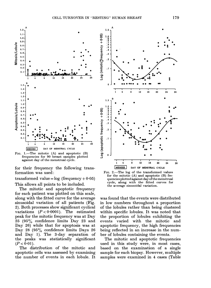
This study examines cell turnover within the lobules of the "resting" human breast and correlates it to the stage of the menstrual cycle. The results are based on the morphological identification of both cell multiplication (mitosis) and cell deletion (apoptosis). It is found that these events undergo significant cyclical changes during the menstrual cycle, with raised levels towards the end of the cycle and during menses. However, in relation to a 28-day menstrual cycle, the position of the mitotic and apoptotic peaks, at Days 25 and 28 respectively, are significantly different. The high values are associated with an increase in the number of lobules showing a slight response rather than a large reaction within a few lobules. It appears that the "resting" breast tissue shows a general, rather than a focal reaction to a given hormonal environment. The possible role of oestrogen and progesterone as effectors of these changes is discussed. Our results show that the menstrual cycle influences cell turnover, though different factors may be affecting mitosis and apoptosis.
Full text
PDF


Selected References
These references are in PubMed. This may not be the complete list of references from this article.
- Carson F. L., Martin J. H., Lynn J. A. Formalin fixation for electron microscopy: a re-evaluation. Am J Clin Pathol. 1973 Mar;59(3):365–373. doi: 10.1093/ajcp/59.3.365. [DOI] [PubMed] [Google Scholar]
- Hopwood D., Levison D. A. Atrophy and apoptosis in the cyclical human endometrium. J Pathol. 1976 Jul;119(3):159–166. doi: 10.1002/path.1711190305. [DOI] [PubMed] [Google Scholar]
- Kerr J. F., Wyllie A. H., Currie A. R. Apoptosis: a basic biological phenomenon with wide-ranging implications in tissue kinetics. Br J Cancer. 1972 Aug;26(4):239–257. doi: 10.1038/bjc.1972.33. [DOI] [PMC free article] [PubMed] [Google Scholar]
- Masters J. R., Drife J. O., Scarisbrick J. J. Cyclic Variation of DNA synthesis in human breast epithelium. J Natl Cancer Inst. 1977 May;58(5):1263–1265. doi: 10.1093/jnci/58.5.1263. [DOI] [PubMed] [Google Scholar]
- Meyer J. S. Cell proliferation in normal human breast ducts, fibroadenomas, and other ductal hyperplasias measured by nuclear labeling with tritiated thymidine. Effects of menstrual phase, age, and oral contraceptive hormones. Hum Pathol. 1977 Jan;8(1):67–81. doi: 10.1016/s0046-8177(77)80066-x. [DOI] [PubMed] [Google Scholar]
- Sandow B. A., West N. B., Norman R. L., Brenner R. M. Hormonal control of apoptosis in hamster uterine luminal epithelium. Am J Anat. 1979 Sep;156(1):15–35. doi: 10.1002/aja.1001560103. [DOI] [PubMed] [Google Scholar]
- West N. B., Norman R. L., Sandow B. A., Brenner R. M. Hormonal control of nuclear estradiol receptor content and the luminal epithelium in the uterus of the golden hamster. Endocrinology. 1978 Nov;103(5):1732–1741. doi: 10.1210/endo-103-5-1732. [DOI] [PubMed] [Google Scholar]
- Wyllie A. H., Kerr J. F., Currie A. R. Cell death: the significance of apoptosis. Int Rev Cytol. 1980;68:251–306. doi: 10.1016/s0074-7696(08)62312-8. [DOI] [PubMed] [Google Scholar]