Abstract
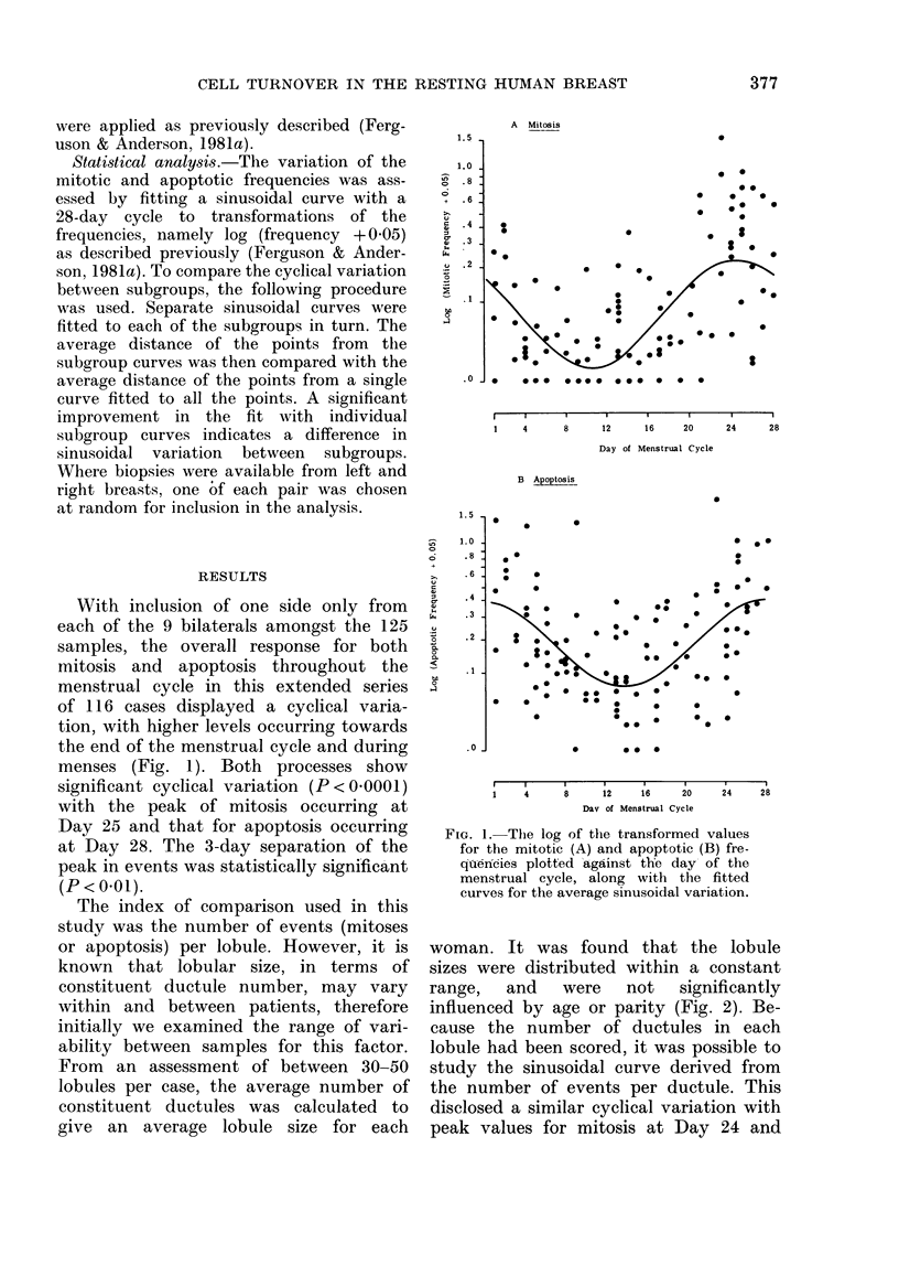
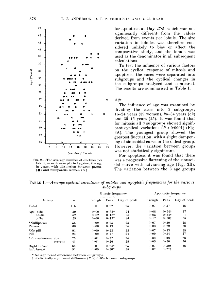
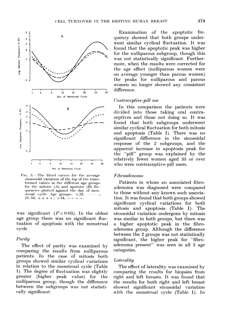
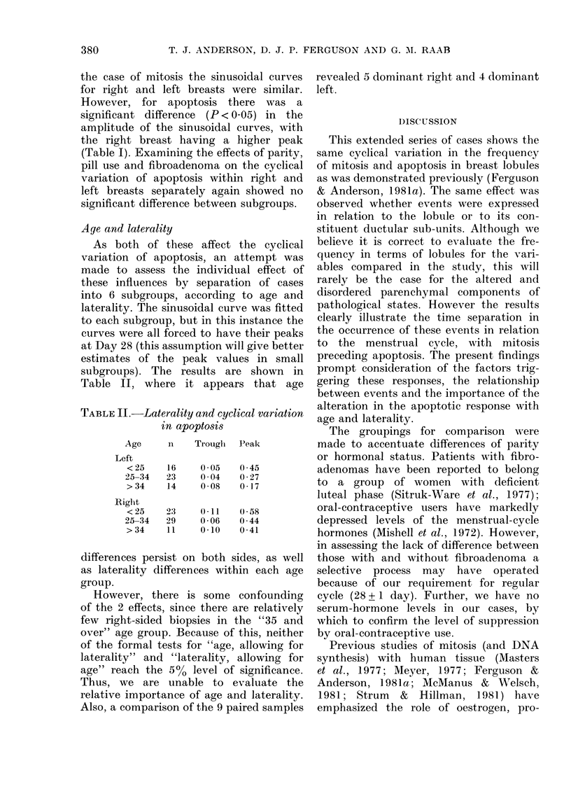
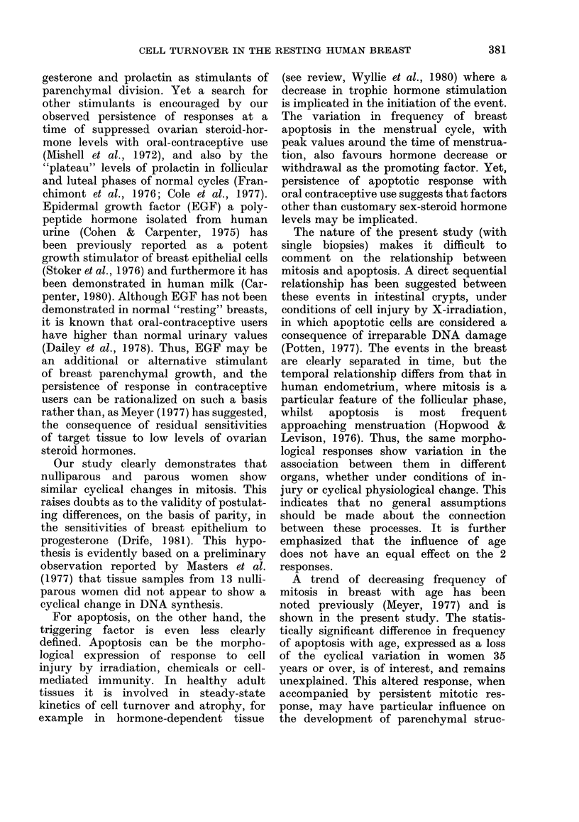
Morphological identification of cell multiplication (mitosis) and cell deletion (apoptosis) within the lobules of the "resting" human breast is used to assess the response of the breast parenchyma to the menstrual cycle. The responses are shown to have a biorhythm in phase with the menstrual cycle, with a 3-day separation of the mitotic and apoptotic peaks. The study fails to demonstrate significant differences in the responses between groups defined according to parity, contraceptive-pill use or presence of fibroadenoma. However, significant differences are found in the apoptotic response according to age and laterality. The results highlight the complexity of modulating influences on breast parenchymal turnover in the "resting" state, and prompt the investigation of other factors as well as steroid hormones and prolactin in the promotion of mitosis. The factors promoting apoptosis in the breast are still not clear.
Full text
PDF
Selected References
These references are in PubMed. This may not be the complete list of references from this article.
- Carpenter G. Epidermal growth factor is a major growth-promoting agent in human milk. Science. 1980 Oct 10;210(4466):198–199. doi: 10.1126/science.6968093. [DOI] [PubMed] [Google Scholar]
- Cohen S., Carpenter G. Human epidermal growth factor: isolation and chemical and biological properties. Proc Natl Acad Sci U S A. 1975 Apr;72(4):1317–1321. doi: 10.1073/pnas.72.4.1317. [DOI] [PMC free article] [PubMed] [Google Scholar]
- Cole E. N., Sellwood R. A., England P. C., Griffiths K. Serum prolactin concentrations in benign breast disease throughout the menstrual cycle. Eur J Cancer. 1977 Jun;13(6):597–603. doi: 10.1016/0014-2964(77)90122-0. [DOI] [PubMed] [Google Scholar]
- Dailey G. E., Kraus J. W., Orth D. N. Homologous radioimmunoassay for human epidermal growth factor (urogastrone). J Clin Endocrinol Metab. 1978 Jun;46(6):929–936. doi: 10.1210/jcem-46-6-929. [DOI] [PubMed] [Google Scholar]
- Drife J. O. Breast cancer, pregnancy, and the pill. Br Med J (Clin Res Ed) 1981 Sep 19;283(6294):778–779. doi: 10.1136/bmj.283.6294.778. [DOI] [PMC free article] [PubMed] [Google Scholar]
- Ferguson D. J., Anderson T. J. Morphological evaluation of cell turnover in relation to the menstrual cycle in the "resting" human breast. Br J Cancer. 1981 Aug;44(2):177–181. doi: 10.1038/bjc.1981.168. [DOI] [PMC free article] [PubMed] [Google Scholar]
- Ferguson D. J., Anderson T. J. Ultrastructural observations on cell death by apoptosis in the "resting" human breast. Virchows Arch A Pathol Anat Histol. 1981;393(2):193–203. doi: 10.1007/BF00431076. [DOI] [PubMed] [Google Scholar]
- Franchimont P., Dourcy C., Legros J. J., Reuter A., Vrindts-Gevaert Y., Van Cauwenberge J. R., Gaspard U. Prolactin levels during the menstrual cycle. Clin Endocrinol (Oxf) 1976 Nov;5(6):643–650. doi: 10.1111/j.1365-2265.1976.tb03867.x. [DOI] [PubMed] [Google Scholar]
- Hopwood D., Levison D. A. Atrophy and apoptosis in the cyclical human endometrium. J Pathol. 1976 Jul;119(3):159–166. doi: 10.1002/path.1711190305. [DOI] [PubMed] [Google Scholar]
- Masters J. R., Drife J. O., Scarisbrick J. J. Cyclic Variation of DNA synthesis in human breast epithelium. J Natl Cancer Inst. 1977 May;58(5):1263–1265. doi: 10.1093/jnci/58.5.1263. [DOI] [PubMed] [Google Scholar]
- McManus M. J., Welsch C. W. Hormone-induced ductal DNA synthesis of human breast tissues maintained in the athymic nude mouse. Cancer Res. 1981 Sep;41(9 Pt 1):3300–3305. [PubMed] [Google Scholar]
- Meyer J. S. Cell proliferation in normal human breast ducts, fibroadenomas, and other ductal hyperplasias measured by nuclear labeling with tritiated thymidine. Effects of menstrual phase, age, and oral contraceptive hormones. Hum Pathol. 1977 Jan;8(1):67–81. doi: 10.1016/s0046-8177(77)80066-x. [DOI] [PubMed] [Google Scholar]
- Mishell D. R., Jr, Thorneycroft I. H., Nakamura R. M., Nagata Y., Stone S. C. Serum estradiol in women ingesting combination oral contraceptive steroids. Am J Obstet Gynecol. 1972 Dec 1;114(7):923–928. doi: 10.1016/0002-9378(72)90098-1. [DOI] [PubMed] [Google Scholar]
- Potten C. S. Extreme sensitivity of some intestinal crypt cells to X and gamma irradiation. Nature. 1977 Oct 6;269(5628):518–521. doi: 10.1038/269518a0. [DOI] [PubMed] [Google Scholar]
- Senie R. T., Rosen P. P., Lesser M. L., Snyder R. E., Schottenfeld D., Duthie K. Epidemiology of breast carcinoma II: factors related to the predominance of left-sided disease. Cancer. 1980 Oct 1;46(7):1705–1713. doi: 10.1002/1097-0142(19801001)46:7<1705::aid-cncr2820460734>3.0.co;2-q. [DOI] [PubMed] [Google Scholar]
- Sitruk-ware L. R., Sterkers N., Mowszowicz I., Mauvais-Jarvis P. Inadequate corpus luteal function in women with benign breast diseases. J Clin Endocrinol Metab. 1977 Apr;44(4):771–774. doi: 10.1210/jcem-44-4-771. [DOI] [PubMed] [Google Scholar]
- Stoker M. G., Pigott D., Taylor-Papadimitriou J. Response to epidermal growth factors of cultured human mammary epithelial cells from benign tumours. Nature. 1976 Dec 23;264(5588):764–767. doi: 10.1038/264764a0. [DOI] [PubMed] [Google Scholar]
- Strum J. M., Hillman E. A. Human breast epithelium in organ culture: effect of hormones on growth and morphology. In Vitro. 1981 Jan;17(1):33–43. doi: 10.1007/BF02618028. [DOI] [PubMed] [Google Scholar]
- Wyllie A. H., Kerr J. F., Currie A. R. Cell death: the significance of apoptosis. Int Rev Cytol. 1980;68:251–306. doi: 10.1016/s0074-7696(08)62312-8. [DOI] [PubMed] [Google Scholar]


