Abstract
Currently available detectors for spores of Bacillus anthracis, the causative agent of anthrax, are inadequate for frontline use and general monitoring. There is a critical need for simple, rugged, and inexpensive detectors capable of accurate and direct identification of B. anthracis spores. Necessary components in such detectors are stable ligands that bind tightly and specifically to target spores. By screening a phage display peptide library, we identified a family of peptides, with the consensus sequence TYPXPXR, that bind selectively to B. anthracis spores. We extended this work by identifying a peptide variant, ATYPLPIR, with enhanced ability to bind to B. anthracis spores and an additional peptide, SLLPGLP, that preferentially binds to spores of species phylogenetically similar to, but distinct from, B. anthracis. These two peptides were used in tandem in simple assays to rapidly and unambiguously identify B. anthracis spores. We envision that these peptides can be used as sensors in economical and portable B. anthracis spore detectors that are essentially free of false-positive signals due to other environmental Bacillus spores.
The gram-positive soil bacterium Bacillus anthracis, the causative agent of anthrax, has been developed into a weapon of mass destruction by numerous foreign governments and terrorist groups (8). The use of B. anthracis as a biological weapon, with severe consequences, was demonstrated in the fall of 2001 in the United States. The threat persists that other releases will occur with even more devastating results. B. anthracis is an effective agent for biological warfare and terrorism primarily because it forms spores. Spores are resistant to extreme temperatures, noxious chemicals, desiccation, and physical damage, which makes them suitable for incorporation into explosive weapons and for concealment in terrorist devices (17). Spores enter the body through skin abrasions or by ingestion or inhalation. Once exposed to internal tissues, the spores germinate and vegetative cell growth ensues, often resulting in the death of the host within several days (9, 24). Natural strains of B. anthracis are sensitive to common antibiotics that can be used to treat anthrax. However, to ensure a successful outcome, treatment must begin within a day or two after exposure to spores (11). Thus, rapid detection of B. anthracis spores is critical in responding to the anthrax threat.
Several detection systems are currently used to identify B. anthracis. The most accurate systems employ either PCR-based assays or traditional phenotyping of cultured bacteria (2, 7, 24). However, these methods are complex, expensive, cumbersome, and slow, typically requiring spore germination and outgrowth of vegetative cells. Other systems, less complex and more portable, are based on antibody binding to spore surface antigens. These systems are relatively fast because they detect spores directly. However, current antibody-based detectors suffer from a lack of accuracy and limited sensitivity, which result in an unacceptably high level of both false-positive and false-negative responses, according to federal government trials (www.gsa/gov/mailpolicy) and other, independent tests (D. King, V. Luna, A. Cannons, J. Cattani, and P. Amuso, Letter, J. Clin. Microbiol. 41:3454-3455, 2003). The lack of accuracy with these systems is compounded by the normal presence in the environment of Bacillus spores that resemble (and share surface antigens with) B. anthracis spores. Particularly problematic are spores of the opportunistic human pathogen B. cereus and the insect pathogen B. thuringiensis, species which, based on genome sequence comparisons, are the most similar to B. anthracis (22). These three species, along with B. mycoides, comprise the phylogenetically similar B. cereus group (1, 20). Therefore, due to the aforementioned limitations and deficiencies, all currently available systems for detecting B. anthracis are inadequate for frontline use by emergency workers and soldiers on the battlefield and for routine monitoring of public areas. Clearly, there is an urgent need for a better detector that can be used where the threat of B. anthracis spore exposure is the greatest.
The desired detector will, in all probability, require simple and hardy ligands capable of tight and specific binding to B. anthracis spores. In this report, we describe the discovery and initial characterization of such ligands: short peptides that bind selectively to spores of B. anthracis. We also identify an unrelated peptide that selectively binds to spores of the Bacillus species most closely related to B. anthracis. We demonstrate that these two classes of peptides permit unambiguous identification of B. anthracis spores via simple fluorescence-based assays. We envision that these peptides can be incorporated into an assortment of platforms to provide simple, rugged, and inexpensive detectors capable of accurate and direct identification of B. anthracis spores.
MATERIALS AND METHODS
Bacterial strains and spores.
The Bacillus strains used in this study and their sources were as follows: the Sterne and ΔAmes strains of B. anthracis, B. cereus T, B. thuringiensis subsp. kurstaki, B. thuringiensis B8, and B. globigii (also called B. atrophaeus and B. subtilis variety niger) were from John Ezzell, U.S. Army Medical Research Institute of Infectious Diseases, Fort Detrick, Md.; B. thuringiensis Al Hakum, B. thuringiensis USDA HD-571, B. cereus 3A (also FRI-41), B. cereus F1-15 (also FRI-43), B. cereus D17 (also FRI-13), and B. cereus S2-8 (also FRI-42) were from Paul Jackson, Los Alamos National Laboratory, Los Alamos, N.Mex.; B. subtilis (trpC2) 1A700 (originally designated 168), B. amyloliquefaciens 10A1 (originally H), B. licheniformis 5A36 (originally ATCC 14580), and B. pumilus 8A3 (originally ATCC 7061) were from the Bacillus Genetic Stock Center, Ohio State University, Columbus; and B. cereus ATCC 4342, B. mycoides ATCC 10206, and B. megaterium ATCC 14581 were from the American Type Culture Collection, Manassas, Va. Spores were produced by cells grown in liquid Difco sporulation medium (18) at 37°C for 48 to 72 h with shaking (except for B. pumilis, which was grown on solid medium at 30°C). Spores were purified by sedimentation through a Renografin step gradient as previously described (5) and were quantitated microscopically using a Petroff-Hausser counting chamber. In the biopanning experiment with B. anthracis ΔAmes spores, the spores were killed by gamma irradiation before use (an initial precautionary measure). In all other biopanning and spore-binding experiments (including those with ΔAmes spores), unirradiated spores were used. Gamma irradiation of spores did not appear to affect peptide binding.
Screening the phage display peptide library.
The New England Biolabs (NEB) Ph.D.-7 Phage Display Peptide Library was biopanned for spore-binding phages, and these phages were analyzed as described previously (26).
Peptide synthesis and fluorochrome conjugation.
Peptides were chemically synthesized and purified by high-performance liquid chromatography; (University of Alabama at Birmingham Peptide Synthesis Core Facility). Peptide molecules were attached to R-phycoerythrin (PE; Prozyme) by using the heterobifunctional cross-linker sulfosuccinimidyl-4-(N-maleimidomethyl)cyclohexane-1-carboxylate (Pierce) (6).
FACS analysis.
Spores (107) were mixed with a peptide-PE conjugate (at a concentration indicated in the text) in 20 μl of phosphate-buffered saline (PBS) (23) and incubated at room temperature for 60 min to ensure complete binding. Unbound conjugate molecules were removed by washing the spores three times in 200-μl volumes of PBS-0.5% Tween 20; after each wash, spores were collected by centrifugation at 820 × g and 4°C for 5 min. Spore-conjugate complexes were resuspended in 200 μl of PBS, and fluorescence was measured by fluorescence-activated cell sorting (FACS) analysis with a FACSCalibur instrument and analyzed with CellQuest Pro software (Becton Dickinson Biosciences). Spore structure was unaffected by this assay as judged by microscopic examination.
Fluorescence microscopy.
Spores (108) were mixed with a peptide-PE conjugate, incubated, and washed essentially as described for the FACS assay. The spore pellet was resuspended in a drop of Fluoromount-G (Electron Microscopy Sciences, Fort Washington, Pa.), and a sample was examined under a Nikon Eclipse E600 microscope equipped with a Y-FL epifluorescence attachment. Fluorescence micrographs were taken with a Spot charge-coupled device camera (Diagnostic Instruments Inc., Sterling Heights, Mich.), using a 5-s exposure time and a gain of 8.
RESULTS
Biopanning a phage display library for peptides that bind B. anthracis spores.
To identify peptides that bind to spores of B. anthracis, we screened (or “biopanned”) the NEB Ph.D.-7 Phage Display Peptide Library for spore-binding phages. In the Ph.D.-7 library, random 7-mer peptides were displayed on the surface of the filamentous coliphage M13 as fusions to the surface-exposed amino terminus of the minor coat protein pIII. Each phage contained five copies of the same peptide-pIII fusion, which was encoded by phage gene III containing a random 21-base insert. The phage display library contained 2 × 10⁹ independent clones. In two separate experiments, we biopanned against purified spores produced by either the ΔAmes or Sterne strain of B. anthracis. The ΔAmes (pXO1−) and Sterne (pXO2−) strains are avirulent due to the absence of a plasmid necessary to produce anthrax toxins or the capsule of the vegetative cell, respectively (16). For each biopanning experiment, spores and phages were mixed to allow binding, spore-phage complexes were collected by centrifugation and washed 10 times, phages were eluted from the complexes, and the eluted phages were amplified by infecting Escherichia coli cells (under conditions similar to those recommended by NEB). The amplified phages were used for a second round of biopanning; in total, four rounds of biopanning were performed, after which the eluted phages (each displaying a putative spore-binding peptide) were plated to obtain single plaques. These plaques (27 and 35 with ΔAmes and Sterne spores, respectively) were used to prepare phage stocks, from which genomic DNA was purified, and the 21-base insert in gene III of each phage was sequenced. The amino acid sequences encoded by the inserts revealed putative spore-binding 7-mer peptides.
Based on related sequences, the peptides were grouped into several families, each defined by a unique consensus sequence. However, only one peptide family was found in both biopanning experiments (i.e., with ΔAmes and Sterne spores). This family, with the consensus sequence TYPXPXR (hereafter referred to as TYP), was the largest in terms of number of phages (19 of 62) and unique peptide sequences (5) (Table 1). Often, more than one phage clone displayed a particular TYP peptide, and this peptide was encoded by the same nucleotide sequence. In one case, the peptide sequence (i.e., TYPLPIR) was encoded by two different nucleotide sequences, as permitted by the degeneracy of the genetic code. Although the TYP consensus sequence was variable at positions 4 and 6, the residues at these positions were typically similar. For example, Leu, Ile, or Val occupied position 4 in all but one unique peptide sequence.
TABLE 1.
Phage display peptides selected for binding to spores of the ΔAmes or Sterne strain of B. anthracis
| Amino acid sequence | Nucleotide sequence | No. of clones/total no. of phages | Target spore |
|---|---|---|---|
| T Y P I P I R | ACT TAT CCT ATT CCG ATT CGT | 3/27 | ΔAmes |
| T Y P I P F R | ACT TAT CCT ATT CCG TTT CGT | 3/27 | ΔAmes |
| T Y P V P H R | ACT TAT CCG GTG CCG CAT CGG | 1/27 | ΔAmes |
| T Y P L P I R | ACG TAT CCG CTT CCG ATT CGG | 8/35 | Sterne |
| T Y P L P I R | ACG TAT CCG CTG CCT ATT AGG | 3/35 | Sterne |
| T Y P P P T R | ACT TAT CCG CCG CCG ACT CGG | 1/35 | Sterne |
Analysis of spore binding by TYP peptides.
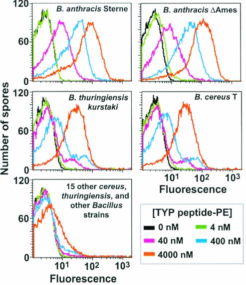
To confirm and analyze the binding of TYP peptides to spores, we employed a FACS assay. This assay required the attachment of a fluorochrome to a test peptide prior to spore binding and analysis of spore-peptide complexes. To this end, we chemically synthesized a representative TYP peptide with the sequence TYPLPIRGGGC; the GGGC extension was included as a carboxy-terminal linker for fluorochrome attachment. Approximately 10 peptide molecules were then attached (using a cross-linker) through their terminal cysteine residues to the ε-amino groups of dispersed lysine residues on one molecule of PE, a 240-kDa highly fluorescent protein. Peptide binding to B. anthracis (Sterne and ΔAmes) spores was then measured by incubating spores with from 4 to 4,000 nM peptide-PE conjugate, removing unbound conjugate by washing, and analyzing spore-peptide complexes by FACS. The results showed essentially identical, concentration-dependent binding of the peptide-PE conjugate to spores of the Sterne and ΔAmes strains (Fig. 1).
FIG. 1.
FACS analysis of the binding of the TYPLPIRGGGC-PE conjugate to selected Bacillus spores. The concentrations of the peptide-PE conjugate (TYP peptide-PE) are indicated. The spore species is indicated in each panel, where possible. The data shown in the bottom panel are for spores of B. cereus ATCC 4342 (representative of the other 15 strains), and the minimal binding at a conjugate concentration of 4,000 nM was not peptide specific. Other details are provided in Materials and Methods.
To examine the specificity of peptide binding, we measured (as described above) the binding of the TYPLPIRGGGC-PE conjugate to spores of 17 other Bacillus strains, including 6 strains of B. cereus (T, ATCC 4342, D17/FRI-13, 3A/FRI-41, S2-8/FRI-42, and F1-15/FRI-43), 4 strains of B. thuringiensis (subsp. kurstaki, B8, Al Hakum, and USDA HD-571), and 1 strain each of B. mycoides, B. pumilus, B. globigii, B. amyloliquefaciens, B. subtilis, B. licheniformis, and B. megaterium. These strains were all members of Bacillus group 1 (of 5), within which B. anthracis, B. cereus, B, thuringiensis, and B. mycoides comprise the closely related B. cereus group. Seven of these strains—i.e., B. thuringiensis strains Al Hakum and USDA HD-57 and all B. cereus strains except T—are human pathogens and nearest neighbors to B. anthracis as determined by amplified fragment length polymorphism analysis (21). The binding assays showed that the peptide-PE conjugate did not bind to 15 of the other Bacillus strains (Fig. 1) (minimal binding at a conjugate concentration of 4,000 nM was due to nonspecific entrapment). Peptide binding was detected for spores of B. cereus T and B. thuringiensis subsp. kurstaki, but this binding was weaker (or less extensive) than that observed with B. anthracis Sterne and ΔAmes spores. These results indicated a high degree of specificity in TYPLPIR binding to spores but revealed that binding was not absolutely restricted to B. anthracis spores.
To control for nonspecific binding in each experiment shown in Fig. 1, several dissimilar 11-mer peptides (for example, HWHHHGHGGGC and ILPRPYTGGGC, the latter being a scrambled version of a TYP peptide) were attached to PE as described above. These conjugates were tested for spore binding. No significant binding was detected. In a related control experiment, we showed that binding of the TYPLPIRGGGC-PE conjugate to B. anthracis spores was not inhibited by inclusion of bovine serum albumin at 10 mg/ml in the binding and wash buffers. Furthermore, we demonstrated that the TYPLPIRGGGC-PE conjugate did not bind to vegetative cells of the Sterne and ΔAmes strains (data not shown).
Enhanced spore binding by ATYPLPIR.
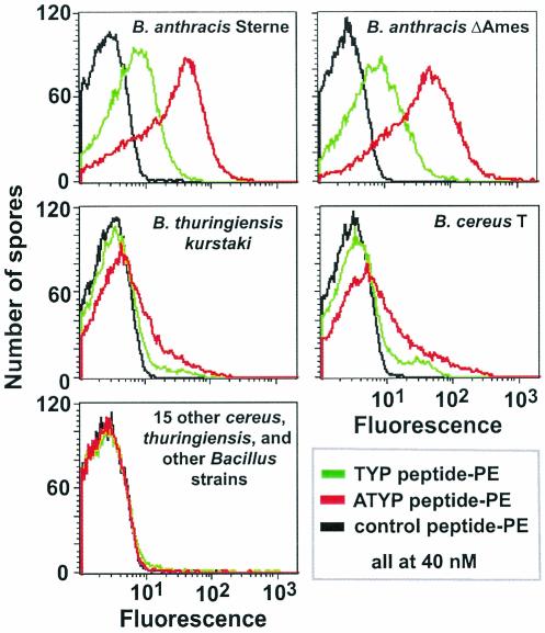
In a separate study, we identified a family of 7-mer or 12-mer peptides that selectively bound B. subtilis spores (12). This family contained a five-residue consensus sequence that permitted spore binding only when present at the amino terminus of a peptide or protein. To determine if TYP also required a free amino terminus for spore binding, we synthesized a peptide with the sequence ATYPLPIRGGGC and attached it to PE as described above. The spore-binding ability of this conjugate was compared to that of TYPLPIRGGGC-PE, with both conjugates used at a concentration of 40 nM (Fig. 2). The results showed clearly that the TYPLPIR sequence did not require a free amino terminus to bind spores. In fact, the addition of an Ala residue permitted nearly 10-fold-enhanced binding to both Sterne and ΔAmes spores. Binding to B. thuringiensis subsp. kurstaki and B. cereus T spores was only slightly enhanced by the Ala addition, while this modification still did not permit detectable binding to spores of all other species examined. These experiments provided us with an improved ligand for B. anthracis spores, namely ATYPLPIR, and suggested that even better peptide ligands can be produced by additional modifications. Presumably, ATYP (i.e., ATYPXPXR) and TYP peptides bind to the same spore receptor, although this remains to be confirmed.
FIG. 2.
FACS analysis comparing the abilities of the TYPLPIRGGGC-PE (TYP peptide-PE) and ATYPLPIRGGGC-PE (ATYP peptide-PE) conjugates to bind to selected Bacillus spores. Also shown are the binding results for a control peptide (HWHHHGHGGGC)-PE conjugate. Spores were mixed with a 40 nM concentration of each peptide-PE conjugate.
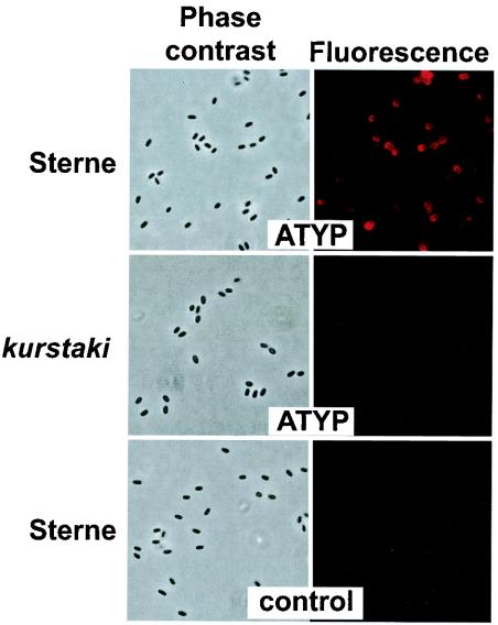
To demonstrate directly that the ATYPLPIR peptide was binding to the spore surface, we examined by fluorescence microscopy the binding of the ATYPLPIRGGGC-PE conjugate to B. anthracis spores. The results showed that when binding occurred at a conjugate concentration of 400 nM and unbound conjugate was removed by washing, every Sterne or ΔAmes spore was completely encircled by fluorescent ligand (data not shown). At this concentration of conjugate, low-level but detectable binding to spores of B. thuringiensis subsp. kurstaki and B. cereus T (but not other strains) was observed. We then attempted to identify conditions under which only spores of B. anthracis would bind enough conjugate to be detectable by fluorescence microscopy. We found that at a conjugate concentration of 40 nM, Sterne and ΔAmes spores were readily detectable, although with somewhat uneven fluorescence, while spores of the other 17 Bacillus strains examined in this study were essentially nonfluorescent (Fig. 3). In addition, we used several control peptide-PE conjugates to confirm that fluorescent labeling of spores required the ATYPLPIR sequence (Fig. 3 and data not shown). The reason for the uneven fluorescence observed with B. anthracis spores at 40 nM ATYPLPIRGGGC-PE is not known, but it appears to be unrelated to spore damage (e.g., loss of the outer spore layer).
FIG. 3.
Fluorescence microscopy showing selective binding of ATYPLPIRGGGC-PE to B. anthracis spores. The indicated spores (B. anthracis Sterne or B. thuringiensis subsp. kurstaki) were mixed with a 40 nM concentration of either ATYPLPIRGGGC-PE (ATYP) or a control peptide (ILPRPYTGGGC)-PE conjugate and examined by either phase-contrast or fluorescence microscopy.
Use of two peptides for unambiguous spore identification.
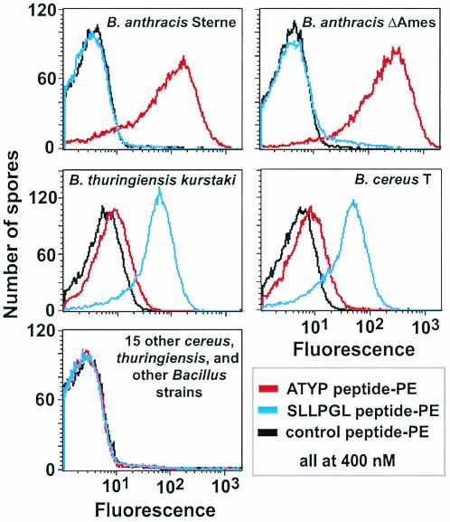
In yet another biopanning experiment, we screened the Ph.D.-7 Phage Display Peptide Library for peptides that bind to spores of an uncharacterized Bacillus strain (probably a strain of B. cereus or B. thuringiensis) that we had isolated from an environmental sample (data not shown). These peptides revealed a single consensus sequence, SLLPGL, which was subsequently shown to bind to spores (but not to vegetative cells) of only two strains in our collection, B. thuringiensis subsp. kurstaki and B. cereus T. The fact that the SLLPGL peptides bind well to spores of B. thuringiensis subsp. kurstaki and B. cereus T, the only non-B. anthracis spores that were found to bind TYP peptides, suggested that SLLPGL peptides could be used in tandem with TYP or ATYP peptides to unambiguously identify B. anthracis spores. To demonstrate this application, we synthesized an SLLPGLPGGGC-PE conjugate that was equivalent to the previously described ATYPLPIRGGGC-PE conjugate. We then used a FACS assay to compare the abilities of the SLLPGL and ATYP conjugates (at 400 nM concentrations) to bind to spores of the 19 Bacillus strains used in this study (Fig. 4). The binding pattern for spores of both strains of B. anthracis was unique, with extensive binding by the ATYP conjugate and no binding by the SLLPGL conjugate. In clear contrast, the binding pattern for spores of B. thuringiensis subsp. kurstaki and B. cereus T was essentially reversed, with extensive binding by the SLLPGL conjugate and limited binding by the ATYP conjugate. No peptide-conjugate binding was observed with the other 15 spore types. Thus, the SLLPGL and ATYP conjugates can be used to clearly distinguish between spores of B. anthracis and the other Bacillus species examined.
FIG. 4.
FACS analysis contrasting the binding of the ATYPLPIRGGGC-PE (ATYP peptide-PE) and SLLPGLPGGGC-PE (SLLPGL peptide-PE) conjugates to selected Bacillus spores. Also shown are the binding results for a control peptide (HWHHHGHGGGC)-PE conjugate. Spores were mixed with a 400 nM concentration of each peptide-PE conjugate.
DISCUSSION
In this study, we identified short peptides (i.e., ATYPLPIR and SLLPGLP) whose differential spore-binding abilities can be used to discriminate between spores of B. anthracis and those of other Bacillus species. For example, the ATYPLPIR peptide binds well to B. anthracis spores but not to other spores, with the exception of weaker binding to spores of an apparently small subset of B. cereus and B. thuringiensis strains. In contrast, the SLLPGLP peptide does not bind B. anthracis (or most other Bacillus species) spores but binds well to the subset of B. cereus and B. thuringiensis spores that bind ATYPLPIR. Thus, by comparing the spore-binding abilities of the two peptides, it is possible to unambiguously identify B. anthracis spores. This discrimination is remarkable because it distinguishes spores of B. anthracis from spores of other members of the closely related B. cereus group. Apparently, spores of this group contain species-specific surface features (e.g., peptide receptors), which may reflect the different ecological niches and/or hosts of these species (20).
The obvious next question is whether the peptides will discriminate between B. anthracis and non-B. anthracis spores when more strains are examined. Answering this question will require the testing of a larger panel of Bacillus (and even non-Bacillus) spores, a study that we will undertake soon. Our first goal will be to examine a large number of virulent B. anthracis strains. The present study included only the avirulent Sterne and ΔAmes strains; however, these strains are likely to adequately represent the species for several reasons. The Sterne (pXO2−) and ΔAmes (pXO1−) strains differ from virulent strains only in the absence of one of two plasmids, neither of which is likely to alter the spore surface (22). In addition, spores produced by the ΔAmes and Sterne strains appear to be essentially identical to spores of virulent strains except for superficial differences in the length of the hair-like nap on the spore surface (13, 15, 25). Finally, B. anthracis strains are highly monomorphic, with genes from different isolates typically having greater than 99% nucleotide sequence identity (19).
If the peptides identified in this study are indeed generally useful in identifying B. anthracis spores, they offer several advantages in detector design. They bind directly to the spore, eliminating the need for extracting spore components or for growing vegetative cells. They can be easily incorporated, covalently if necessary, into detectors presently employing antibodies or into detection platforms that cannot accommodate antibodies because of size limitations or denaturing conditions. They can be easily and differentially labeled with assayable tags, such as luminescent quantum dots that provide a signal sufficient to detect a single spore (3). They can be produced rapidly and inexpensively. Finally, the use of two peptides should eliminate or greatly reduce the incidence of false-positive signals. We expect that the peptides described in this paper can be used as the probe for B. anthracis spores in simple, inexpensive, and portable detectors based on an assortment of analytical platforms. In our study we employed assays based on increased fluorescence, but binding of peptides to spores can be detected by many other analytical techniques (10, 14).
We have not yet identified the sites on the B. anthracis spore surface to which ATYP and TYP peptides bind. However, the most likely binding sites are components of the exosporium, a prominent, loose-fitting, balloon-like layer that encloses the spores of B. cereus group strains. The exosporium, which is composed of a basal layer and an external hair-like nap, serves as a primary permeability barrier that would exclude the M13 phage and PE (4). Preliminary experiments performed in our laboratory indicate that the binding sites for ATYP and TYP peptides are on the basal layer of the B. anthracis exosporium. In addition, we have not yet determined whether SLLPGL peptides fail to bind spores of B. anthracis because of the absence of receptors or because access to these receptors is blocked. Further characterization of peptide receptors and factors that influence peptide-receptor interactions is in progress.
Acknowledgments
We thank R. Brice Vinson for contributing to this work, John Kearney for valuable discussions, and Millie Donlon for continuous support.
D.D.W. was supported by the Medical Scientist Training Program at the University of Alabama at Birmingham. This work was funded by DARPA grant MDA972-01-1-0030 and ARO grant DAAD19-00-1-0032.
REFERENCES
- 1.Ash, C., J. A. E. Farrow, S. Wallbanks, and M. D. Collins. 1991. Phylogenetic heterogeneity of the genus Bacillus revealed by comparative analysis of small-subunit-ribosomal RNA sequences. Lett. Appl. Microbiol. 13:202-206. [Google Scholar]
- 2.Bell, C. A., J. R. Uhl, T. L. Hadfield, J. C. David, R. F. Meyer, T. F. Smith, and F. R. Cockerill III. 2002. Detection of Bacillus anthracis DNA by LightCycler PCR. J. Clin. Microbiol. 40:2897-2902. [DOI] [PMC free article] [PubMed] [Google Scholar]
- 3.Chan, W. C., D. J. Maxwell, X. Gao, R. E. Bailey, M. Han, and S. Nie. 2002. Luminescent quantum dots for multiplexed biological detection and imaging. Curr. Opin. Biotechnol. 13:40-46. [DOI] [PubMed] [Google Scholar]
- 4.Gerhardt, P. 1967. Cytology of Bacillus anthracis. Fed. Proc. 26:1504-1517. [PubMed] [Google Scholar]
- 5.Henriques, A. O., B. W. Beall, K. Roland, and C. P. Moran, Jr. 1995. Characterization of cotJ, a σE-controlled operon affecting the polypeptide composition of the coat of Bacillus subtilis spores. J. Bacteriol. 177:3394-3406. [DOI] [PMC free article] [PubMed] [Google Scholar]
- 6.Hermanson, G. T. 1995. Bioconjugate techniques. Academic Press, San Diego, Calif.
- 7.Higgins, J. A., M. Cooper, L. Schroeder-Tucker, S. Black, D. Miller, J. S. Karns, E. Manthey, R. Breeze, and M. L. Perdue. 2003. A field investigation of Bacillus anthracis contamination of U.S. Department of Agriculture and other Washington, D.C., buildings during the anthrax attack of October 2001. Appl. Environ. Microbiol. 69:593-599. [DOI] [PMC free article] [PubMed] [Google Scholar]
- 8.Inglesby, T. V., D. A. Henderson, J. G. Bartlett, M. S. Ascher, E. Eitzen, A. M. Friedlander, J. Hauer, J. McDade, M. T. Osterholm, T. O'Toole, G. Parker, T. M. Perl, P. K. Russell, and K. Tonat. 1999. Anthrax as a biological weapon: medical and public health management. JAMA 281:1735-1745. [DOI] [PubMed] [Google Scholar]
- 9.Inglesby, T. V., T. O'Toole, D. A. Henderson, J. G. Bartlett, M. S. Ascher, E. Eitzen, A. M. Friedlander, J. Gerberding, J. Hauer, J. Hughes, J. McDade, M. T. Osterholm, G. Parker, T. M. Perl, P. K. Russell, and K. Tonat. 2002. Anthrax as a biological weapon, 2002: updated recommendations for management. JAMA 287:2236-2252. [DOI] [PubMed] [Google Scholar]
- 10.Ivnitski, D., I. Abdel-Hamid, P. Atanasov, and E. Wilkins. 1999. Biosensors for detection of pathogenic bacteria. Biosens. Bioelectron. 14:599-624. [DOI] [PubMed] [Google Scholar]
- 11.Jernigan, J. A., D. S. Stephens, D. A. Ashford, C. Omenaca, M. S. Topiel, M. Galbraith, M. Tapper, T. L. Fisk, S. Zaki, T. Popovic, R. F. Meyer, C. P. Quinn, S. A. Harper, S. K. Fridkin, J. J. Sejvar, C. W. Shepard, M. McConnell, J. Guarner, W. J. Shieh, J. M. Malecki, J. L. Gerberding, J. M. Hughes, and B. A. Perkins. 2001. Bioterrorism-related inhalational anthrax: the first 10 cases reported in the United States. Emerg. Infect. Dis. 7:933-944. [DOI] [PMC free article] [PubMed] [Google Scholar]
- 12.Knurr, J., O. Benedek, J. Heslop, R. B. Vinson, J. A. Boydston, J. McAndrew, J. F. Kearney, and C. L. Turnbough, Jr. Peptide ligands that bind selectively to spores of Bacillus subtilis and closely related species. Appl. Environ. Microbiol., in press. [DOI] [PMC free article] [PubMed]
- 13.Kramer, M. J., and I. L. Roth. 1968. Ultrastructural differences in the exosporium of the Sterne and Vollum strains of Bacillus anthracis. Can. J. Microbiol. 14:1297-1299. [DOI] [PubMed] [Google Scholar]
- 14.Luppa, P. B., L. J. Sokoll, and D. W. Chan. 2001. Immunosensors—principles and applications to clinical chemistry. Clin. Chim. Acta 314:1-26. [DOI] [PubMed] [Google Scholar]
- 15.Mikesell, P., B. E. Ivins, J. D. Ristroph, and T. M. Dreier. 1983. Evidence for plasmid-mediated toxin production in Bacillus anthracis. Infect. Immun. 39:371-376. [DOI] [PMC free article] [PubMed] [Google Scholar]
- 16.Mock, M., and A. Fouet. 2001. Anthrax. Annu. Rev. Microbiol. 55:647-671. [DOI] [PubMed] [Google Scholar]
- 17.Nicholson, W. L., N. Munakata, G. Horneck, H. J. Melosh, and P. Setlow. 2000. Resistance of Bacillus endospores to extreme terrestrial and extraterrestrial environments. Microbiol. Mol. Biol. Rev. 64:548-572. [DOI] [PMC free article] [PubMed] [Google Scholar]
- 18.Nicholson, W. L., and P. Setlow. 1990. Sporulation, germination and outgrowth, p. 391-450. In C. R. Harwood and S. M. Cutting (ed.), Molecular biological methods for Bacillus. John Wiley & Sons, Ltd., West Sussex, United Kingdom .
- 19.Price, L. B., M. Hugh-Jones, P. J. Jackson, and P. Keim. 1999. Genetic diversity in the protective antigen gene of Bacillus anthracis. J. Bacteriol. 181:2358-2362. [DOI] [PMC free article] [PubMed] [Google Scholar]
- 20.Priest, F. G. 1993. Systematics and ecology of Bacillus, p. 3-16. In A. L. Sonenshein, J. A. Hoch, and R. Losick (ed.), Bacillus subtilis and other gram-positive bacteria: biochemistry, physiology, and molecular biology. American Society for Microbiology, Washington, D.C.
- 21.Radnedge, L., P. G. Agron, K. K. Hill, P. J. Jackson, L. O. Ticknor, P. Keim, and G. L. Andersen. 2003. Genome differences that distinguish Bacillus anthracis from Bacillus cereus and Bacillus thuringiensis. Appl. Environ. Microbiol. 69:2755-2764. [DOI] [PMC free article] [PubMed] [Google Scholar]
- 22.Read, T. D., S. N. Peterson, N. Tourasse, L. W. Baillie, I. T. Paulsen, K. E. Nelson, H. Tettelin, D. E. Fouts, J. A. Eisen, S. R. Gill, E. K. Holtzapple, O. A. Okstad, E. Helgason, J. Rilstone, M. Wu, J. F. Kolonay, M. J. Beanan, R. J. Dodson, L. M. Brinkac, M. Gwinn, R. T. DeBoy, R. Madpu, S. C. Daugherty, A. S. Durkin, D. H. Haft, W. C. Nelson, J. D. Peterson, M. Pop, H. M. Khouri, D. Radune, J. L. Benton, Y. Mahamoud, L. Jiang, I. R. Hance, J. F. Weidman, K. J. Berry, R. D. Plaut, A. M. Wolf, K. L. Watkins, W. C. Nierman, A. Hazen, R. Cline, C. Redmond, J. E. Thwaite, O. White, S. L. Salzberg, B. Thomason, A. M. Friedlander, T. M. Koehler, P. C. Hanna, A. B. Kolsto, and C. M. Fraser. 2003. The genome sequence of Bacillus anthracis Ames and comparison to closely related bacteria. Nature 423:81-86. [DOI] [PubMed] [Google Scholar]
- 23.Sambrook, J., E. F. Fritsch, and T. Maniatis. 1989. Molecular cloning: a laboratory manual, 2nd ed. Cold Spring Harbor Laboratory, Cold Spring Harbor, N.Y.
- 24.Swartz, M. N. 2001. Recognition and management of anthrax—an update. N. Engl. J. Med. 345:1621-1626. [DOI] [PubMed] [Google Scholar]
- 25.Sylvestre, P., E. Couture-Tosi, and M. Mock. 2003. Polymorphism in the collagen-like region of the Bacillus anthracis BclA protein leads to variation in exosporium filament length. J. Bacteriol. 185:1555-1563. [DOI] [PMC free article] [PubMed] [Google Scholar]
- 26.Turnbough, C. L., Jr. 2003. Discovery of phage display peptide ligands for species-specific detection of Bacillus spores. J. Microbiol. Methods 53:263-271. [DOI] [PubMed] [Google Scholar]


