

Abstract
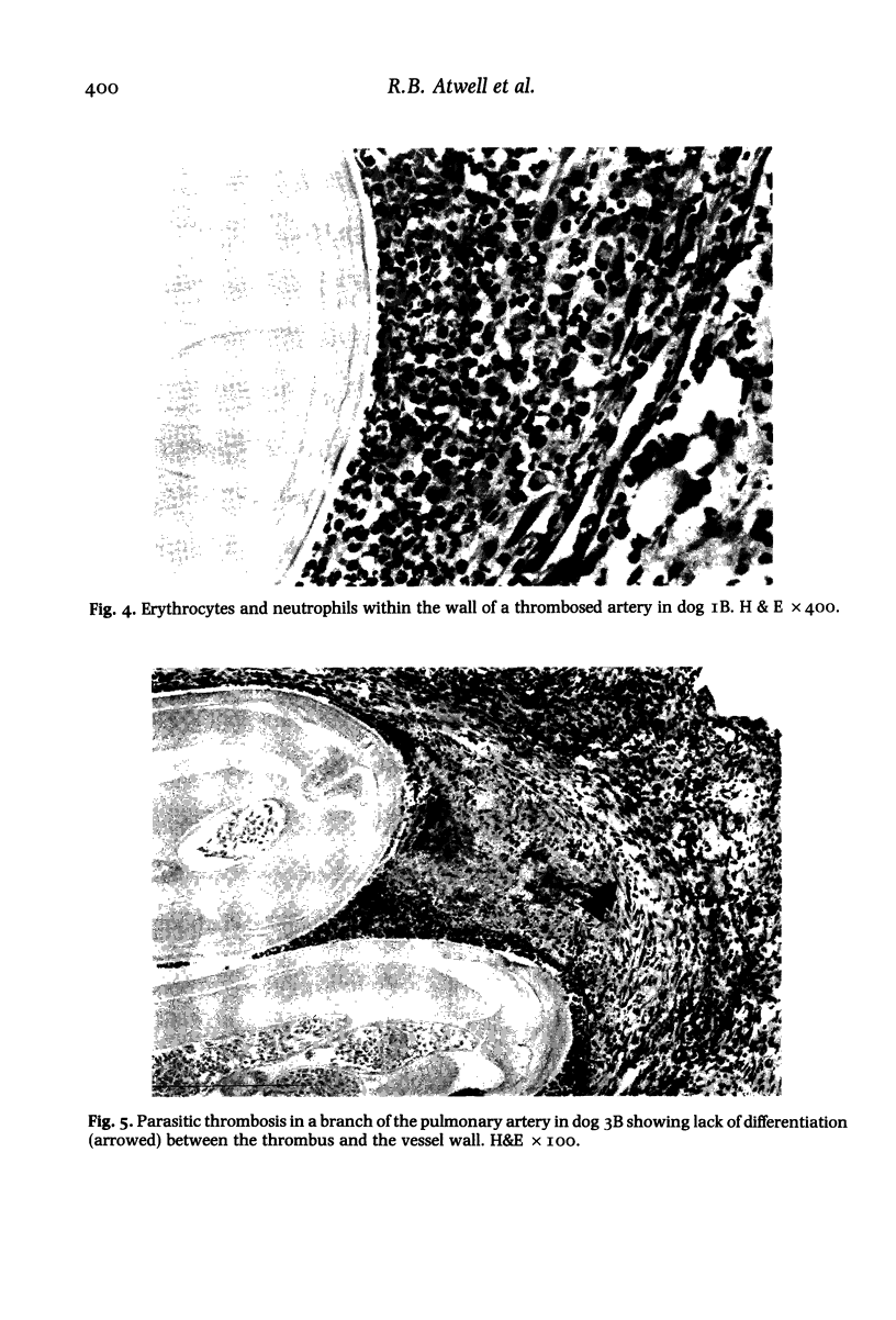
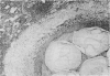
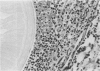
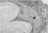
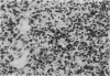
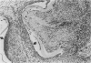
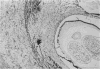
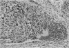
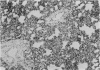
Dead adult D. immitis were inserted into the pulmonary arteries of 10 healthy pups, five of which were given a crude antigenic extract of D. immitis subcutaneously. Two dogs, one from each group were then killed 1, 3, 5, 7 and 9 days after the insertion of filariae and the associated pulmonary artery pathology examined. Microscopically, haemorrhage was evident at day 1, followed by suppuration and organization at days 5 to 7, with organization, fibrosis and focal pleural retraction evident on days 7 to 9. Within arteries haemorrhage and thrombosis was evident, particularly on days 1, 3 and 5. Histology confirmed the extent of haemorrhage evident grossly and, as no rupture was evident, suggested it to be due to diapedesis. Wall disruption occurred due to the local pressure effects of impacted filariae, but also due to inflammation, which was initially suppurative, later being replaced by plasma cell and lymphocyte involvement. Maximal wall disruption occurred at day 5 followed by organization and fibrosis. After day 5 there was more intense organization in the dogs given antigen, in association with the development of interstitial thickening, a finding previously reported in dogs with similar antigenic exposure.
Full text
PDF


Images in this article

Selected References
These references are in PubMed. This may not be the complete list of references from this article.
- Atwell R. B., Buoro I. B., Sutton R. H. Experimental production of lesions in canine pulmonary arteries similar to those produced by Dirofilaria immitis infection. Vet Rec. 1985 May 18;116(20):539–541. doi: 10.1136/vr.116.20.539. [DOI] [PubMed] [Google Scholar]
- Atwell R. B., Sutton R. H., Moodie E. W. Preliminary report on the pulmonary pathology associated with subcutaneous injections of Dirofilaria immitis antigen. Vet Res Commun. 1983 Jan;6(1):59–62. doi: 10.1007/BF02214897. [DOI] [PubMed] [Google Scholar]
- Sutton R. H., Atwell R. B. Lesions of pulmonary pleura associated with canine heartworm disease. Vet Pathol. 1985 Nov;22(6):637–639. doi: 10.1177/030098588502200620. [DOI] [PubMed] [Google Scholar]