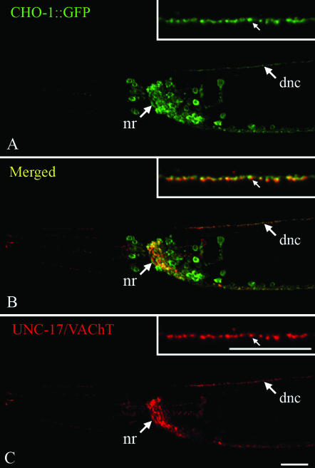
Figure 3.—
A functional CHO-1∷GFP fusion is localized to cholinergic synapses. Transgenic animals were stained with anti-GFP (green, A and B) and anti-UNC-17/VAChT (red, B and C) antibodies. Head region, anterior is to the left, ventral is down, and the bar is ∼10 μm. Insets are magnified sections of the dorsal nerve cord and correspond to ∼20 μm. The positions of the nerve ring (nr) and dorsal nerve cord (dnc) are indicated. The CHO-1∷GFP fusion is localized to cholinergic synapses and is slightly offset from the pool of VAChT-containing synaptic vesicles.

