Abstract
New methods that allow, for the first time, genetic analysis in Archaea of the genus Methanosarcina are presented. First, several autonomously replicating plasmid shuttle vectors have been constructed based on the naturally occurring plasmid pC2A from Methanosarcina acetivorans. These vectors replicate in 9 of 11 Methanosarcina strains tested and in Escherichia coli. Second, a highly efficient transformation system based upon introduction of DNA by liposomes has been developed. This method allows transformation frequencies of as high as 2 × 10⁸ transformants per microgram of DNA per 109 cells or ≈20% of the recipient population. During the course of this work, the complete 5467-bp DNA sequence of pC2A was determined. The implications of these findings for the future of methanoarchaeal research are also discussed.
Methanoarchaea play a key role in the global carbon cycle by recycling organic carbon from anaerobic environments into the atmosphere as methane gas. One of the most significant achievements in microbiology in recent years has been the recognition that Archaea, including the methanoarchaea, constitute a truly unique form of life, evolutionarily distinct from both eukaryotes and eubacteria (1, 2). Biochemical study of the methanogenic pathways of methanoarchaea has provided a wealth of information on novel enzyme mechanisms and cofactors (3, 4). However, apart from the methanogenic pathways, very little is known about these organisms. Because there is a dearth of genetic methods available for examination of methanoarchaea, the study of these organisms has been largely confined to biochemical and physiological approaches. Thus, relatively little is known about other aspects of their metabolism, including the pathways by which they synthesize their unique cofactors and other cellular components, as well as how they regulate gene expression in response to changes in their environment.
Development of methods for genetic analysis of methanoarchaea is crucial for increasing our understanding of how they adapt and survive in their limited environments. The recently published sequence of the Methanococcus jannaschii genome illustrates this point (5). Many unexpected genes were observed, whereas other, expected genes were absent. Close to 60% of the putative coding regions in the Methanococcus jannaschii genome were not significantly similar to anything previously known. Without methods for analyzing the function of these unknown genes and for identifying which of the genes are involved in known pathways, these observations will remain unexplained. Genetic analysis is the most direct method for addressing these questions.
After decades of effort, genetic analysis of methanoarchaea remains fairly primitive relative to the analyses of eubacteria and many eukaryotes. This is primarily due to the lack of tools commonly used for analysis of more tractable organisms. Because of their unique properties, there are few antibiotic, or other, selections that are effective on methanoarchaea (6). Thus, although both plasmids and transposons are known to exist in methanoarchaea, no usable plasmid vectors or selectable transposons have been developed (5, 7, 8). A transducing phage has been identified for Methanobacterium thermoautotrophicum, but it is impractical for routine use due to a very low burst size (about six phage per cell; ref. 9). Further, conjugation is not known to occur among the methanoarchaea. Lastly, transformation or transfection with purified DNA has yet to be demonstrated in most methanoarchaea and is usually inefficient in those where it has been shown (10–12).
Despite these drawbacks, some progress has been made in developing methods of genetic analysis applicable to methanoarchaea. The antibiotics puromycin, pseudomonic acid, and neomycin have been shown to be active against some methanoarchaea (13–15). The puromycin-resistance (PurR) gene of Streptomyces alboniger, pac, and the aminoglycoside phosphotransferase genes aphI and aphII have been modified to allow expression in Methanococcus species (15, 16). Both cassettes have subsequently been used in reverse genetic approaches to construct Methanococcus mutants (15–17). This method has been significantly improved by development of a highly efficient polyethylene glycol (PEG)-mediated transformation protocol for Methanococcus maripaludis (18).
It has long been known that traditional methods of chemical mutagenesis are effective on methanoarchaea. With these methods, a variety of mutants have been isolated in diverse organisms (11, 19–21). Unfortunately, both the reverse genetic and the chemical mutagenesis methods have significant limitations. The former method requires the gene of interest to be identified and cloned before mutagenesis, as well as an efficient transformation method; the latter method does not allow identification of the effected gene once a mutant is obtained. Development of a functional cloning vehicle would greatly facilitate mutant analysis, and, in addition, would allow other experiments, such as identification of gene regulatory elements. Such a vector was recently reported for use in Methanococcus maripaludis§. In this paper, we report the construction of a shuttle vector for gene cloning and analysis that is functional in a number of Methanosarcina species. Using this vector, we have developed a highly efficient protocol for liposome-mediated transformation of Methanosarcina that is applicable to both plasmid transformation and reverse genetic experiments.
MATERIALS AND METHODS
Bacterial Strains, Media, and Growth Conditions.
Standard conditions were used for growth of Escherichia coli strains (22). DH5α and DH5α/λpir (23) were from S. Maloy (University of Illinois). The former was used for plasmid constructions with pBluescript or pTZ18R vectors, and the latter was used for constructions of pir-dependent replicons. Methanosarcina acetivorans C2A (DSM 2834), Methanosarcina barkeri Fusaro (DSM 804), Methanosarcina barkeri MS (DSM 800), Methanosarcina barkeri W, Methanosarcina mazei C-16 (DSM 3318), Methanosarcina mazei S-6 (DSM 2053), Methanosarcina mazei LYC (DSM 4556), Methanosarcina thermophila TM-1 (DSM 1825), Methanosarcina siciliae C2J, Methanosarcina spp. WH1 (DSM 4659), and Methanosarcina spp. WH2 were from laboratory stocks. Methanosarcina strains were grown in single cell morphology (24) at 35°C in HS-methanol-acetate or T100-trimethylamine broth media under strictly anaerobic conditions (25). Plating of Methanosarcina strains was essentially as described (24, 26). In some cases HS-methanol-acetate medium solidified by addition of noble agar at either 1% (bottom agar) or 0.5% (top agar) was used. Puromycin was used at 1 μg/ml for Methanosarcina spp. All plating manipulations were carried out under strictly anaerobic conditions in an anaerobic glove box.
Plasmids.
Plasmid pBluescript KS(+) was from Stratagene, pTZ18R and pSL1180 were from Pharmacia, pMip1 (16) was from J. Konisky (Rice University, Houston), pGP704 (23) was from J. Slauch (University of Illinois), and pXS2 (27) was from our laboratory stock. Plasmid pC2A is a naturally occurring plasmid from Methanosarcina acetivorans. Standard methods were used for isolation and manipulation of plasmid DNA from E. coli (28). Plasmid DNA was isolated from Methanosarcina species by a modification of the standard alkaline lysis method (28) in which the cell pellet was resuspended in either growth medium or 20 mM Tris·HCl (pH 8.0) with 0.85 M sucrose rather than the standard lysis buffer. Plasmid pJK1 was constructed by ligation of a 1773-bp EcoRI fragment from pMip1 into EcoRI-digested pBluescript KS(+) such that the pac gene was in the same orientation as the lacZα peptide. Plasmid pJK3 was constructed by ligation of three fragments: (i) ApaI- and NotI-digested pBluescript KS(+), (ii) a 427-bp ApaI and RcaI-digested PCR fragment amplified from pJK1 using the primers 5′-GCTTGTACTCGGTCATGAGAATCACTCC-3′ and 5′-AGCGGATAACAATTTACACAGG-3′, and (iii) a 1001-bp RcaI and NotI-digested PCR fragment amplified from pJK1 using the primers 5′-GGAGTGATTCTCATGACCGAGTACAAGC-3′ and 5′-GTTTTCCCAGTCACGAC-3′. Plasmid pWM223 was constructed by ligation of BglII-digested pC2A into the BamHI site of pBluescript KS(+). Plasmids pWM224 and pWM225 were constructed by ligation of EcoRI-digested pC2A into the EcoRI site of pBluescript KS(+). The two plasmids differ with respect to the orientation of the pC2A insert. In addition to the pC2A insert, pWM225 also carries an 80-bp EcoRI fragment of unknown origin. Plasmid pWM241 was constructed by ligation of SpeI-digested pC2A into the SpeI site of pBluescript KS(+). Plasmid pJK8 was constructed by ligation of the BamHI to XhoI pac cassette of pJK3 into the same sites in pWM241. Plasmid pJK21 carries the KpnI to EcoRV pac cassette of pJK8 ligated with HincII- and KpnI-digested pTZ18R. Plasmid pJK24 was made by self-ligation of a 3523-bp AscI-digested PCR fragment amplified from pJK21 with the primers 5′-CCCGGCGCGCCTCTAGAGGATGATTAATTTTAAG-3′ and 5′-CCCGGCGCGCCAGGTGGCACTTTTCGGGGAAATG-3′. Plasmid pWM303 was made by ligation of a 420-bp EcoRI to BamHI fragment of pGP704 to a 2329-bp MfeI- and BglII-digested PCR fragment amplified from pJK24 with the primers 5′-CCGAGATCTAAAAAAAAGCCCGCTCATTAGGCGGGCTGACAGTTACCAATGCTTAATC-3′ and 5′-CCGCCGCAATTGCCCCCAGTGAATTAAAAATATATAAAAAAAGG-3′. Plasmid pWM307 was constructed by ligation of the 5467-bp SpeI fragment from pJK8 with XbaI-digested pWM303. Plasmids pWM309, pWM311, pWM313, pWM315, pWM317, pWM319, and pWM321 were constructed by ligation of AscI-digested pWM307 with AscI-digested PCR fragments carrying the lacZα and polylinker regions amplified from pMTL20, pMTL21, pMTL22, pMTL23, pMTL24, pMTL25, and pSL1180, respectively, (29), using the primers 5′-GCCGGCGCGCCTTAACCATTCGCCATTCAGGCTGC-3′ and 5′-GCCGGCGCGCCAATACGCAAACCGCCTCTCC-3′.
DNA Sequencing, Analysis, and Hybridization.
The complete DNA sequence of pC2A was determined from double-stranded templates by automated dye terminator sequencing. Standard primers were used to generate junction sequences from pWM223, pWM224, and pWM225. Internal sequences were derived from deletion derivatives of pWM224 and pWM225 constructed using the Exo III/Mung Bean Deletion Kit (Stratagene). The pC2A junction sequences in pWM223, pWM224, and pWM225 were verified by sequencing of pC2A with synthetic oligonucleotide primers. Gaps in the sequence were determined from pWM224 or pWM225 with synthetic oligonucleotide primers. DNA sequencing and oligonucleotide synthesis were performed at the Genetic Engineering Facility (University of Illinois). DNA sequences were compiled and analyzed using the gcg package (Version 8, September 1994, Genetics Computer Group, Madison, WI). Chromosomal DNA isolation and DNA hybridization experiments were performed as described (28).
Transformation.
E. coli was transformed by electroporation using an E. coli Gene Pulser (Bio-Rad) as recommended. Electroporation of Methanosarcina in high-resistance medium was essentially identical to the method used for E. coli except that 0.85 M sucrose was used as the electroporation buffer. Electroporation of protoplasts, PEG-lithium acetate-mediated, and PEG-mediated transformations were performed essentially as described (12, 18, 30) except that the buffers were made isoosmotic to the growth medium by addition of sucrose. Natural transformation was performed by mixing cells with DNA followed by incubation for various time periods. For liposome-mediated transformation, cells from log-phase cultures (OD650 between 0.2 and 0.5) were collected by centrifugation and resuspended in 0.85 M sucrose at a density of ≈1 × 10⁹ cells per milliliter. DNA:liposome complexes were formed by mixing 2–25 μl DOTAP (Boehringer Mannheim) in 100 μl of 20 mM Hepes (pH 7.4) with 2 μg of plasmid DNA in 50 μl of 20 mM Hepes (pH 7.4), followed by a 15-min incubation at room temperature. A 1.0-ml portion of the resuspended cells was added to the DNA:liposome suspension and incubated for 4 hr at room temperature. With these cell and DNA concentrations, the maximum transformation frequency of Methanosarcina acetivorans C2A was achieved with 15 μl of DOTAP reagent. For all methods, cells were transferred to 10 ml of broth medium after transformation, incubated at 35°C for 12–16 hr, and then plated on medium with puromycin.
RESULTS
Cloning and Sequence Analysis of pC2A.
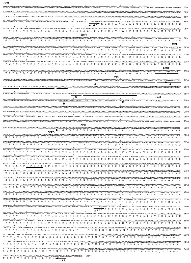
The complete 5467-bp DNA sequence of the naturally occurring Methanosarcina acetivorans plasmid pC2A was determined to provide a rational basis for design of an Methanosarcina–E. coli shuttle vector (Fig. 1). The sequence is in agreement with the previously determined restriction map of pC2A (7). Four ORFs of greater than 120 aa were identified, and their putative products were examined for homology with other proteins in the GenBank and EMBL databases. Only one showed strong homology with other proteins. This putative protein shares extensive homology with a family of known site-specific recombinases (32). Its gene was therefore designated ssrA. A second ORF, designated repA, encodes a putative protein that shares limited homology with the replication initiation proteins from a family of phage and plasmids that replicate by a rolling-circle mechanism (31). The homologous region includes one of three conserved motifs believed to be directly involved in DNA replication (see Fig. 1). An inspection of the sequence surrounding the repA gene suggested that it may be translated using CTG as the initiation codon. This CTG codon is preceded by a consensus ribosome binding site (AGGAA), whereas alternate initiation codons are much further downstream and are not preceded by consensus ribosome binding sites. The putative proteins encoded by the other two ORFs, orf1 and orf2, did not share significant homology with any other proteins in the databases. Three structural features that may be relevant for plasmid replication were noted in the pC2A DNA sequence. First, two very long direct repeats, 64 and 56 bp, were observed in the intergenic region between ssrA and repA. Second, a 20-bp perfect inverted repeat was observed immediately following the SsrA coding sequence.
Figure 1.
The DNA sequence of pC2A. The complete 5467-bp DNA sequence is shown (also see Fig. 2). Selected restriction sites are underlined. Four ORFs, ssrA, repA, orf1, and orf2, of greater than 120 aa were observed in the sequence. Their orientation is shown by the small arrows. The amino acid sequences of the putative proteins encoded by these ORFs are shown below the corresponding DNA sequences. The HUHUU (U = bulky hydrophobic residues) motif conserved among the Rep proteins of rolling circle plasmids (31) was identified in the RepA protein (heavy underline). Two long direct repeats of 64 bp (A) and 56 bp (B), and a 20-bp perfect inverted repeat which may play a role in plasmid replication were noted in the intergenic region between ssrA and repA.
Replication of pC2A Derivatives in Methanosarcina.
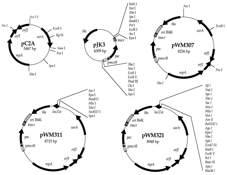
A series of hybrid plasmids were constructed as potential Methanosarcina–E. coli shuttle vectors. The PurR gene of Streptomyces alboniger, pac, was chosen as selectable marker for these plasmids because (i) it confers PurR upon other methanoarchaea (16), (ii) a variety of Methanosarcina tested showed complete growth inhibition by puromycin at 0.5 μg/ml (data not shown), and (iii) a gene cassette with pac transcribed from a strong methanoarchaeal promoter is available (16). To facilitate further constructions, we modified this cassette by removal of numerous extraneous restriction sites, fusion of the pac gene directly to the start codon of mcrB, and introduction of additional flanking restriction sites. The resulting construct, pJK3, should be generally useful in methanoarchaea (Fig. 2).
Figure 2.
Plasmids used in the study. pC2A is a naturally occurring plasmid from Methanosarcina acetivorans. The genes shown are described in the text. pJK3 is a pBluescript derivative carrying a modified pac gene cassette that confers PurR upon methanoarchaea. The promoter (pmcrB) and terminator (tmcr) of the Methanococcus voltae methyl reductase operon regulate expression of the puromycin acetyltransferase gene (pac) from Streptomyces alboniger in methanoarchaea. pWM307, pWM311, and pWM321 are Methanosarcina–E. coli shuttle vectors. The origin of replication from the plasmid R6K allows maintenance in E. coli, and manipulation of copy number by choice of an appropriate host strain. Each also carries the entire pC2A replicon for replication in Methanosarcina. Plasmids pWM309, pWM313, pWM315, pWM317, and pWM319 (not shown) are like pWM311 but carry different polylinkers within the lacZα gene. The lacZα gene allows blue-white screening of recombinant clones in E. coli for pWM309, pWM311, pWM313, pWM315, pWM317, and pWM319, but not in pWM321. The β-lactamase gene (bla) encodes resistance to penicillin derivatives in E. coli.
Potential shuttle vectors were constructed by cloning pC2A in its entirety into the E. coli plasmid pBluescript KS(+). Each was disrupted at a different region of the pC2A replicon in the hope that at least one of the disruptions would not interfere with essential replication functions of the plasmid. The pac cassette from pJK3 was then added to each to provide a selectable marker for transformation experiments.
Initially, we attempted to transform both Methanosarcina acetivorans and Methanosarcina barkeri Fusaro by electroporation under a variety of conditions using several of the shuttle vector candidates. One of these experiments yielded a few PurR transformants of Methanosarcina acetivorans with the plasmid pJK8, in which pC2A is disrupted at the unique SpeI site upstream of repA. We were able to isolate intact pJK8 plasmid DNA from these transformants, and with it we could retransform both E. coli and Methanosarcina acetivorans. These data indicate that the PurR strains obtained in this experiment were true transformants and that pJK8 can replicate as a plasmid in either host.
Construction of Improved Shuttle Vectors and Optimization of Transformation Conditions.
Plasmid pJK8 lacks many of the features desirable in a cloning vector. We modified pJK8 as described to generate pWM307, pWM309, pWM311, pWM313, pWM315, pWM317, pWM319, and pWM321 (Fig. 2). These plasmids provide a variety of useful features, including blue-white screening for recombinant clones (pWM309, pWM311, pWM313, pWM315, pWM317, and pWM319), symmetrical polylinkers (pWM313 and pWM315), and a large variety of unique restriction sites (pJK21). Also, because these plasmids use the pir-dependent R6K γ replication origin, their copy number can be modified from low to very high by using appropriate E. coli strains as hosts (33).
Using electroporation in high-resistance medium, the transformation frequency of Methanosarcina acetivorans with pJK8 was ≈102 per microgram of DNA per 2 × 10⁸ cells. A variety of modifications were tried to increase this frequency. These included varying the voltage, pulse length, and buffer composition, as well as the growth state and number of recipient cells. For these experiments, pWM307 was used because of its smaller size and fewer restriction endonuclease recognition sites. Despite these modifications, we were unable to increase the transformation frequency above ≈103 per microgram of DNA per 2 × 10⁸ cells (data not shown). Attempts to modify the PEG-mediated protocol used with Methanococcus maripaludis, and the PEG-lithium acetate-mediated protocol used with yeast were even less successful (<101 per microgram of DNA per 108 cells). Natural transformation never yielded transformants.
An alternative method, liposome-mediated transformation, resulted in a dramatic improvement in transformation frequency. Although there was significant variability in the exact number of transformants obtained in each experiment (which we believe is due to the difficulties inherent to plating these extremely oxygen-sensitive anaerobes), we were reproducibly able to achieve at least 107 transformants per microgram of DNA per 109 cells using Methanosarcina acetivorans as host. In some experiments, the transformation frequency was as high as 2 × 10⁸ transformants per microgram of DNA per 109 cells, or ca. 20% of the recipient population.
Transformation of Other Methanosarcina Species by pWM307.
Because a variety of Methanosarcina species are in routine use, we attempted to transform Methanosarcina barkeri Fusaro, Methanosarcina barkeri MS, Methanosarcina barkeri W, Methanosarcina mazei C-16, Methanosarcina mazei S-6, Methanosarcina mazei LYC, Methanosarcina thermophila TM-1, Methanosarcina siciliae C2J, Methanosarcina spp. WH1, and Methanosarcina spp. WH2 with pWM307. Eight of the 10 strains tested gave PurR transformants using the optimum liposome-mediated conditions determined with Methanosarcina acetivorans as host. Including Methanosarcina acetivorans, this represents strains from four of the five known Methanosarcina species. Only Methanosarcina mazei strains C-16 and LYC failed to yield PurR transformants. The transformation frequency in these strains was not determined, although it was clearly much lower than that achieved with Methanosarcina acetivorans. However, no attempt was made to optimize transformation conditions for these species.
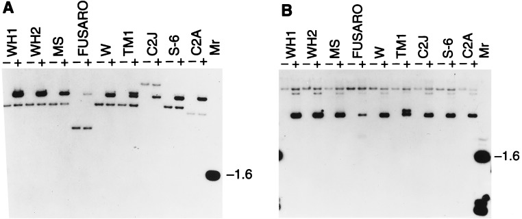
To verify that these PurR strains were pWM307 transformants, we isolated total DNA from selected transformants and from the untransformed parental strains, and tested them for hybridization with pXS2. Plasmid pXS2 caries the serC gene from Methanosarcina barkeri Fusaro, which was shown to hybridize to all Methanosarcina tested (27), as well as the same β-lactamase (bla) gene present on pWM307. Therefore, pXS2 will hybridize to both the chromosome and shuttle vector present in each strain. As shown in Fig. 3, a common band corresponding to the chromosomal serC locus is seen in both transformed and untransformed isolates of each strain. In the PurR transformants only, a second band corresponding to pWM307 is also seen. This second band is the only one seen with pWM307 as probe, a result that verifies the identity of this second band as the pM307 shuttle vector (data not shown). In undigested total DNA, the pWM307 band has a much greater mobility than the chromosomal serC band, indicating that pWM307 is replicating as a plasmid in these strains. In addition to the pWM307 band, Methanosarcina thermophila shows a second plasmid band of higher molecular weight. The nature of this second band is unclear at this time; however, we were able to isolate intact pWM307 from each transformant, including Methanosarcina thermophila, indicating that unmodified pWM307 was present in each strain tested.
Figure 3.
Transformation of Methanosarcina species by pWM307. Total DNA was isolated from each strain and from PurR clones obtained from each after transformation with pWM307. EcoRI-digested (A) or undigested (B) DNA was electrophoresed, blotted, and hybridized to labeled pXS2 as described. Plasmid pXS2 hybridizes to both the chromosomal serC gene and the pWM307 bla gene. A band corresponding to the chromosomal serC locus is seen in both transformed and untransformed strains, whereas a band corresponding to pWM307 is seen only in the transformed strains. The pWM307 band migrates with a mobility much higher than the chromosomal serC band in undigested DNA, indicating that pWM307 is replicating as a plasmid in these transformants. The strains examined were Methanosarcina spp. WH1 (WH1), Methanosarcina spp. WH2 (WH2), Methanosarcina barkeri MS (MS), Methanosarcina barkeri Fusaro (Fusaro), Methanosarcina barkeri W (W), Methanosarcina thermophila TM1 (TM1), Methanosarcina siciliae C2J (C2J), Methanosarcina mazei S-6 (S-6), and Methanosarcina acetivorans C2A (C2A). +, PurR clones; −, untransformed parental strains. The 1.6-kbp band of the molecular weight markers (Mr) hybridizes to pXS2 vector sequences.
DISCUSSION
Development of genetic techniques for routine analysis of methanoarchaea has been a long-time goal of researchers in the field. With the methods presented here, that goal has been achieved. The availability of a functional cloning vehicle and the application of efficient liposome-mediated DNA delivery methodology will allow genetic experiments that were not previously possible in methanoarchaea. The shuttle vectors reported here possess a variety of features desirable in a cloning vector. Because these vectors function in a number of Methanosarcina species, they should be usable without modification by laboratories working with different species within this genus. The shuttle vectors can be used for cloning Methanosarcina genes by complementation, and for identification of gene regulatory elements. These applications, however, represent only a small fraction of those possible with these vectors. A myriad of techniques involving plasmids are commonplace in modern molecular genetics. Most of these techniques should now be adaptable to methanoarchaeal research.
Equally important is the finding that liposome-mediated transformation can be highly efficient in methanoarchaea. The use of liposomes as a DNA delivery vehicle in Archaea has not previously been reported to our knowledge. This method is not restricted to transformation by autonomously replicating plasmids. In our experiments with Methanosarcina acetivorans, as many as 20% of the recipient cells were transformed by pWM307. Thus, experiments involving relatively infrequent events, such as homologous recombination or transposition, can be performed. We have recently used this method to construct Methanosarcina mutants by a reverse genetic approach.
It is likely that liposome-mediated transformation of Methanosarcina requires their growth in a single cell morphology (24). Methanosarcina species growing as single cells are bound solely by a membrane with a protein coat known as the S-layer. When these cells are suspended in an isoosmotic, Mg2+-free medium, the S-layer is lost as well, exposing the cell membrane. Such exposed membranes probably promote liposome fusion and DNA delivery. We have achieved transformation without resuspending the cells in Mg2+-free medium; however, under these conditions, the frequency is significantly reduced (data not shown). Liposome-mediated transformation may also be applicable to members of the genus Methanococcus. Like the single cell morphology of Methanosarcina, Methanococcus species have a cell structure composed of a membrane and S-layer protein coat, which can be removed by resuspension in appropriate buffer (12).
In combination, these methods represent a functional genetic system for the genus Methanosarcina. With the achievement of this goal, wide areas of the unique metabolism and physiology of methanoarchaea are now amenable to genetic analysis.
Acknowledgments
We thank S. Maloy, J. Konisky, and J. Slauch for providing bacterial strains and plasmids. This work was supported by National Institutes of Health Grant GM51334 and Department of Energy Grants DE-FG02–87ER13651 (R.S.W) and DE-FG02–93ER20106 (K.R.S.). W.W.M. is supported by a National Research Service Award postdoctoral fellowship (1 F32 GM16504–01A1).
ABBREVIATIONS
- PEG
polyethylene glycol
- PurR
puromycin resistance
Footnotes
References
- 1.Balch W E, Magrum L J, Fox G E, Wolfe R S, Woese C R. J Mol Evol. 1977;9:305–311. doi: 10.1007/BF01796092. [DOI] [PubMed] [Google Scholar]
- 2.Woese C R, Kandler O, Wheelis M L. Proc Natl Acad Sci USA. 1990;87:4576–4579. doi: 10.1073/pnas.87.12.4576. [DOI] [PMC free article] [PubMed] [Google Scholar]
- 3.Deppenmeier U, Muller V, Gottschalk G. Arch Microbiol. 1996;165:149–163. [Google Scholar]
- 4.DiMarco A A, Bobik T A, Wolfe R S. Annu Rev Biochem. 1990;59:355–394. doi: 10.1146/annurev.bi.59.070190.002035. [DOI] [PubMed] [Google Scholar]
- 5.Bult C J, White O, Olsen G J, Zhou L, Fleischmann R D, et al. Science. 1996;273:1017–1140. [Google Scholar]
- 6.Hilpert R, Winter J, Hammes W, Kandler O. Zentralbl Bakteriol Mikrobiol Hyg Abt 1 Orig C. 1981;2:11–12. [Google Scholar]
- 7.Sowers K J, Gunsalus R P. J Bacteriol. 1988;170:4979–4982. doi: 10.1128/jb.170.10.4979-4982.1988. [DOI] [PMC free article] [PubMed] [Google Scholar]
- 8.Hamilton P T, Reeve J N. Mol Gen Genet. 1985;200:47–59. doi: 10.1007/BF00383311. [DOI] [PubMed] [Google Scholar]
- 9.Meile L, Abendschein P, Leisinger T. J Bacteriol. 1990;172:3507–3508. doi: 10.1128/jb.172.6.3507-3508.1990. [DOI] [PMC free article] [PubMed] [Google Scholar]
- 10.Bertani G, Baresi L. J Bacteriol. 1987;169:2730–2738. doi: 10.1128/jb.169.6.2730-2738.1987. [DOI] [PMC free article] [PubMed] [Google Scholar]
- 11.Micheletti P A, Sment K A, Konisky J. J Bacteriol. 1991;173:3414–3418. doi: 10.1128/jb.173.11.3414-3418.1991. [DOI] [PMC free article] [PubMed] [Google Scholar]
- 12.Patel G B, Nash J H, Agnew B J, Sprott G D. Appl Environ Microbiol. 1994;60:903–907. doi: 10.1128/aem.60.3.903-907.1994. [DOI] [PMC free article] [PubMed] [Google Scholar]
- 13.Possot O, Gernhardt P, Klein A, Sibold L. Appl Environ Microbiol. 1988;54:734–740. doi: 10.1128/aem.54.3.734-740.1988. [DOI] [PMC free article] [PubMed] [Google Scholar]
- 14.Kiener A, Rechsteiner T, Leisinger T. FEMS Microbiol Lett. 1986;33:15–18. [Google Scholar]
- 15.Argyle J L, Tumbula D L, Leigh J A. Appl Environ Microbiol. 1996;62:4233–4237. doi: 10.1128/aem.62.11.4233-4237.1996. [DOI] [PMC free article] [PubMed] [Google Scholar]
- 16.Gernhardt P, Possot O, Foglino M, Sibold L, Klein A. Mol Gen Genet. 1990;221:273–279. doi: 10.1007/BF00261731. [DOI] [PubMed] [Google Scholar]
- 17.Berghoffer Y, Klein A. Appl Environ Microbiol. 1996;61:1770–1775. doi: 10.1128/aem.61.5.1770-1775.1995. [DOI] [PMC free article] [PubMed] [Google Scholar]
- 18.Tumbula D L, Makula R A, Whitman W B. FEMS Microbiol Lett. 1994;121:309–314. [Google Scholar]
- 19.Ladapo J, Whitman W B. Proc Natl Acad Sci USA. 1990;87:5598–5602. doi: 10.1073/pnas.87.15.5598. [DOI] [PMC free article] [PubMed] [Google Scholar]
- 20.Jain M K, Zeikus J G. Appl Environ Microbiol. 1987;53:1387–1390. doi: 10.1128/aem.53.6.1387-1390.1987. [DOI] [PMC free article] [PubMed] [Google Scholar]
- 21.Kiener A, Holliger C, Leisinger T. Arch Microbiol. 1984;139:87–90. [Google Scholar]
- 22.Wanner B L. J Mol Biol. 1986;191:39–58. doi: 10.1016/0022-2836(86)90421-3. [DOI] [PubMed] [Google Scholar]
- 23.Miller V L, Mekalanos J J. J Bacteriol. 1988;170:2575–2583. doi: 10.1128/jb.170.6.2575-2583.1988. [DOI] [PMC free article] [PubMed] [Google Scholar]
- 24.Sowers K R, Boone J, Gunsalus R P. Appl Environ Microbiol. 1993;59:3832–3839. doi: 10.1128/aem.59.11.3832-3839.1993. [DOI] [PMC free article] [PubMed] [Google Scholar]
- 25.Sowers K R, Schreier H J. In: Archaea: A Laboratory Manual. Robb F T, Place A R, Sowers K R, Schreier H J, DasSarma S, Fleischmann E M, editors. Vol. 2. Plainview, NY: Cold Spring Harbor Lab.; 1995. [Google Scholar]
- 26.Apolinario E A, Sowers K R. FEMS Microbiol Lett. 1996;145:131–137. [Google Scholar]
- 27.Metcalf W W, Zhang J-K, Shi X, Wolfe R S. J Bacteriol. 1996;178:5797–5802. doi: 10.1128/jb.178.19.5797-5802.1996. [DOI] [PMC free article] [PubMed] [Google Scholar]
- 28.Ausubel F M, Brent R, Kingston R E, Moore D D, Seidman J G, Smith J A, Struhl K, editors. Current Protocols in Molecular Biology. New York: Wiley; 1992. Vols. 1 and 2. [Google Scholar]
- 29.Chambers S P, Prior S E, Barstow D A, Minton N P. Gene. 1988;68:139–149. doi: 10.1016/0378-1119(88)90606-3. [DOI] [PubMed] [Google Scholar]
- 30.Elble R. BioTechniques. 1992;13:18–20. [PubMed] [Google Scholar]
- 31.Argos P, Landy A, Abremski K, Egan J B, Haggard-Ljungquist E, Hoess R H, Kahn M L, Kalionis B, Narayana S V L, Pierson L S, Sternberg N, Leong J M. EMBO J. 1986;5:433–440. doi: 10.1002/j.1460-2075.1986.tb04229.x. [DOI] [PMC free article] [PubMed] [Google Scholar]
- 32.Ilyina T V, Koonin E V. Nucleic Acids Res. 1992;20:3279–3285. doi: 10.1093/nar/20.13.3279. [DOI] [PMC free article] [PubMed] [Google Scholar]
- 33.Metcalf W W, Jiang W, Wanner B L. Gene. 1994;138:1–7. doi: 10.1016/0378-1119(94)90776-5. [DOI] [PubMed] [Google Scholar]


