Abstract
Aims
To determine the influence of the hand circulation on the determination of venous distensibility with venous occlusion plethysmography.
Methods
In a randomised study, duplicate measurements of forearm venous distensibility, with and without a wrist cuff, were made over occlusion periods of 3 and 12 min in eight volunteers. Treatments were compared with paired Student's t-tests and differences are presented as 95% confidence intervals (CI). Intra-subject variability was assessed with analysis of variance.
Results
Non-significant differences in increases in forearm volume between the occlusions with and without wrist cuff were found for the 3 min occlusion (CI: −0.4,+0.2%) and the 12 min occlusion period (CI: −0.7, +0.2%). However, the coefficient of variation was lower with the use of a wrist cuff; after 3 min occlusion (12% vs 19%) and after 12 min of occlusion (14% vs 24%). Forearm volume after 12 min of venous occlusion was 0.5% (CI:+0.4,+0.7) higher than after 3 min.
Conclusions
Although venous distensibility was equal when assessed with and without wrist cuff, exclusion of the hand circulation reduces intraindividual variability. Equilibrium in forearm volume is not reached after 3 min period of venous occlusion, as often assumed. The magnitude of the additional increase after prolonged occlusion stresses the need for well-controlled studies.
Keywords: forearm, plethysmography, vascular capacitance, veins
Introduction
Mercury-strain-gauge venous occlusion plethysmography is the most widely used method to investigate blood flow and venous distensibility. It has been shown that, for accurate measurement of (arterial) forearm blood flow, a wrist cuff inflated to supra-systolic pressure is mandatory. Exclusion of the circulation of the hand leads to lower and more reproducible results in basal conditions and after interventions [1]. However, whether a wrist cuff is also necessary to assess venous distensibility has never been investigated. Since venous distensibility is commonly measured using a short or an extended period of venous occlusion [2], an experiment was carried out in which the venous distensibility of the human forearm was measured with or without wrist cuff over a 3 min and a 12 min occlusion period. The design also allowed the investigation of whether venous occlusion for 3 or 12 min results in equivalent venous distensibility.
Methods
Eight apparently healthy normotensive male volunteers (age range: 23–28 years) not using any medication participated in the study after giving written informed consent. The study protocol was approved by the Medical Ethics Committee of Leiden University Medical Center and was executed in accordance with the principles outlined in the Declaration of Helsinki.
Trial design and treatments
The study was conducted using an open, randomised study design. The following treatments were applied in two series: (1) a 3 min venous occlusion with or without a wrist cuff and (2) a 12 min venous occlusion with or without a wrist cuff. Each treatment sequence was randomised according to 4 × 4 Latin squares balanced for first order carry-over. Subsequent measurements were separated by 15 min after completion of the preceding treatment.
Methods
After an overnight fast, subjects were studied in a semirecumbent position with the arm used for the measurements slightly elevated above heart level. After installation and application of the occlusion cuff(s) and a mercury-strain gauge, a period of at least 15 min was allowed for equilibration. Venous occlusion plethysmo-graphy was performed using an electronically calibrated plethysmograph (Hokanson type EC-4, Issaquah, Washington, USA) equipped with a rapid upper arm cuff inflator set at 30 mmHg (Hokanson type E20) connected to a thermal array recorder (model WS-628G Nihon Kohden, Amsterdam, The Netherlands). The paper speed of the recorder was 1 mm s−1 during the venous occlusion period and 10 mm s−1 at the start and the end of the occlusion period to allow for accurate measurements. Each measurement was preceded by calibration of the plethysmograph. The increase in forearm volume was assessed by manual measurement of the difference between baseline and plateau level of the tracing at the end of each occlusion using an electronic calliper. When appropriate, a wrist cuff was inflated manually to 180 mmHg from 1 min prior until the measurement was completed (3 or 12 min). All measurements were performed at room temperature and in a quiet environment. The room temperature varied on average less than 1°C. Between measurements the subjects were allowed slight movements of the arm.
Data analysis
The mean increase in forearm volume for each treatment was calculated from the duplicate measurements. The difference and their 95% confidence intervals (CI) between forearm volume for comparable occlusion periods with and without wrist cuff was calculated and were compared using paired Student's t-tests. To compare the increase in forearm volume between the short and extended occlusion period, pooled averages were calculated for the 3 and 12 min measurements using all data for an occlusion period regardless of the use of a wrist cuff. Paired t-tests were performed to compare the short and extended occlusion period. Furthermore, analysis of variance was done to determine the within-subject variability for the different treatments. The analysis was performed using SPSS software. Variation in the measured values was graphically displayed according to the Bland-Altman method where differences in two measurements are plotted against the average of two measurements [3].
Results
Adequate measurements could be obtained for all subjects. The difference in forearm volume between the treatments with and without wrist cuff was 0.1% (CI: –0.4,+0.2) for the 3 min occlusion and 0.3% (CI: −0.7,+ 0.2) for the 12 min occlusion (Table 1). The within-subject coefficient of variation after the 3 min occlusion was 19.0% without wrist cuff and 12.3% with use of the wrist cuff. After 12 min of occlusion the coefficients of variation were 23.7% and 14.1% for the measurements without and with wrist cuff, respectively.
Table 1.
Individual and average (s.d.) percentage increase in forearm volume (%FV) including within-subject coefficient of variation (CV).
| %FV after 3 min without wrist cuff | %FV after 3 min with wrist cuff | %FV after 12 min without wrist cuff | %FV after 12 min with wrist cuft | |||||||||
|---|---|---|---|---|---|---|---|---|---|---|---|---|
| Subject | a | b | Mean | a | b | Mean | a | b | Mean | a | b | Mean |
| 1 | 1.84 | 2.24 | 2.04 | 2.06 | 2.34 | 2.20 | 2.51 | 3.12 | 2.82 | 2.73 | 3.72 | 3.23 |
| 2 | 1.85 | 2.91 | 2.38 | 1.41 | 2.10 | 1.76 | 3.25 | 1.60 | 2.43 | 2.69 | 1.74 | 2.22 |
| 3 | 1.64 | 1.38 | 1.51 | 1.91 | 1.81 | 1.86 | 2.89 | 1.53 | 2.21 | 2.13 | 2.25 | 2.19 |
| 4 | 1.49 | 2.03 | 1.76 | 1.78 | 1.85 | 1.82 | 2.26 | 1.93 | 2.10 | 1.77 | 2.33 | 2.05 |
| 5 | 2.15 | 2.70 | 2.43 | 1.92 | 2.64 | 2.28 | - | 2.15 | 2.15 | 3.46 | 3.62 | 3.54 |
| 6 | 2.40 | 1.98 | 2.19 | 2.47 | 2.40 | 2.44 | 3.39 | 2.75 | 3.09 | 3.07 | 3.11 | 2.67 |
| 7 | 3.22 | 2.37 | 2.80 | 3.00 | 3.28 | 3.14 | 3.63 | 3.05 | 3.34 | 4.18 | 3.59 | 3.89 |
| 8 | 3.31 | 2.92 | 3.12 | 3.61 | 3.20 | 3.41 | 3.58 | 3.68 | 3.63 | 4.27 | 3.95 | 4.11 |
| Average | 2.27 | 2.36 | 2.76 | 3.01 | ||||||||
| (s.d.) | (0.53) | (0.62) | (0.62) | (0.81) | ||||||||
| CV (%) | 19.0 | 12.3 | 23.7 | 14.1 | ||||||||
a/b: first/second measurement.
The difference in forearm volume between the 3 and 12 min occlusion period was 0.5% (CI: +0.4, +0.7).
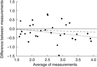
The Bland-Altman plots (Figure 1) for the difference between the measurements with and without wrist cuff plotted against the average value of the measurements indicated no systematic trend. The coefficients of correlation (r2 = 0.01 for 3 min occlusion period and r2 = 0.06 for 12 min occlusion) were nonsignificant.
Figure 1.
Difference between the measurements with and without wrist cuff against the average value of the measurements without and with a wrist cuff after a 3 min (•) and after a 12 min (▪) occlusion period. The broken line indicates the regression line for the values after the 3 min occlusion (r2 = 0.01; NS) and the dashed line indicates the regression line for the values after the 12 min occlusion (r2 = 0.06; NS).
Discussion
This experiment showed that for assessment of venous distensibility of the human forearm the absolute values of measured forearm volume are not influenced by exclusion of the hand circulation. However, the intrasubject variability of measurements was lower during exclusion of the hand circulation. When this difference in intraindividual variability is translated into number of subjects necessary to detect a 25% increase (with a P<0.05 and 80% power) smaller sample sizes are needed when a wrist cuff is used; (3 min occlusion with/without; 6/13 subjects; 12 min occlusion with/without; 8/10 subjects).
This experiment also showed that the venous disten-sibility was significantly increased after an extended period of venous occlusion. This finding, although not new [4], is not often recognized, since many experiments investigating the influence of interventions on the forearm venous system assume that a steady state in forearm volume is reached after a relative short period of venous occlusion [5–7]. Changes in forearm volume after the initial plateau following an intervention are then ascribed to the intervention as for instance nitroglycerin [6]. Ignoring the additional increase due the prolonged occlusion can then erroneously be interpreted and attributed to nitroglycerin, age, and/or other factors, which is unjustified. The extension of the period of venous occlusion accounted for an additional 0.55% (95% CI: 0.37–0.72) increase in forearm volume to the increase at 3 min (2.3%), which appears minor but represents approximately 25% of the assumed equilibrium. Interestingly, the increase in forearm volume ascribed to nitroglycerin in the above mentioned studies was in the order of 11–16% [6, 7], which is well within the range of the increase due to the extended occlusion.
This experiment does not allow to define the optimal occlusion period to be used in the investigation of drug effects on the venous system. This would require another means to determine the period during which the increase of venous diameter and pooling occurs, without extravasation. It is conceivable that ultrasound measurements of venous diameter during venous occlusion may be helpful in this respect [8].
In conclusion, exclusion of the hand circulation during the plethysmographic measurement of venous forearm distensibility decreases variability allowing for smaller treatment groups. Furthermore, this study showed that equilibrium in forearm volume is not reached after a short period of venous occlusion, illustrating the necessity of properly controlled experiments when investigating the influence of interventions on the forearm venous system.
References
- 1.Lenders J, Janssen G-J, Smits P, Thien T. Role of the wrist cuff in forearm plethysmography. Clin Sci. 1991;80:413–417. doi: 10.1042/cs0800413. [DOI] [PubMed] [Google Scholar]
- 2.Mason DT, Braunwald E. The effects of nitroglycerin and amyl nitrite on arteriolar and venous tone in the human forearm. Circulation. 1965;32:755–766. doi: 10.1161/01.cir.32.5.755. [DOI] [PubMed] [Google Scholar]
- 3.Bland JM, Altman DG. Statistical methods for assessing agreement between two methods of clinical measurement. Lancet. 1986;i:307–310. [PubMed] [Google Scholar]
- 4.Dittrich HC, Peck WW, Slutsky RA. Sustained venous occlusion plethysmography: effects on protein osmotic pressure, intravascular volume, and capillary filtration. Am Heart J. 1984;108:548–553. doi: 10.1016/0002-8703(84)90422-8. [DOI] [PubMed] [Google Scholar]
- 5.Zelis R, Mason DT. Comparison of the reflex reactivity of skin and muscle veins in the human forearm. J Clin Invest. 1969;48:1870–1877. doi: 10.1172/JCI106153. [DOI] [PMC free article] [PubMed] [Google Scholar]
- 6.Gascho JA, Fanelli C, Zelis R. Aging reduces venous distensibility and the venodilatory respons to nitroglycerin in normal subjects. Am J Cardiol. 1989;63:1267–1270. doi: 10.1016/0002-9149(89)90188-4. [DOI] [PubMed] [Google Scholar]
- 7.Fanelli C, Zelis R, Gascho JA. Comparison of venodilatory effects of nitroglycerin spray and tablets in healthy volunteers. Am J Cardiol. 1989;63:637–639. doi: 10.1016/0002-9149(89)90918-1. [DOI] [PubMed] [Google Scholar]
- 8.Burggraaf J, van den Berg R, Schipper J, Breimer DD, Cohen AF. Comparison of Duplex scanning and venous occlusion plethysmography in the assessment of the reactivity of the forearm venous system in man. Br J Clin Pharmacol. 1991;31:584. [Google Scholar]


