Abstract
Pathogen recognition at the plant cell surface typically results in the initiation of a multicomponent defense response. Transient influx of Ca2+ across the plasma membrane is postulated to be part of the signaling chain leading to pathogen resistance. Patch-clamp analysis of parsley protoplasts revealed a novel Ca2+-permeable, La3+-sensitive plasma membrane ion channel of large conductance (309 pS in 240 mM CaCl2). At an extracellular Ca2+ concentration of 1 mM, which is representative of the plant cell apoplast, unitary channel conductance was determined to be 80 pS. This ion channel (LEAC, for large conductance elicitor-activated ion channel) is reversibly activated upon treatment of parsley protoplasts with an oligopeptide elicitor derived from a cell wall protein of Phytophthora sojae. Structural features of the elicitor found previously to be essential for receptor binding, induction of defense-related gene expression, and phytoalexin formation are identical to those required for activation of LEAC. Thus, receptor-mediated stimulation of this channel appears to be causally involved in the signaling cascade triggering pathogen defense in parsley.
Keywords: patch-clamp, peptide elicitor, Petroselinum crispum, phytoalexins, signal transduction
Plants use a large arsenal of defense reactions to resist invading microbial pathogens (1–4). The molecular basis of pathogen recognition at the plant cell surface and of signaling cascades leading to the initiation of plant defense responses, however, is largely unknown. Perception of fungal pathogen-derived signals, referred to as elicitors, is believed to be mediated by specific receptors residing in the plant plasma membrane (5–8). Intracellular signal conversion and transduction include changes in the ion permeability of the plasma membrane, generation of reactive oxygen species, and alterations in the phosphorylation status of various proteins, giving rise to signal-specific responses of the plant (9–12).
The nonhost resistance response of parsley (Petroselinum crispum) leaves to infection with zoospores of the phytopathogenic fungus, P. sojae, has been found to be closely mimicked in parsley cell cultures upon treatment with fungus-derived elicitors (13, 14). An oligopeptide (Pep-13) originating from a cell wall glycoprotein of the fungus induces transcriptional activation of defense-related genes and phytoalexin production in parsley cells and protoplasts (15, 16). Recognition of the elicitor by its receptor, a 91-kDa plasma membrane protein, rapidly stimulates large, transient influxes of Ca2+ and H+ and effluxes of K+ and Cl− (15, 17, 18). Pharmacological studies revealed that this pattern of ion fluxes, and in particular extracellular Ca2+, is necessary for the production of reactive oxygen species (oxidative burst), defense-related gene activation, and phytoalexin production (18). Both omission of Ca2+ from the extracellular medium and inhibitors of animal slow-type Ca2+ channels abolished these plant responses (15, 19). Furthermore, identical structural features of the elicitor were found to be essential for receptor binding and initiation of all plant responses analyzed, indicating a sequence of events that may constitute part of a signaling cascade triggering pathogen defense in plants (15).
Ca2+ channels in the plasma membrane have been suggested to provide a major pathway for Ca2+ influx into higher plant cells (20–22). To identify plasma membrane ion channels that mediate Ca2+ influxes and thereby contribute to elevated levels of cytosolic Ca2+ in elicitor-treated parsley cells (23), we applied the patch-clamp technique to parsley protoplasts. Here we report the electrophysiological identification of a large conductance Ca2+-permeable ion channel, which was specifically activated upon addition of elicitor.
MATERIALS AND METHODS
Plant Cell Culture/Protoplast Preparation.
Cell suspension cultures of parsley (P. crispum) were maintained as described (24). Parsley protoplasts were isolated from 5-day-old cultured cells (25).
Elicitor Treatment/Inhibitor Studies.
Elicitors and inhibitors were applied as stock solutions at concentrations given in the text. Elicitor-stimulated production of furanocoumarin phytoalexins was routinely tested (25) for each protoplast preparation used in patch-clamp experiments. The elicitor-induced oxidative burst and phytoalexin production in cultured parsley cells were quantified as described in ref. 15. Viability of cultured parsley cells was checked 24 hr after addition of inhibitor (11).
Patch-Clamp Experiments.
Patch-clamp experiments with freshly prepared parsley protoplasts were performed using standard protocols (26) and the experimental setup as described (27). Digitized data (VR10, Instrutech, Elmont, NY) were stored on videotape and analyzed using a TL-1 DMA interface and patch-clamp software pclamp 5.5.1 (Axon Instruments, Foster City, CA). Unless stated otherwise, the bath solution was 240 mM CaCl2, 10 mM Mes/Tris (pH 5.5) and the pipette solution was 150 mM KCl, 2 mM MgCl2, 2 mM ATP, 10 mM Mes/Tris (pH 6.8). In both solutions osmolality was adjusted to 640 mosmol with d-sorbitol as was done in experiments with reduced extracellular Ca2+ concentrations. Removal of elicitor or inhibitor from the bath solution was achieved by perfusing the recording chamber with 5 ml of fresh bath solution (10-fold chamber volume, flow rate 1–2 ml/min). To assure establishment of whole-cell configuration, the accessibility of the protoplast interior (series resistance, whole-cell capacitance) was regularly checked throughout each experiment. Membrane voltage values were corrected for the liquid junction potential as described (28). Channel activity was quantified as NpO = Σn=1N npn, where pO is the open probability of the single channel, pn is the probability that n channels are open simultaneously, and N is the apparent number of channels. The value of pn was calculated according to ref. 29. NpO was calculated from 60 sec of recording obtained between 5 and 6 min after onset of any treatment. Elicitor-induced increase in large conductance elicitor-activated ion channel (LEAC) activity (NpO) was expressed as the ratio between the activities in the elicited and in the nonelicited state for each individual protoplast analyzed. Mechanosensitive ion channel activity was evoked as described (30).
RESULTS AND DISCUSSION
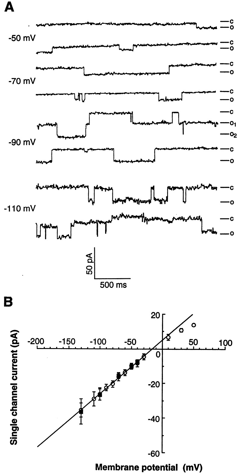
To identify plasma membrane ion channels that may contribute to macroscopic Ca2+ influxes observed in elicitor-treated parsley cells (23), we applied the patch-clamp technique to parsley protoplasts. Under asymmetric ionic conditions designed to resolve Ca2+-inward currents, we were able to detect a channel (LEAC) that exhibited openings often lasting for some hundred milliseconds or even seconds (Fig. 1A). The high Ca2+ concentration in the bath solution, corresponding to the ion concentration used during protoplast isolation (25), greatly facilitated resolution of single channels mediating Ca2+-inward currents. In addition, long open times, a limited number of channels that opened simultaneously, and a large current amplitude of this channel enabled us to detect single channel openings in whole-cell configuration (Fig. 1A). The single channel conductance determined in whole-cell configuration and in excised outside–out membrane patches was 309 ± 24 pS and 325 ± 35 pS, respectively (Fig. 1B). Expectedly, the number of active channels in excised membrane patches was much lower than that observed in whole-cell configuration (not shown). The activity of this channel did not significantly depend on the membrane potential within the physiologically relevant voltage range (−30 to −150 mV). Current amplitudes smaller than those mediated by LEAC were detected at higher membrane potentials (see Vm = −110 mV, Fig. 1A), which may either represent different channel activities or sublevels of LEAC (Fig. 1A).
Figure 1.
Activity of a large conductance ion channel in the plasma membrane of parsley protoplasts. (A) Single channel recordings in the whole-cell configuration with the membrane potential (Vm) clamped to −50, −70, −90, and −110 mV. (B) I–V plot of unitary currents from recordings in whole-cell (○, n = 15) and outside–out configuration (▪, n = 6), respectively. Freshly prepared parsley protoplasts were used for patch-clamp analyses under the conditions described in Materials and Methods.
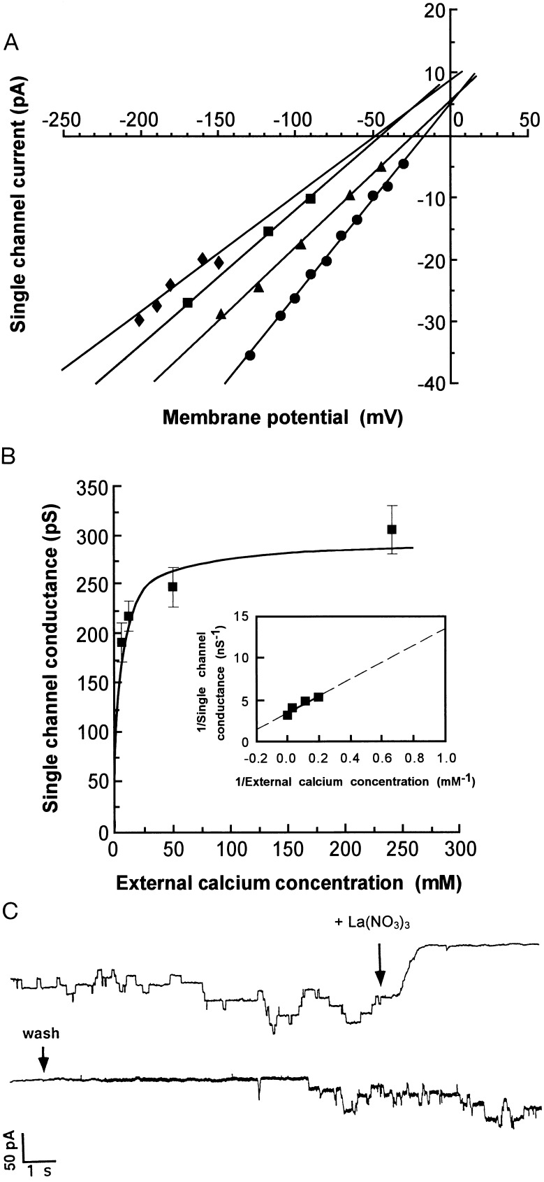
Reduction of the extracellular concentration of CaCl2 from 240 mM to 5 mM (the minimum concentration at which unitary LEAC currents could be resolved in whole-cell configuration) resulted in both a shift of the reversal potential toward more negative voltages (Δ−34 mV, Fig. 2A) and a decrease in single channel conductance (Fig. 2B). Single channel conductances determined from mean current amplitudes at 240 mM, 50 mM, 10 mM, and 5 mM CaCl2 were 309 pS (n = 15), 245 pS (n = 5), 216 pS (n = 4), and 186 pS (n = 3), respectively. A negative shift of the reversal potential (against a positive shift of the reversal potential for Cl−) indicates a preferential cation permeability of LEAC. Therefore, under our experimental conditions, Ca2+ (influx) and K+ (efflux) rather than Cl− represent major charge carriers of this channel. To explicitly rule out the existence of Cl− efflux, KCl in the pipette solution was substituted by K+-gluconate for which the anion is considered to be incapable of permeating ion channels. Channel amplitude and channel open probability remained unchanged under these conditions, demonstrating that LEAC did not mediate Cl− efflux. The reversal potential of the channel, as determined by linear regression (Fig. 2A), did not correspond to that of one particular cation, suggesting that both Ca2+ and K+ were transported. Under biionic conditions (given that LEAC is impermeable to anions and disregarding alterations of the internal Ca2+ concentration) the relative permeability ratio for Ca2+ to K+ (PCa2+/PK+) at 240 mM extracellular CaCl2 was 0.16 as calculated by the Goldman–Hodgkin–Katz equation (Erev = RT/2F ln 4PCa2+[Ca2+]out/PK+[K+]in) (29). Assuming constant permeability for the K+-outward current, as well as regarding variable Ca2+-inward currents at varying extracellular Ca2+ concentrations, nonlinear fits of both currents revealed increasing PCa2+/PK+ at decreasing extracellular Ca2+ concentrations (not shown). Thus, a reduction of the external Ca2+ concentration toward physiological concentrations would result in an increased Ca2+ permeability of LEAC. In addition, in the physiological range of plant membrane potentials (more negative than the reversal potential of LEAC), currents mediated by LEAC would largely correspond to Ca2+ influx.
Figure 2.
Cation permeability of LEAC. (A) I–V plot of unitary currents recorded in whole-cell mode at various extracellular Ca2+ concentrations [240 mM (•), 50 mM (▴), 10 mM (▪), and 5 mM (♦), respectively]. (B) Single channel conductance of LEAC as a function of extracellular Ca2+ concentration. Single channel conductances from A were plotted vs. extracellular Ca2+ concentration. The graph was fitted to Michaelis–Menten kinetics according to γ = γmaxCa2+out/(Kd + Ca2+out) with γ being LEAC conductance. (Inset) Lineweaver–Burk plot of the data shown in B. The broken line may facilitate determination of single channel conductances at lower external Ca2+ concentrations than those tested. (C) Inhibition of LEAC by the Ca2+ channel inhibitor La(NO3)3. Channel activity was recorded in whole-cell mode (Vm = −50 mV) in standard bath solution either in the absence or presence of 1.5 mM La(NO3)3. Arrows indicate time points of addition of inhibitor [La(NO3)3] and removal of inhibitor (wash) by perfusion of the recording chamber.
LEAC unitary conductance was saturated at higher external Ca2+ concentrations (Fig. 2B). A Lineweaver–Burk plot of the Michaelis–Menten kinetics (Fig. 2B Inset) revealed a unitary channel conductance of 80 pS at an extracellular Ca2+ concentration of 1 mM, which is representative of the plant cell apoplast (31). Taken together, an unusually large Ca2+ conductance associated with the particular gating behavior characterize LEAC as a new type of plant Ca2+-permeable ion channel. To our knowledge, there is no report yet on a plant ion channel with comparably large Ca2+ conductance.
LEAC activity could be completely and reversibly inhibited within seconds upon the addition of La(NO3)3 (1.5 mM, n = 5) (Fig. 2C) and GdCl3 (1 mM, n = 6) (not shown), which are inhibitors of a wide range of Ca2+ channels (31, 32). Similar inhibitory effects of both La(NO3)3 and GdCl3 were also observed at concentrations of 125 μM (n = 8) and 50 μM (n = 4), respectively. Both inhibitors also blocked the elicitor-induced production of reactive oxygen species and phytoalexins in parsley cells when added at these concentrations (not shown), whereas viability of parsley cells was not significantly affected by treatment with either inhibitor.
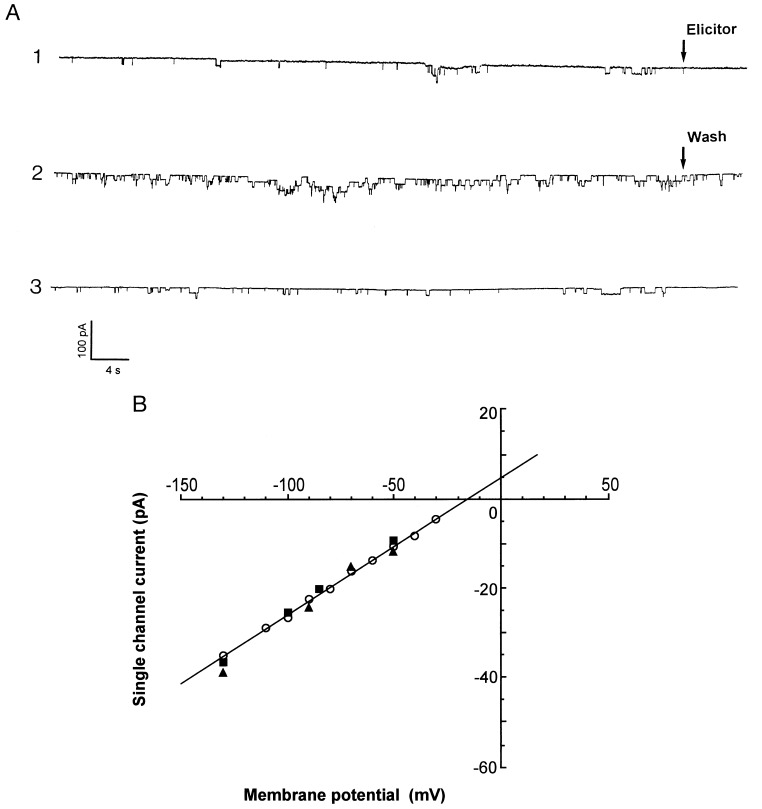
LEAC could be activated by P. sojae-derived elicitors, such as the peptide elicitor Pep-13 (Figs. 3A and 4) and a fungal cell wall preparation (Fig. 4). This activation was solely attributable to an increased channel activity (NpO, ref. 29), since the unitary channel conductance remained virtually unchanged. Conductances were 343 ± 32 pS (n = 4) and 365 ± 45 pS (n = 4) for Pep-13 and the fungal cell wall elicitor, respectively, and 309 ± 24 pS (n = 15) in nonelicited protoplasts (Fig. 3B). Addition of water did not activate LEAC (not shown).
Figure 3.
Activation of LEAC by the oligopeptide elicitor Pep-13. (A) Whole-cell recordings of LEAC activity (Vm = −50 mV) under standard conditions before addition (trace 1), 5 min after addition of 100 nM Pep-13 (trace 2), and 5 min after removal of Pep-13 by perfusion (trace 3). Arrows indicate time points of addition of elicitor (trace 1, Elicitor) and start of perfusion (trace 2, Wash). Traces represent recordings of reversible activation by elicitor of LEAC for an individual protoplast (n = 4). Stimulation by elicitor of LEAC activity was 15-fold in this particular experiment. Channel activity was quantified as described in Materials and Methods. (B) I–V plot of unitary currents recorded in whole-cell mode without elicitor (○, taken from Fig. 1B, n = 15), and after addition of 50 μg/ml of the fungal cell wall elicitor (▴, n = 4) or 100 nM Pep-13 (▪, n = 4), respectively.
Figure 4.
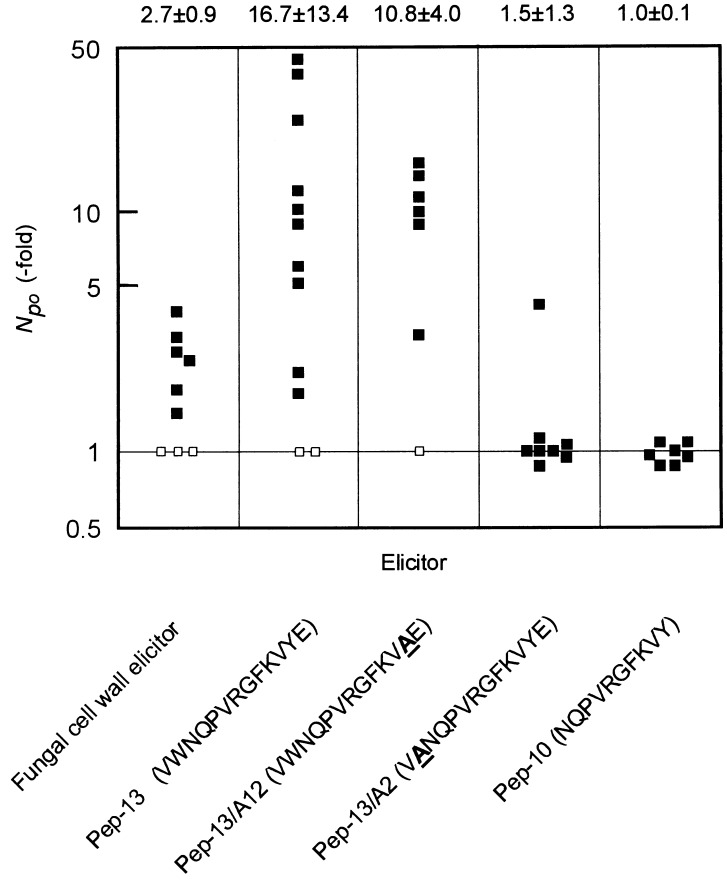
Increase in LEAC open probability (NpO) induced by fungal cell wall elicitor (n = 6), Pep-13 (n = 10), and structural derivatives of Pep-13 (n = 6, Pep-13A/12; n = 8, Pep-13/A2; n = 7, Pep-10, respectively). NpO was calculated from 60 s of recording obtained between 5 and 6 min after addition of elicitor as described in Materials and Methods and normalized to background activity recorded before addition of elicitor. Note the logarithmic scale used to plot increase in LEAC open probability. Data obtained from nonresponsive parsley protoplasts (□, see text for explanation) are included. Mean values ± SD, excluding those for nonresponsive protoplasts, are given. Elicitor concentrations used in patch-clamp experiments were 50 μg/ml fungal cell wall elicitor, 100 nM (Pep-13, Pep-13/A12), and 1 μM (Pep-13/A2, Pep-10), respectively. Amino acid sequences of Pep-13 and its structural derivatives are given in one-letter code. Underlined boldface letters represent alanine substitution sites within Pep-13.
A heterogeneous ligand sensitivity of LEAC was observed because only 70% of the protoplasts analyzed were elicitor-responsive, a situation comparable to the abscisic acid-mediated activation of Ca2+ and K+ permeable channels in Vicia faba guard cells (33). In addition, relatively large variations of LEAC elicitor responsiveness in individual protoplasts (ranging for example from 2- to 45-fold for Pep-13) were observed, which is typical for single-cell analyses with cells from a nonsynchronously growing cell culture (see SD values in Fig. 4). Both findings, however, may as well reflect differences in the physiological fitness of individual protoplasts caused by protoplast preparation.
Activation of LEAC by either elicitor could be observed only in whole-cell configuration but not in membrane patches in outside–out configuration (not shown, n = 7). This, as well as a delay in channel activation of 2–5 min with respect to addition of elicitor, suggests that the elicitor did not activate LEAC directly but through components mediating signal transfer between the elicitor receptor and the ion channel. Removal of the elicitor from the bath solution resulted in a decline of channel activity, indicating that LEAC activation is reversible (Fig. 3A).
As summarized in Fig. 4, LEAC could be efficiently activated by those elicitors that were previously shown to strongly induce macroscopic Ca2+ influx and phytoalexin production in parsley cells (15). A structural derivative of Pep-13, in which the tyrosine residue at position 12 was replaced by alanine (Pep-13/A12), retained its capacity to efficiently stimulate all three responses. In contrast, another single amino acid exchange within Pep-13 (tryptophan by alanine at position 2, Pep-13/A2) rendered this analog largely inactive with respect to LEAC activation, corresponding to observed losses of stimulation of Ca2+ uptake and phytoalexin formation (15), even when this derivative was applied at 10-fold higher concentrations than Pep-13. Similarly, deletion of one C-terminal and two N-terminal amino acid residues from Pep-13 (Pep-10) completely abolished both the ability of this derivative to stimulate LEAC and phytoalexin production. Since identical structural characteristics of the elicitor were also found to be responsible for specific interaction of Pep-13 with its receptor (15), our findings provide strong evidence that LEAC activation by fungal elicitor is a receptor-mediated process. Furthermore, stimulation with the same signal specificity of a multifacetted plant defense response comprising ion fluxes, oxidative burst, ethylene biosynthesis, activation of defense-related genes, and phytoalexin formation (15) indicates that LEAC constitutes a key element of the signal transduction chain initiating pathogen defense in parsley. This is further substantiated by the fact that Ca2+ channel inhibitors, which efficiently inhibit LEAC, block elicitor-induced oxidative burst and phytoalexin production as well.
A mechanosensitive ion channel activity in the parsley plasma membrane was evoked upon suction, which exhibited very similar gating behavior, single channel conductance (320 ± 24 pS in 240 mM CaCl2), and sensitivity toward La(NO3)3 and GdCl3 as LEAC (not shown). However, whether LEAC and the mechanosensitive ion channel are identical or represent distinct channels remains to be elucidated. Stretch-operated channels have been identified on the plasma membrane of several plants and found to be not highly selective for Ca2+, but also allow K+ to permeate (30, 31). Complex behaviors of adaptation and linkage have been ascribed to these channels, which suggest multiple pathways of regulation, e.g., in mediating the plant response to pathogen infection (31, 34). Furthermore, a mechanically activated oxidative burst, which may be mediated by stretch-sensitive channels has been reported from plant cells (35).
LEAC, the first plant plasma membrane ion channel shown to be activated by a phytopathogen-derived signal, is very likely to contribute to the elicitor-induced macroscopic ion fluxes observed in parsley (15, 18). Long open times of LEAC, its cation permeability, a large unitary Ca2+ conductance at physiological extracellular Ca2+ concentrations, and the elicitor inducibility of LEAC may account for the significant increase in cytoplasmic Ca2+ concentration (23) in elicitor-treated parsley cells. Alternatively, membrane depolarization by Ca2+ influx through LEAC could activate Ca2+ and/or voltage-dependent anion channels as well as outward-rectifying K+ channels (36). Since similar macroscopic ion fluxes have been detected in other plants upon elicitor treatment (10, 37–41), the existence of functional homologs of LEAC in these species can be anticipated. A Ca2+-permeable ion channel with similar unitary conductance and gating behavior was found to reside in the plasma membrane of tobacco protoplasts (unpublished results). However, activation of this channel by fungal elicitor remains to be analyzed.
Activation of LEAC by a fungal elicitor exemplifies the hypothesis that the plasma membrane of higher plants harbors a number of Ca2+-permeable ion channels, of which a major part are quiescent but can be rapidly recruited for Ca2+-dependent signal transduction. Complementary to the functional analysis of plant disease resistance genes and of plant mutants impaired in pathogen defense, structural analysis of signal transduction intermediates, such as plant ion channels, will substantially broaden our knowledge on the organization of signaling cascades involved in plant pathogen defense.
Acknowledgments
We are grateful to Drs. H. Barbier-Brygoo, J. L. Dangl, W. Knogge, I. E. Somssich, and S. Thomine for critical reading of the manuscript. We thank the Max-Planck-Society for substantial support. This work was supported by Grants Sche 235/3-1 and Sche 235/3-2 to D.S. and He 1640/1-3 to R.H. from the Deutsche Forschungsgemeinschaft, by Grant CHRX-CT93-0168 from the European Community Human Capital and Mobility Program to D.S., and by a grant from the European Community Biotech Program to J.G. J.-M.F. received an Alexander-von Humboldt-Stiftung postdoctoral fellowship.
ABBREVIATION
- LEAC
large conductance elicitor-activated ion channel
References
- 1.Godiard L, Grant M R, Dietrich R A, Kiedrowski S, Dangl J L. Curr Opin Genet Dev. 1994;4:662–671. doi: 10.1016/0959-437x(94)90132-m. [DOI] [PubMed] [Google Scholar]
- 2.Dixon R A, Harrison M J, Lamb C J. Annu Rev Phytopathol. 1994;32:479–501. [Google Scholar]
- 3.Kombrink E, Somssich I E. Adv Plant Pathol. 1995;21:1–34. [Google Scholar]
- 4.Baron C, Zambryski P C. Annu Rev Genet. 1995;29:107–129. doi: 10.1146/annurev.ge.29.120195.000543. [DOI] [PubMed] [Google Scholar]
- 5.Ebel J, Scheel D. In: Genes Involved in Plant Defense. Boller T, Meins F Jr, editors. Wien, Germany: Springer; 1992. pp. 184–205. [Google Scholar]
- 6.Atkinson M M. Adv Plant Pathol. 1993;10:35–64. [Google Scholar]
- 7.Ebel J, Cosio E G. Int Rev Cytol. 1994;148:1–36. [Google Scholar]
- 8.Boller T. Annu Rev Plant Physiol Plant Mol Biol. 1995;46:189–214. [Google Scholar]
- 9.Dietrich A, Mayer J A, Hahlbrock K. J Biol Chem. 1990;265:6360–6368. [PubMed] [Google Scholar]
- 10.Mathieu Y, Kurkdjian A, Xia H, Guern J, Koller A, Spiro M D, Albersheim P, Darvill A. Plant J. 1991;1:333–343. doi: 10.1046/j.1365-313X.1991.t01-10-00999.x. [DOI] [PubMed] [Google Scholar]
- 11.Renelt A, Colling C, Hahlbrock K, Nürnberger T, Parker J E, Sacks W R, Scheel D. J Exp Bot. 1993;44:257–268. [Google Scholar]
- 12.Suzuki K, Shinshi H. Plant Cell. 1995;7:639–647. doi: 10.1105/tpc.7.5.639. [DOI] [PMC free article] [PubMed] [Google Scholar]
- 13.Jahnen W, Hahlbrock K. Planta. 1988;173:197–204. doi: 10.1007/BF00403011. [DOI] [PubMed] [Google Scholar]
- 14.Schmelzer E, Krüger-Lebus S, Hahlbrock K. Plant Cell. 1989;1:993–1001. doi: 10.1105/tpc.1.10.993. [DOI] [PMC free article] [PubMed] [Google Scholar]
- 15.Nürnberger T, Nennstiel D, Jabs T, Sacks W R, Hahlbrock K, Scheel D. Cell. 1994;79:449–460. doi: 10.1016/0092-8674(94)90423-5. [DOI] [PubMed] [Google Scholar]
- 16.Sacks W R, Nürnberger T, Hahlbrock K, Scheel D. Mol Gen Genet. 1995;246:45–55. doi: 10.1007/BF00290132. [DOI] [PubMed] [Google Scholar]
- 17.Nürnberger T, Nennstiel D, Hahlbrock H, Scheel D. Proc Natl Acad Sci USA. 1995;92:2338–2342. doi: 10.1073/pnas.92.6.2338. [DOI] [PMC free article] [PubMed] [Google Scholar]
- 18.Hahlbrock K, Scheel D, Logemann E, Nürnberger T, Parniske M, Reinold S, Sacks W R, Schmelzer E. Proc Natl Acad Sci USA. 1995;92:4150–4157. doi: 10.1073/pnas.92.10.4150. [DOI] [PMC free article] [PubMed] [Google Scholar]
- 19.Nürnberger T, Colling C, Hahlbrock K, Jabs T, Renelt A, Sacks W R, Scheel D. Biochem Soc Symp. 1994;60:173–182. [PubMed] [Google Scholar]
- 20.Hetherington M A, Graziana A, Mazars C, Thuleau P, Ranjeva R. Philos Trans R Soc London. 1992;338:91–106. [Google Scholar]
- 21.Assmann S M. Annu Rev Cell Biol. 1993;9:345–375. doi: 10.1146/annurev.cb.09.110193.002021. [DOI] [PubMed] [Google Scholar]
- 22.Ward M M, Pei Z-M, Schroeder J I. Plant Cell. 1995;7:833–844. doi: 10.1105/tpc.7.7.833. [DOI] [PMC free article] [PubMed] [Google Scholar]
- 23.Sacks W R, Ferreira P, Hahlbrock K, Jabs T, Nürnberger T, Renelt A, Scheel D. In: Advances in Molecular Genetics of Plant-Microbe Interactions. Nester E W, Verma D P S, editors. Dordrecht, The Netherlands: Kluwer; 1993. pp. 485–495. [Google Scholar]
- 24.Kombrink E, Hahlbrock K. Plant Physiol. 1986;81:216–221. doi: 10.1104/pp.81.1.216. [DOI] [PMC free article] [PubMed] [Google Scholar]
- 25.Dangl J L, Hauffe K D, Lipphardt S, Hahlbrock K, Scheel D. EMBO J. 1987;6:2551–2556. doi: 10.1002/j.1460-2075.1987.tb02543.x. [DOI] [PMC free article] [PubMed] [Google Scholar]
- 26.Hedrich R. In: Single-Channel Recording. Sakmann B, Neher E, editors. New York: Plenum; 1995. pp. 277–305. [Google Scholar]
- 27.Zimmermann S, Thomine S, Guern J, Barbier-Brygoo H. Plant J. 1994;6:707–716. [Google Scholar]
- 28.Neher E. Methods Enzymol. 1992;207:123–131. doi: 10.1016/0076-6879(92)07008-c. [DOI] [PubMed] [Google Scholar]
- 29.Hille B. Ionic Channels of Excitable Membrane. Sunderland, MA: Sinauer; 1992. [Google Scholar]
- 30.Cosgrove D, Hedrich R. Planta. 1991;186:143–153. doi: 10.1007/BF00201510. [DOI] [PubMed] [Google Scholar]
- 31.Bush D S. Annu Rev Plant Physiol Plant Mol Biol. 1995;46:95–122. [Google Scholar]
- 32.Klüsener B, Boheim G, Liss H, Engelberth J, Weiler E W. EMBO J. 1995;14:2708–2714. doi: 10.1002/j.1460-2075.1995.tb07271.x. [DOI] [PMC free article] [PubMed] [Google Scholar]
- 33.Schroeder J I, Hagiwara S. Proc Natl Acad Sci USA. 1990;87:9305–9309. doi: 10.1073/pnas.87.23.9305. [DOI] [PMC free article] [PubMed] [Google Scholar]
- 34.Pickard B G, Ding J P. Aust J Plant Physiol. 1993;20:439–459. doi: 10.1071/pp9930439. [DOI] [PubMed] [Google Scholar]
- 35.Yahraus T, Chandra S, Legendre L, Low P S. Plant Physiol. 1995;109:1259–1266. doi: 10.1104/pp.109.4.1259. [DOI] [PMC free article] [PubMed] [Google Scholar]
- 36.Hedrich R. In: Current Topics on Membranes. Guggino W B, editor. San Diego: Academic; 1994. pp. 1–34. [Google Scholar]
- 37.Atkinson M M, Keppler L D, Orlandi E W, Baker C J, Mischke C F. Plant Physiol. 1990;92:215–221. doi: 10.1104/pp.92.1.215. [DOI] [PMC free article] [PubMed] [Google Scholar]
- 38.Blein J-P, Milat M-L, Ricci P. Plant Physiol. 1991;95:486–491. doi: 10.1104/pp.95.2.486. [DOI] [PMC free article] [PubMed] [Google Scholar]
- 39.Wei Z M, Laby R J, Zumoff C H, Bauer D W, He S Y, Collmer A, Beer S V. Science. 1992;257:85–88. doi: 10.1126/science.1621099. [DOI] [PubMed] [Google Scholar]
- 40.Bach M, Schnitzler J-P, Seitz H U. Plant Physiol. 1993;103:407–412. doi: 10.1104/pp.103.2.407. [DOI] [PMC free article] [PubMed] [Google Scholar]
- 41.Viard M-P, Martin F, Pugin A, Ricci P, Blein J-P. Plant Physiol. 1994;104:1245–1249. doi: 10.1104/pp.104.4.1245. [DOI] [PMC free article] [PubMed] [Google Scholar]


