A woman with no previous cardiac history was admitted to hospital because of dyspnoea and chest discomfort lasting for 3 days since she had celebrated her 70th birthday. An ECG showed T‐wave inversions in leads II, III, aVF and V2–V6. Troponin I was positive, creatine kinase, creatine kinase MB and D‐dimer concentrations were slightly raised.
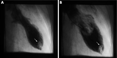
An acute coronary syndrome was assumed and cardiac catheterisation performed. The left ventricular angiogram showed an apical thrombus and severe apical hypokinesis with hyperkinesis of basal segments (panels A and B). Coronary angiography showed <50% diameter stenosis of the mid left anterior descending artery (LAD). Since the LAD was not dominant and perfused only a small part of the apical region, temporal coronary occlusion or coronary spasm could not explain the “apical ballooning”. Because the norepinephrine plasma level was more than three times the upper normal limit, stress‐induced cardiomyopathy, so called “tako‐tsubo cardiomyopathy”, was diagnosed. Despite starting effective anticoagulation within 2 hours of catheterisation, a short transient ischaemic attack of sensoric aphasia occurred.
The patient was discharged without symptoms under phenprocoumon and a β‐blocking agent. Three months later the ECG and wall‐motion abnormalities had normalised and left ventricular thrombus had completely resolved. Only a few cases of “tako‐tsubo cardiomyopathy” complicated by a left ventricular thrombus have been reported. Further research is needed to determine the true incidence of left ventricular thrombus and the role of short‐term anticoagulant treatment in this disorder.


