

Abstract
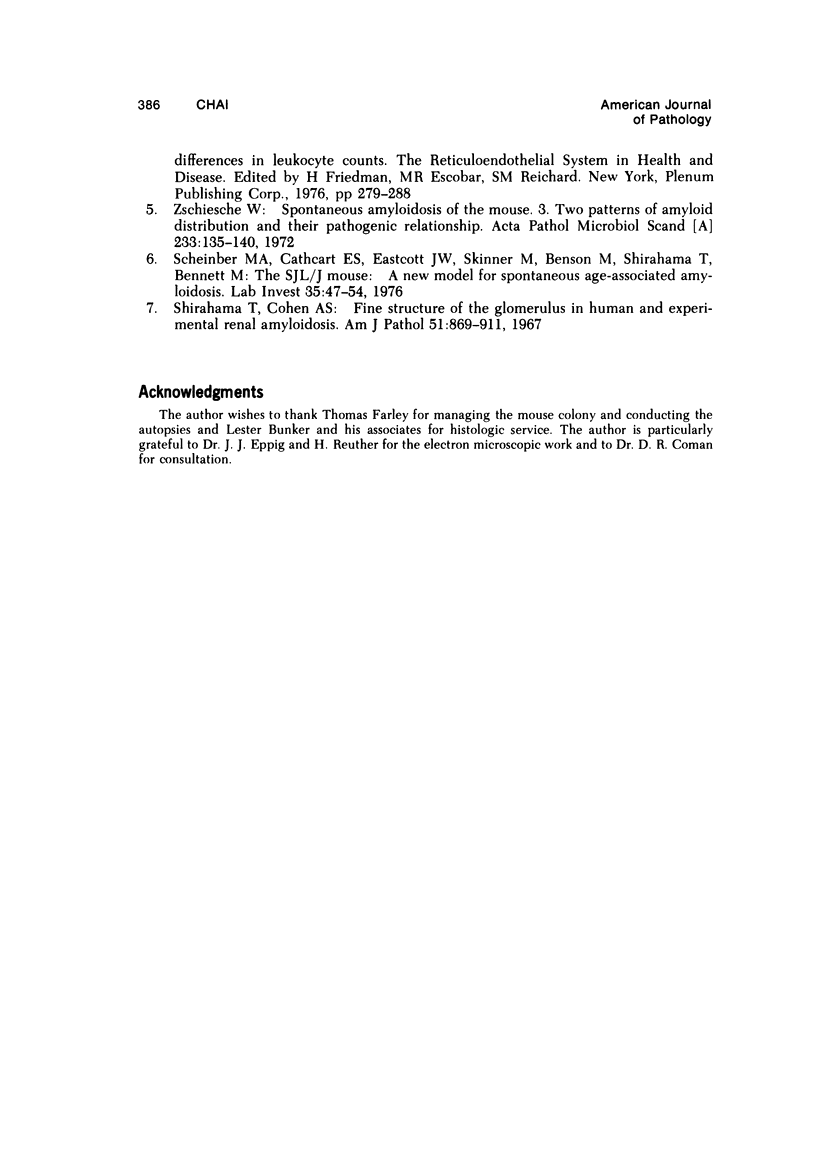
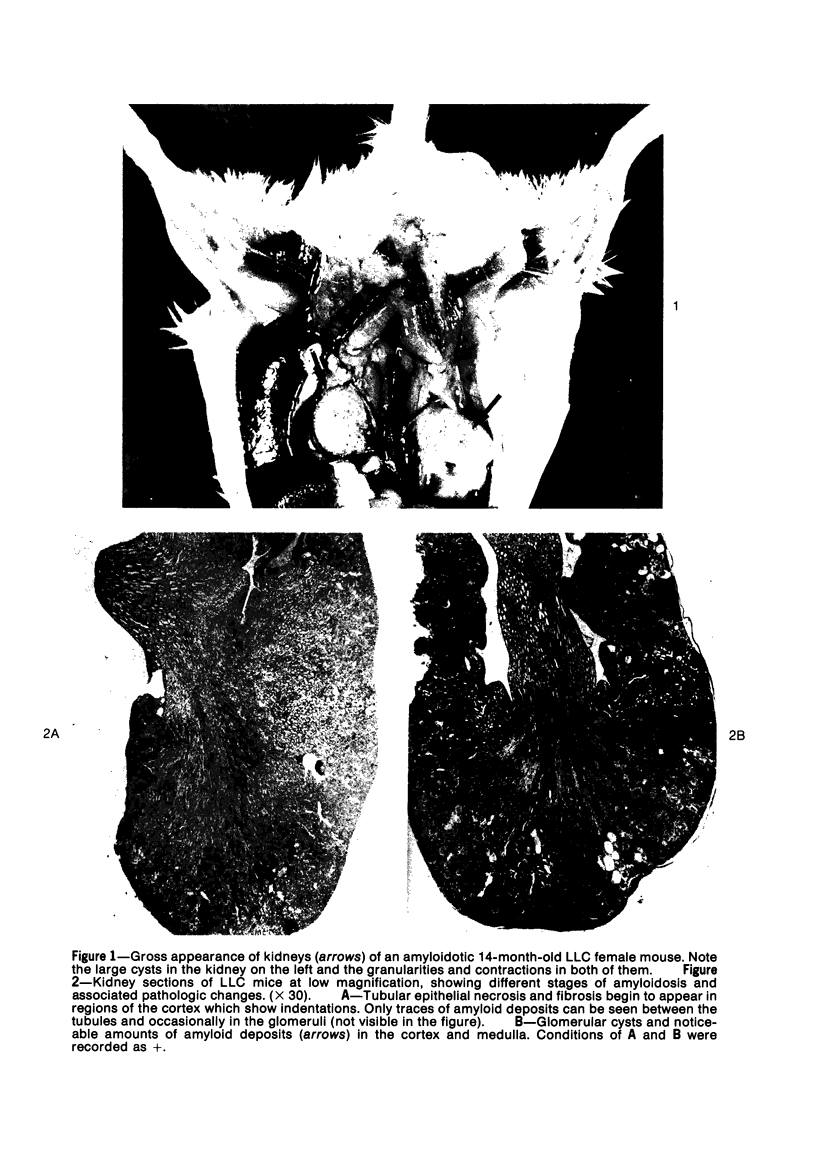
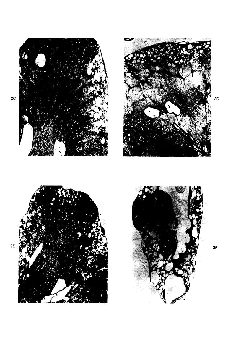
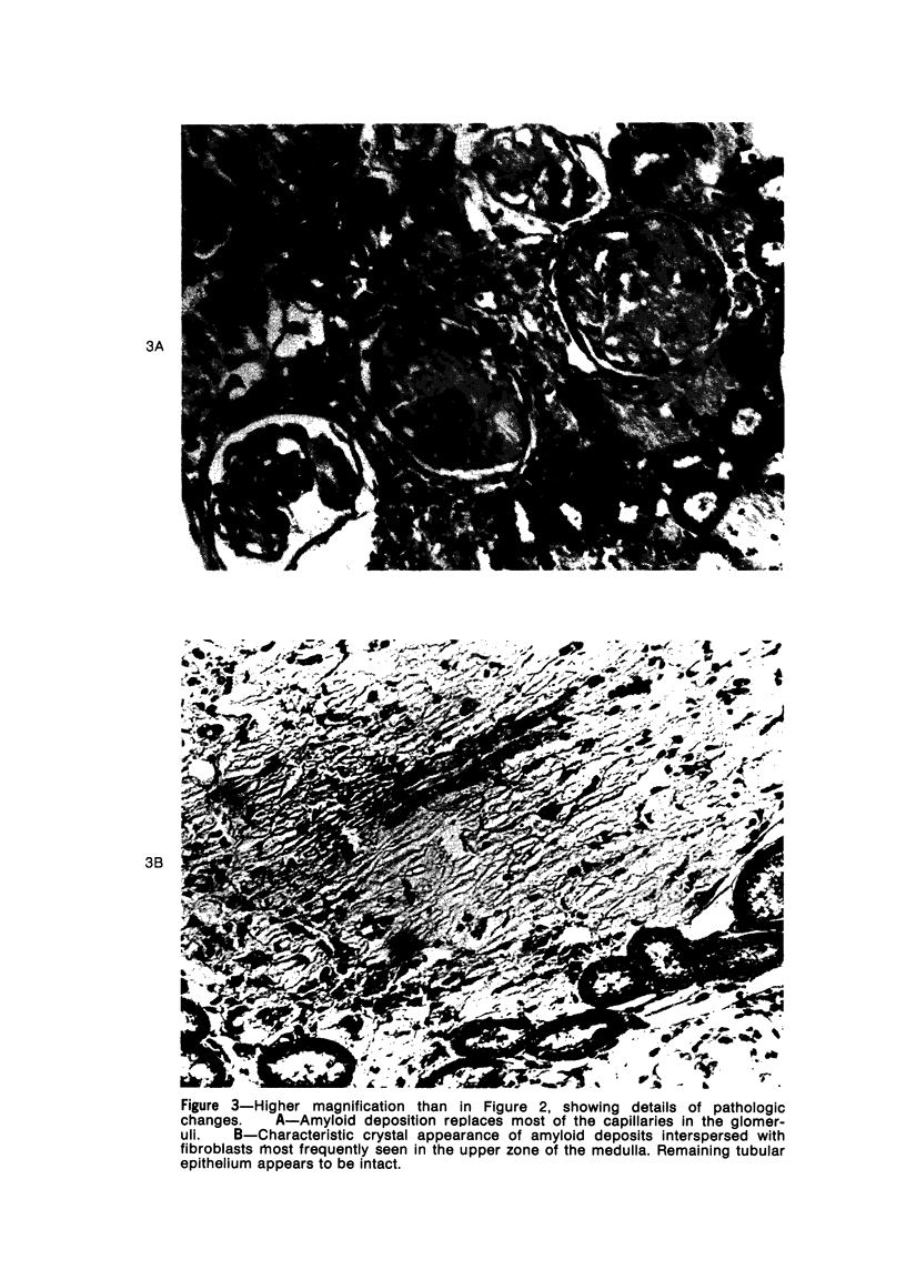
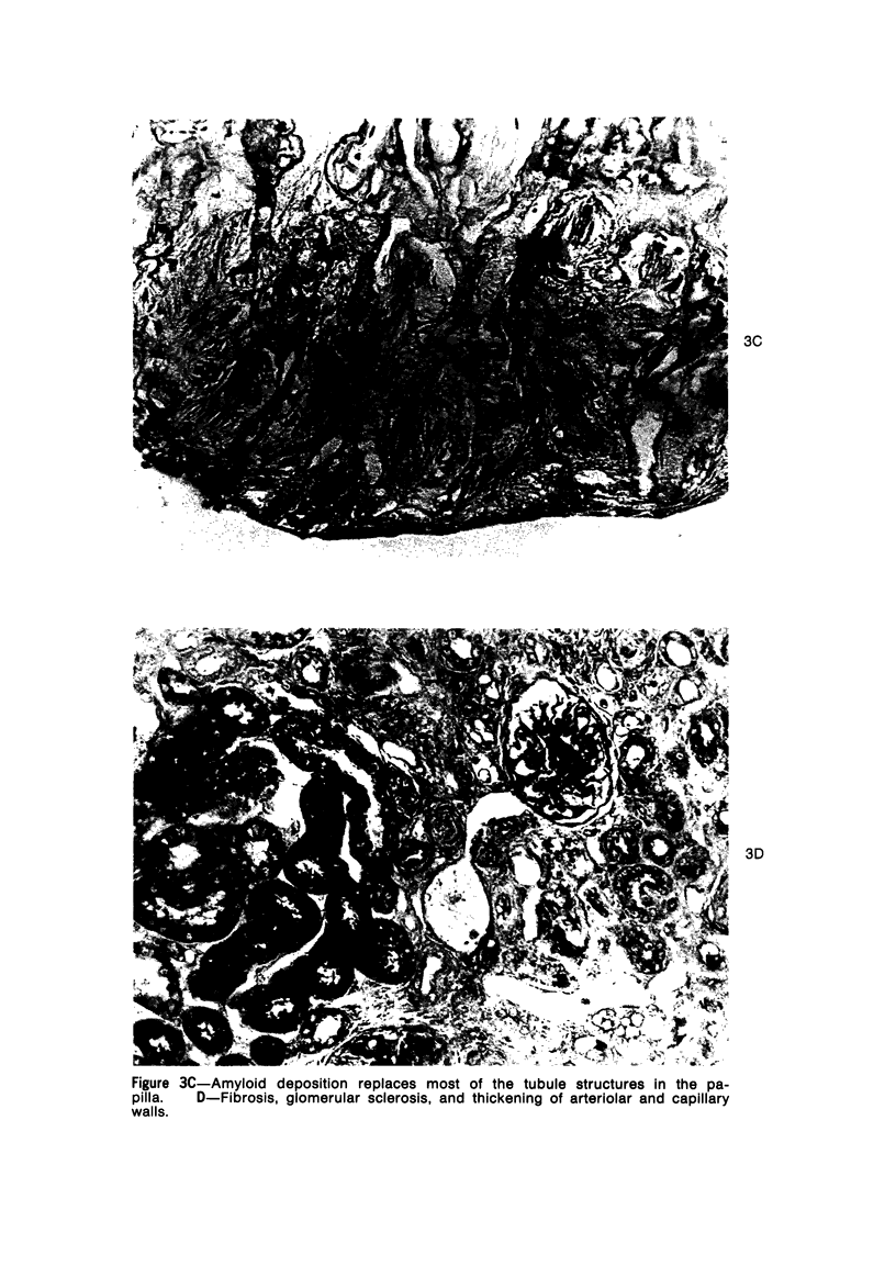
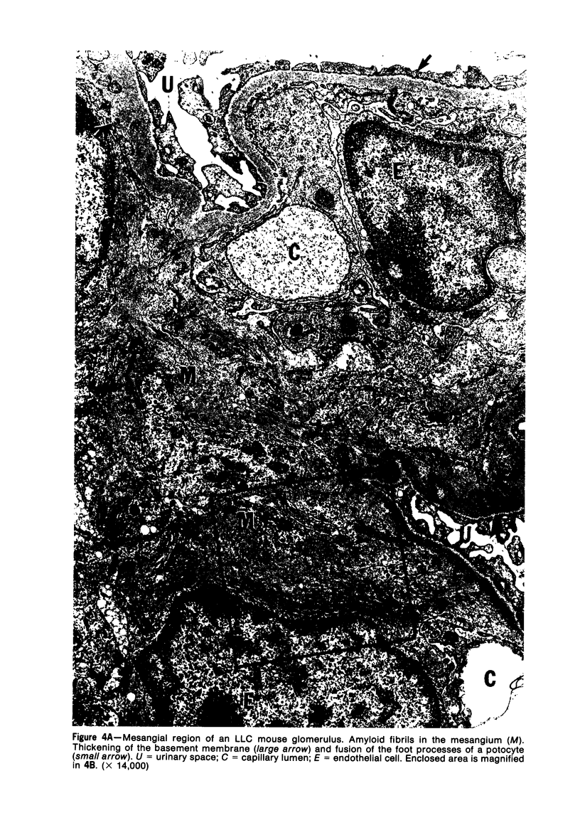
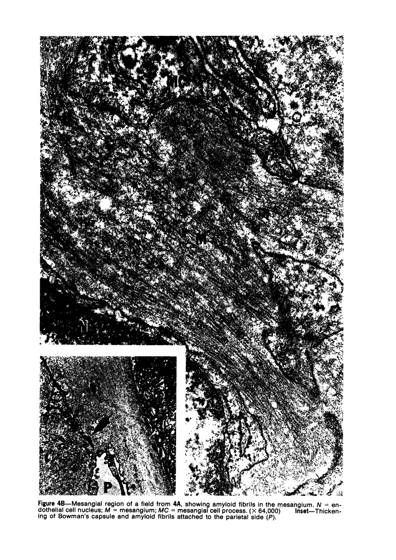
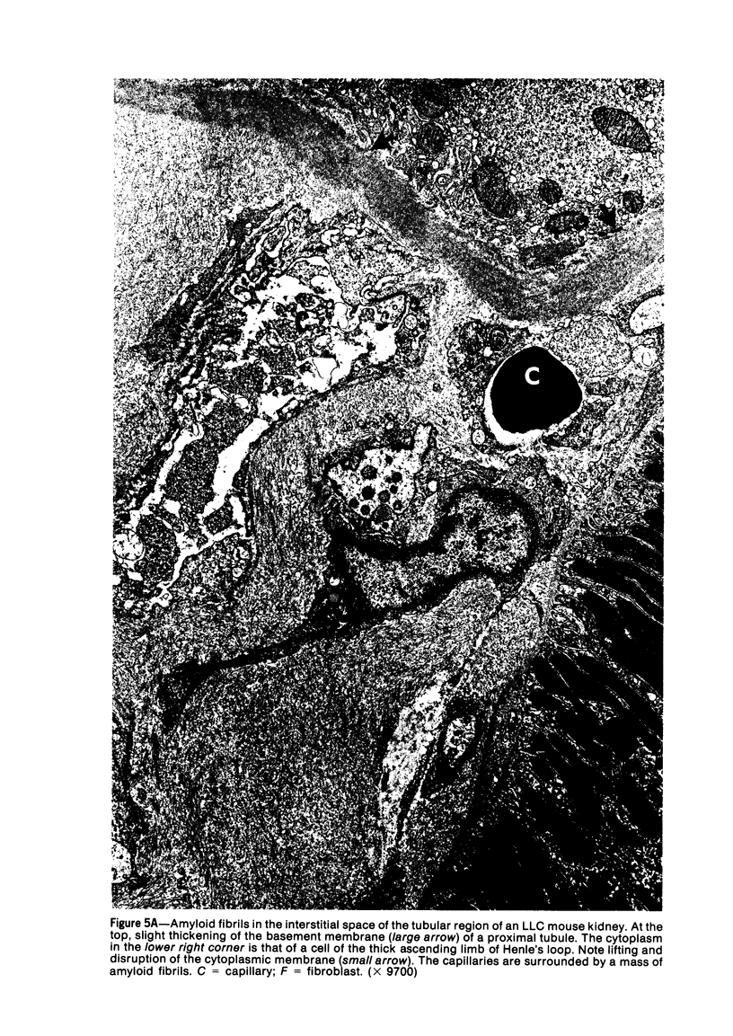
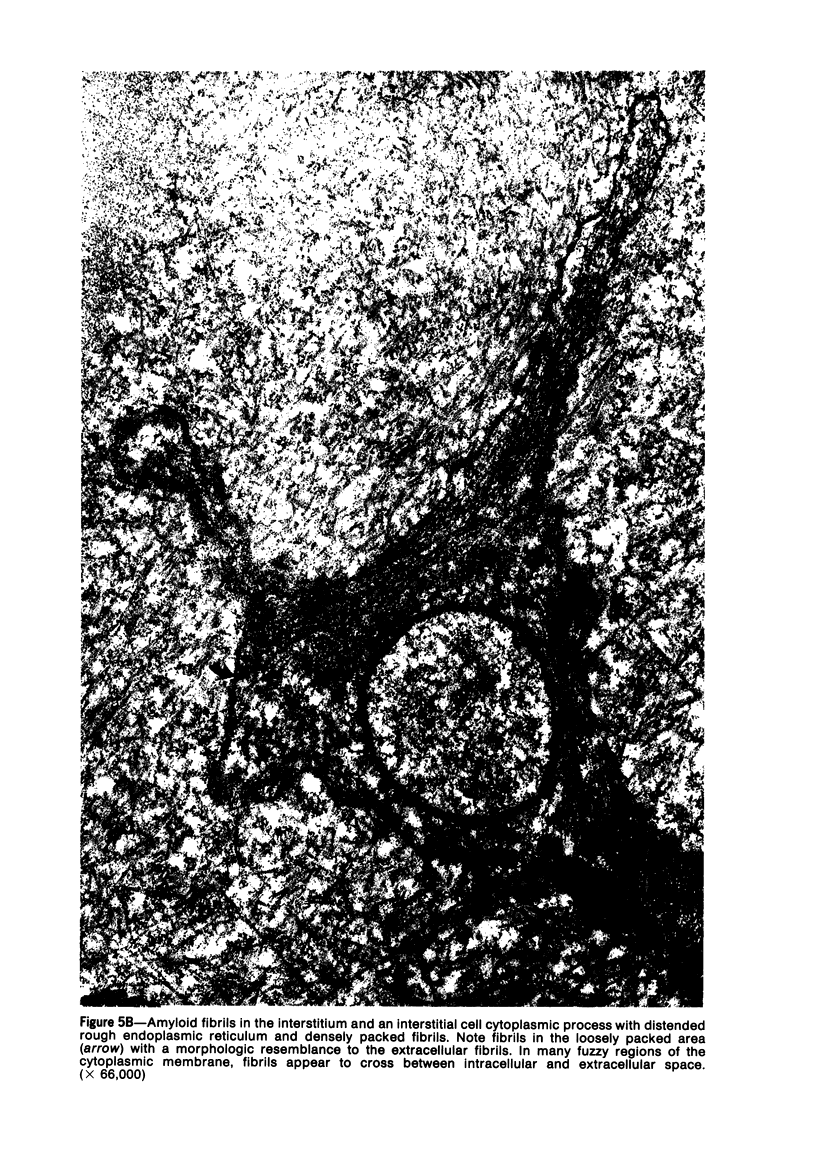
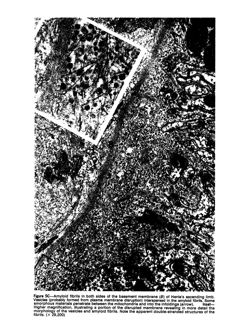
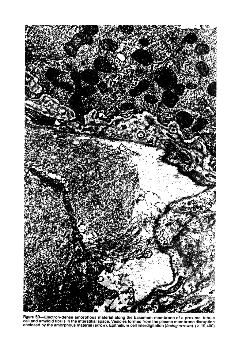
Practically all low leukocyte count (LLC) mice over 1 year of age develop renal amyloidosis. Renal amyloid is deposited in the glomeruli and in the interstitium between the convoluted as well as collecting tubules, with consequent development of cysts and necrosis. LLC mice die of chronic renal failure. Electron microscopic studies reveal amyloid fibrils in the mesangium, a thickening of the basement membranes, and fusion of the foot processes in the glomeruli. Massive amounts of amyloid fibrils are also present in the interstitium, where intracellular fibrils in the fibroblasts as well as in the tubular epithelium cells are found. Vesicles, which are probably formed from membrane disruption, and amorphous materials are seen along the basement membranes. LLC mouse amyloidosis is discussed with regard to its potential as a model for studies on amyloidosis as well as the etiology and origin of amyloid fibrils.
Full text
PDF
Images in this article
Selected References
These references are in PubMed. This may not be the complete list of references from this article.
- Chai C. K. Reticular cell hyperplasia and amyloidosis in a line of mice with low leukocyte counts. Am J Pathol. 1976 Oct;85(1):49–72. [PMC free article] [PubMed] [Google Scholar]
- Chai C. K. Selection for leukocyte counts in mice. Genet Res. 1966 Oct;8(2):125–142. doi: 10.1017/s0016672300010004. [DOI] [PubMed] [Google Scholar]
- Jao W., Pirani C. L. Renal amyloidosis: electron microscopic observations. Acta Pathol Microbiol Scand Suppl. 1972;233:217–227. [PubMed] [Google Scholar]
- Scheinberg M. A., Cathcart E. S., Eastcott J. W., Skinner M., Benson M., Shirahama T. The SJL/J mouse: a new model for spontaneous age-associated amyloidosis. I. Morphologic and immunochemical aspects. Lab Invest. 1976 Jul;35(1):47–54. [PubMed] [Google Scholar]
- Shirahama T., Cohen A. S. Fine structure of the glomerulus in human and experimental renal amyloidosis. Am J Pathol. 1967 Nov;51(5):869–911. [PMC free article] [PubMed] [Google Scholar]
- Zschiesche W. Spontaneous amyloidosis of the mouse. 3. Two patterns of amyloid distribution and their pathogenic relationship. Acta Pathol Microbiol Scand Suppl. 1972;233:135–140. [PubMed] [Google Scholar]