Abstract
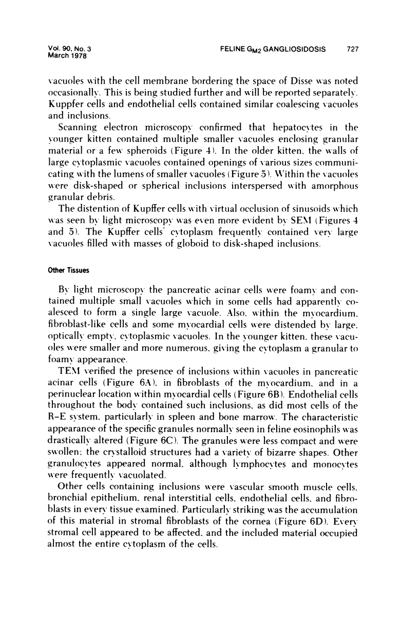
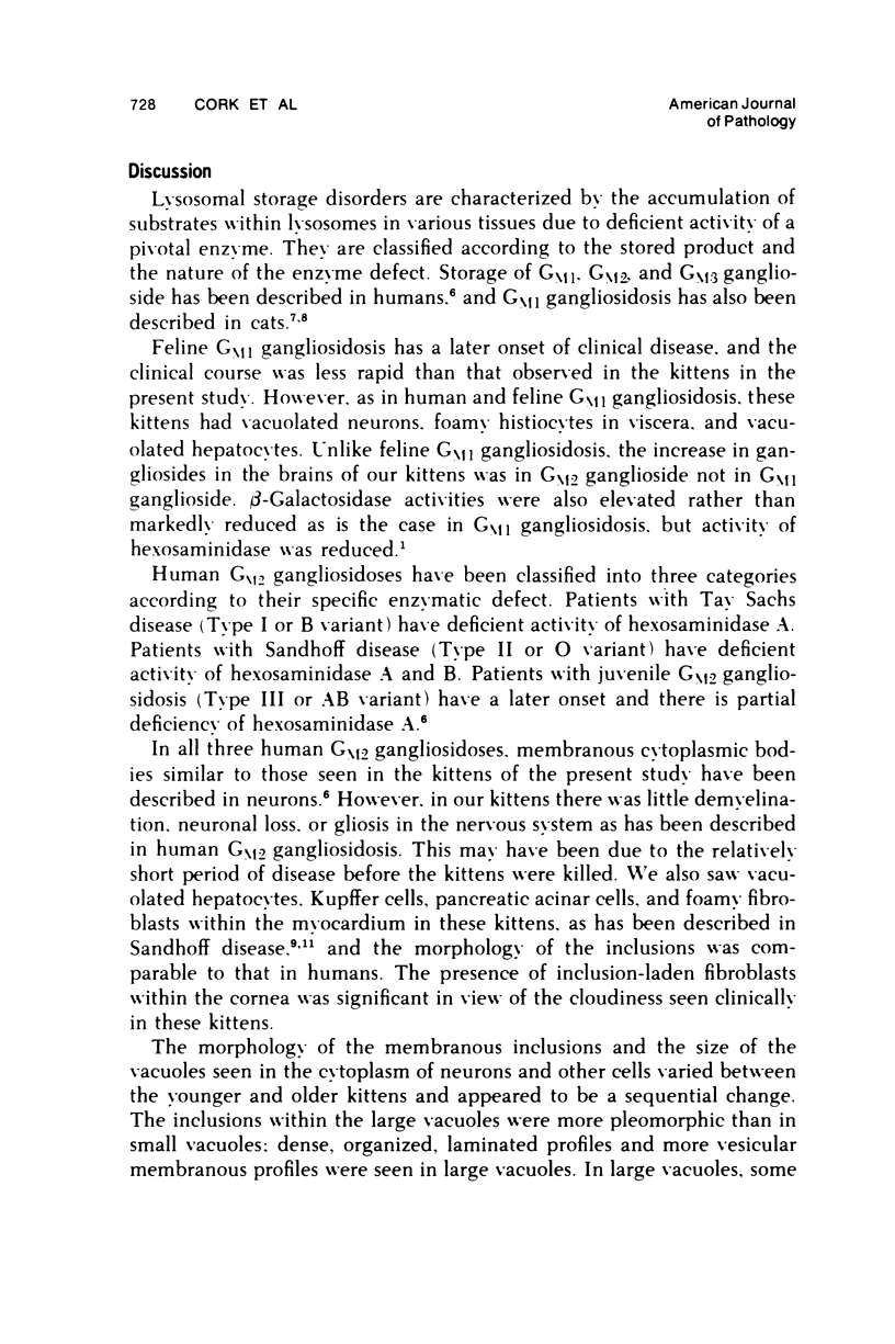
An 11-week-old and a 6-month-old kitten with feline GM2 gangliosidosis and deficiency in both A and B isoenzymes of beta-D-N-acetyl hexosaminidase were studied by light transmission (TEM), and scanning electron microscopy (SEM). Neurons throughout the nervous system contained cytoplasmic, membrane-bound inclusions which were PAS-positive at the fine structure level these inclusions were composed of membranous arrays in whorls, vesicles, or multilaminated stacks. Fusion of the bounding membranes of adjacent inclusions resulted in large inclusion-containing vacuoles. Hepatocytes and Kupffer cells contained inclusions slightly different from those in the central nervous system. SEM of cryofractured liver demonstrated their coalescence to form larger composite vacuoles. Vacuoles with inclusions were also seen in pancreatic acinar cells, endothelial cells, vascular smooth muscle, fibroblasts, myocardial cells, renal interstitial cells, corneal stromal cells, and R-E cells of bone marrow and spleen. The specific granules of eosinophils were swollen and took on bizarre forms. Pathologic manifestations of feline GM2 gangliosidosis differ from those seen in feline GM1 gangliosidosis but closely resemble those of Sandhoff disease in humans.
Full text
PDF


Images in this article
Selected References
These references are in PubMed. This may not be the complete list of references from this article.
- Cork L. C., Munnell J. F., Lorenz M. D., Murphy J. V., Baker H. J., Rattazzi M. C. GM2 ganglioside lysosomal storage disease in cats with beta-hexosaminidase deficiency. Science. 1977 May 27;196(4293):1014–1017. doi: 10.1126/science.404709. [DOI] [PubMed] [Google Scholar]
- Dolman C. L., Chang E., Duke R. J. Pathologic findings in Sandhoff disease. Arch Pathol. 1973 Oct;96(4):272–275. [PubMed] [Google Scholar]
- Farrell D. F., Baker H. J., Herndon R. M., Lindsey J. R., McKhann G. M. Feline GM 1 gangliosidosis: biochemical and ultrastructural comparisons with the disease in man. J Neuropathol Exp Neurol. 1973 Jan;32(1):1–18. [PubMed] [Google Scholar]
- Lampert P. W. A comparative electron microscopic study of reactive, degenerating, regenerating, and dystrophic axons. J Neuropathol Exp Neurol. 1967 Jul;26(3):345–368. doi: 10.1097/00005072-196707000-00001. [DOI] [PubMed] [Google Scholar]
- Spurr A. R. A low-viscosity epoxy resin embedding medium for electron microscopy. J Ultrastruct Res. 1969 Jan;26(1):31–43. doi: 10.1016/s0022-5320(69)90033-1. [DOI] [PubMed] [Google Scholar]


