

Abstract
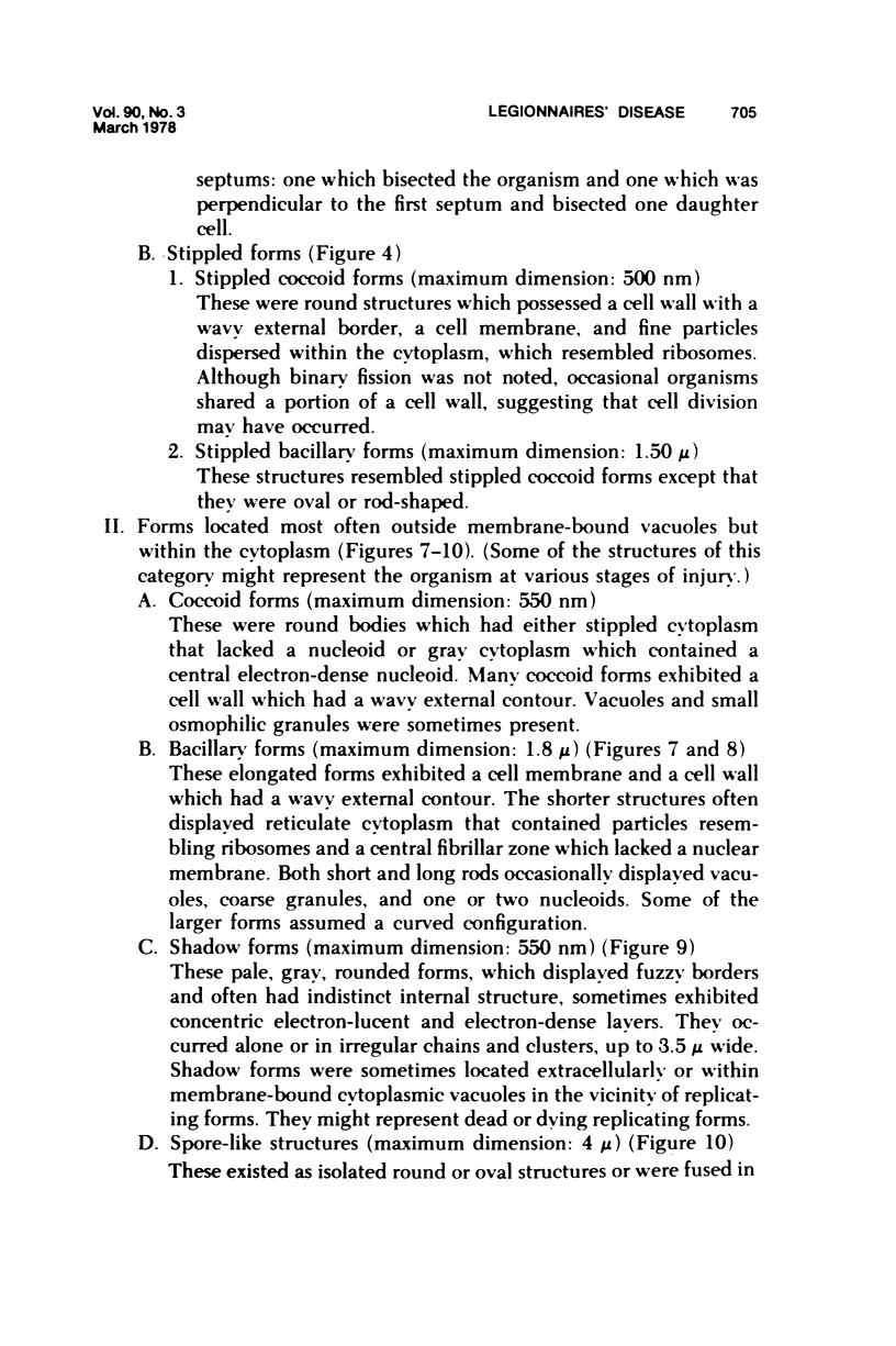
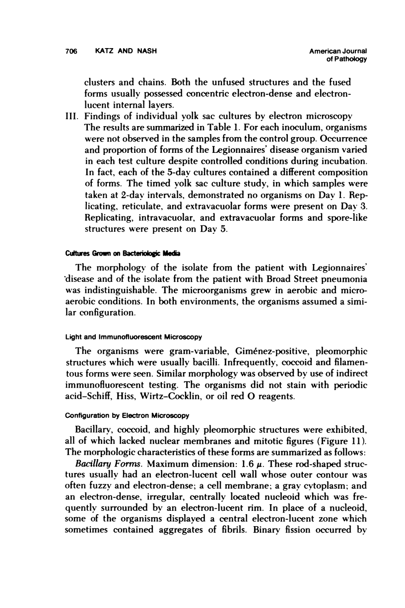
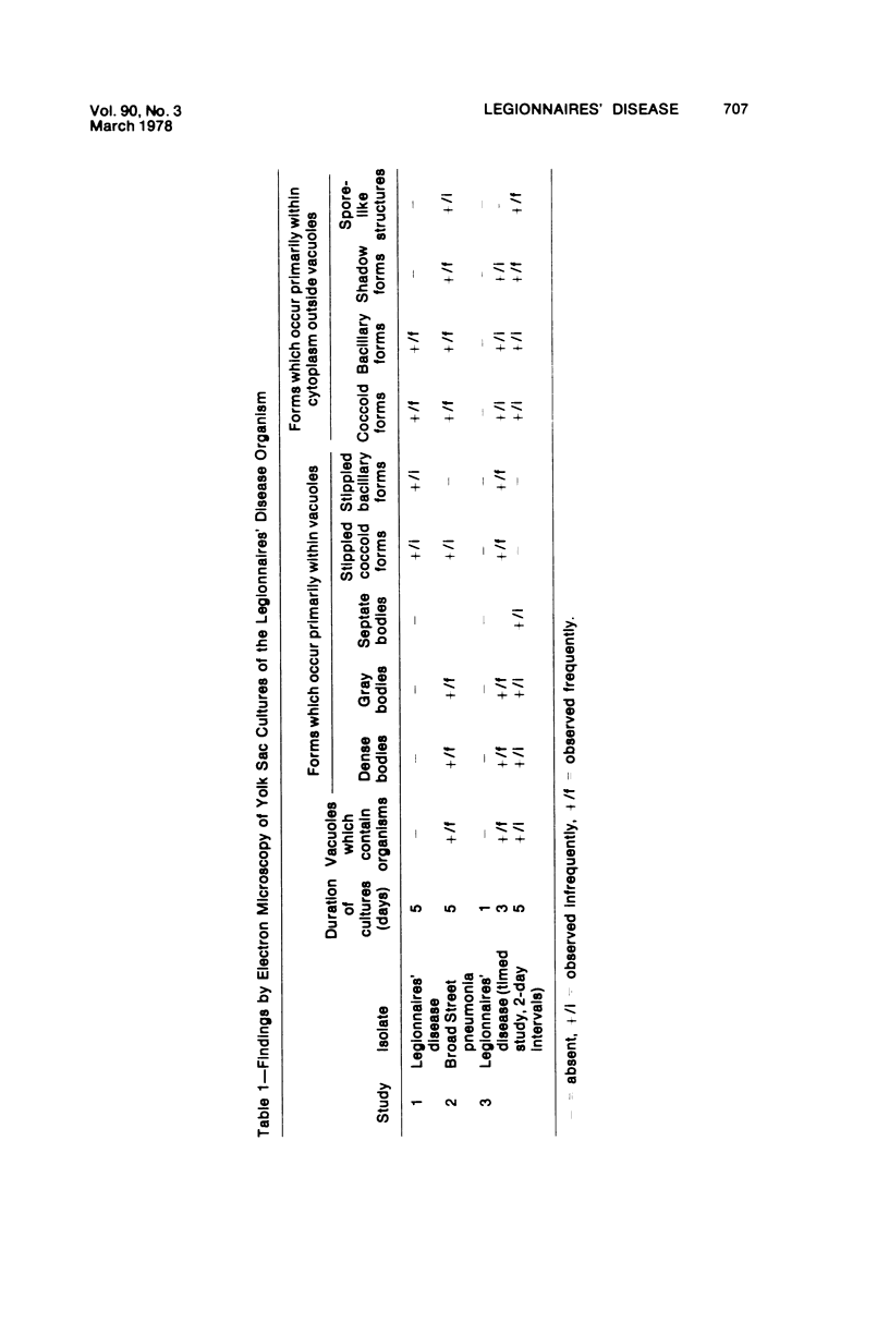
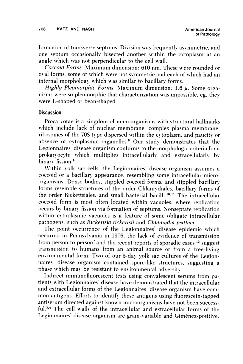
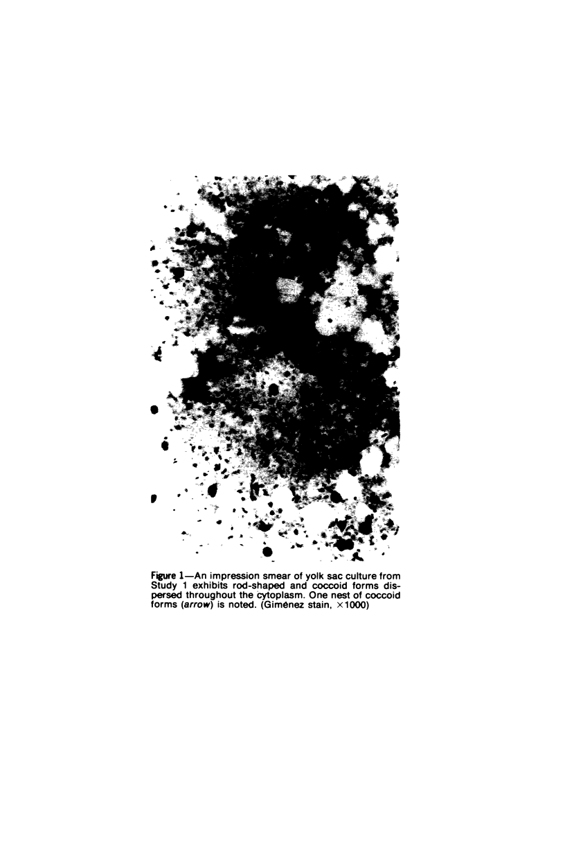
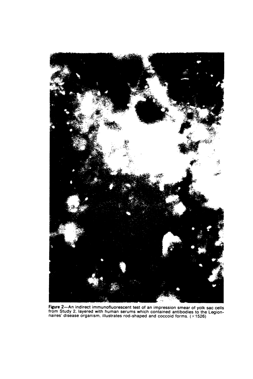
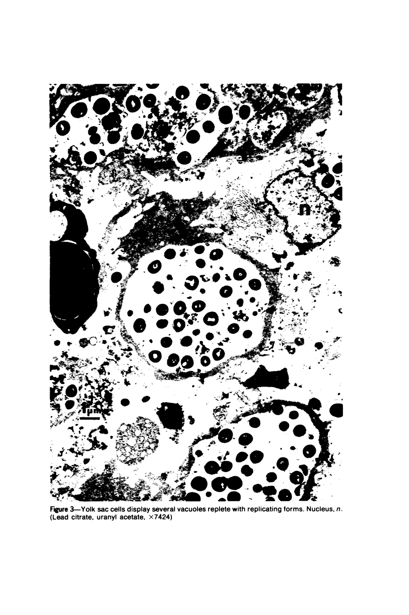
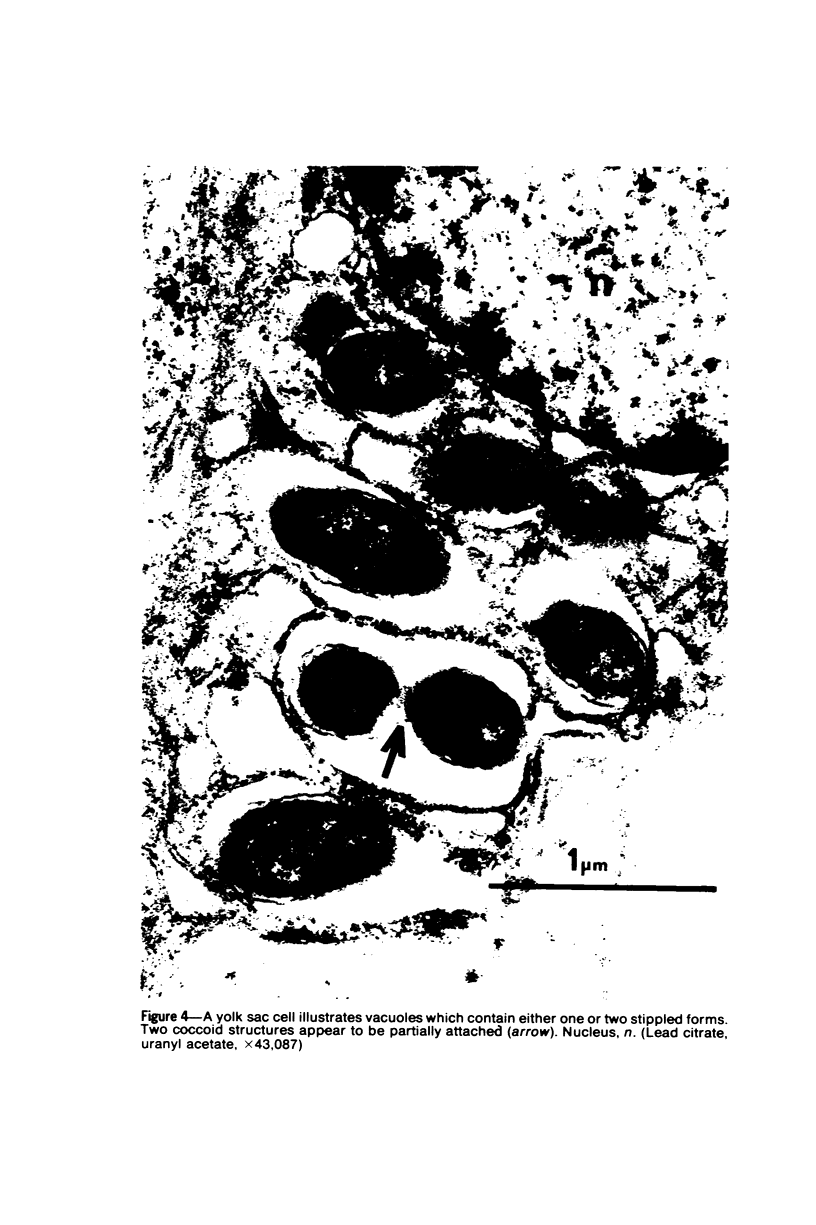
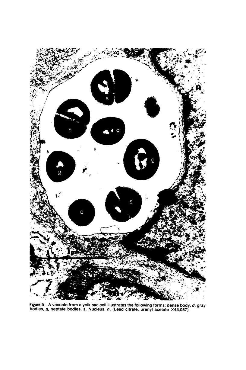
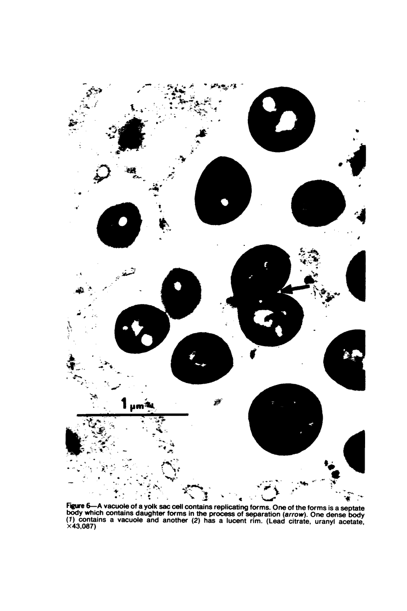
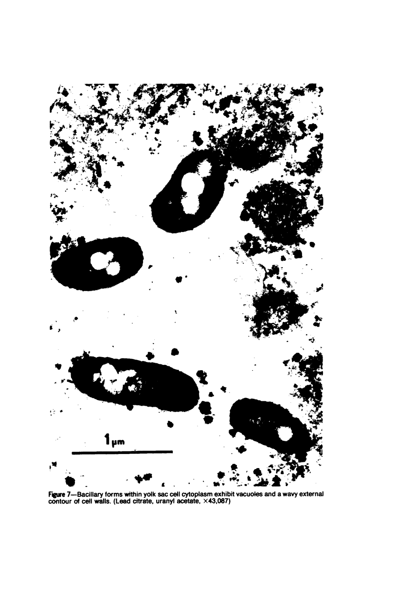
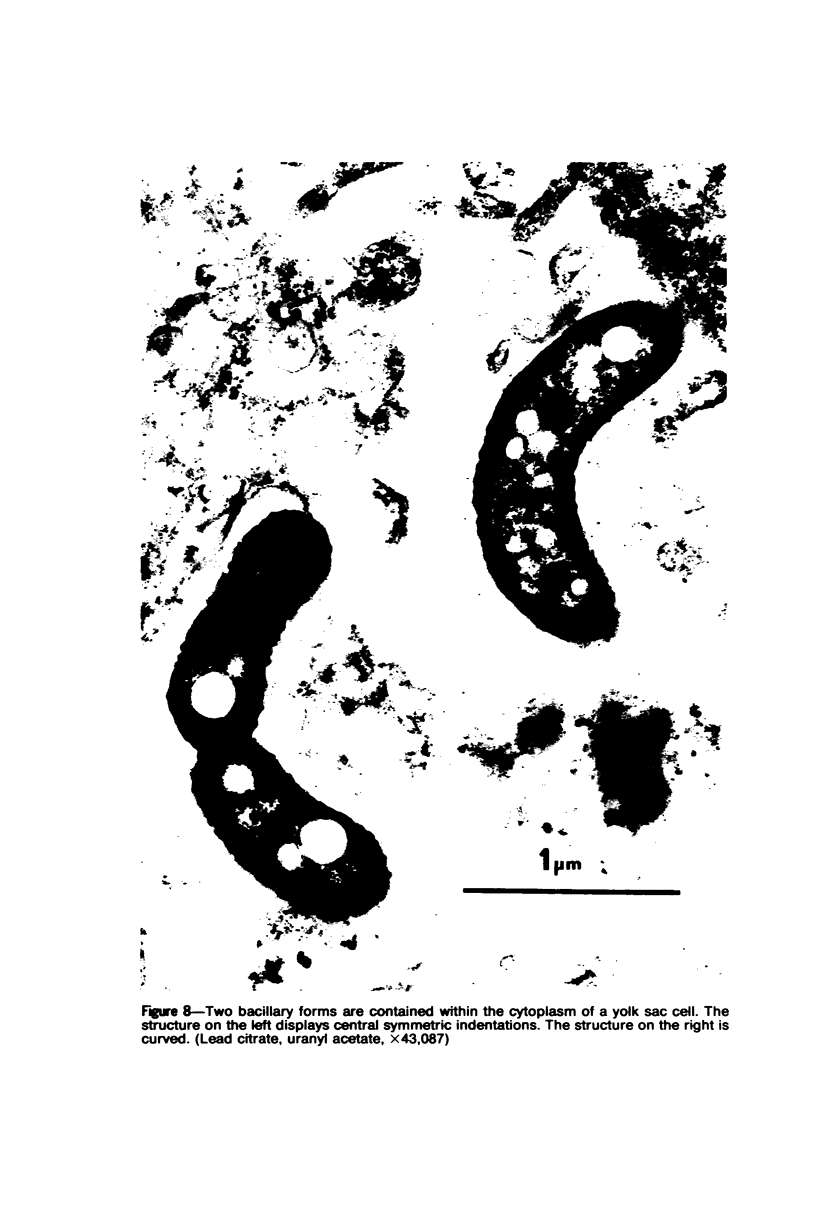
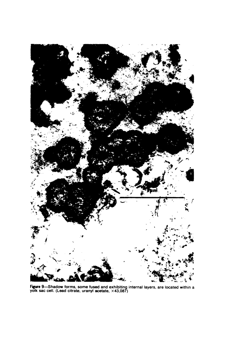
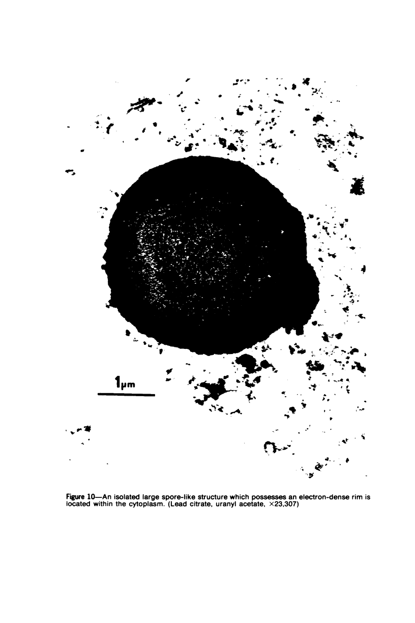
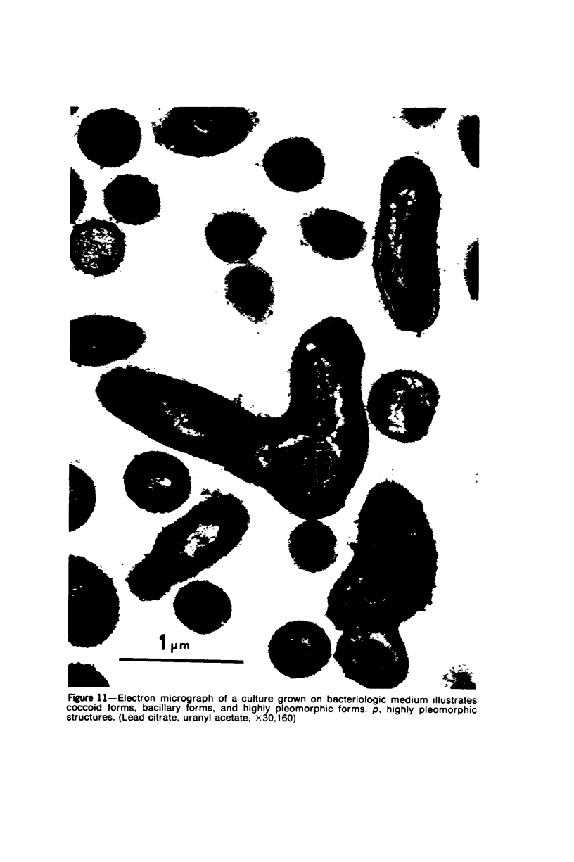
The Pennsylvania Department of Health, Bureau of Laboratories, has isolated the Legionnaires disease organism from two patients with Legionnaires' disease proved by serologic techniques. We have studied the morphology which the isolate assumes in yolk sac tissue and on bacteriologic media. The organism was Giménez-positive and gram-variable. Using an indirect immunofluorescent procedure, it was shown to react with convalescent serum samples taken from patients who had Legionnaires' disease. The organism multiplies by binary fission extracellularly and intracellulary; is both coccoid and bacillary in form; and contains characteristic cytoplasm, nucleoids, a cytoplasmic membrane, and a small cell wall of variable size. It may produce spores of unusual appearance. Intracellular replication characteristically occurs within vacuoles. The Legionnaires' disease organism conforms to the morphologic criteria for a prokaryocyte.
Full text
PDF
Images in this article
Selected References
These references are in PubMed. This may not be the complete list of references from this article.
- BOVARNICK M. R., MILLER J. C., SNYDER J. C. The influence of certain salts, amino acids, sugars, and proteins on the stability of rickettsiae. J Bacteriol. 1950 Apr;59(4):509–522. doi: 10.1128/jb.59.4.509-522.1950. [DOI] [PMC free article] [PubMed] [Google Scholar]
- GIMENEZ D. F. STAINING RICKETTSIAE IN YOLK-SAC CULTURES. Stain Technol. 1964 May;39:135–140. doi: 10.3109/10520296409061219. [DOI] [PubMed] [Google Scholar]