

Abstract
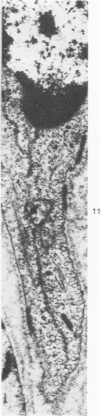
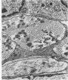
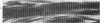
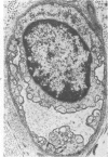
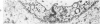
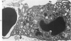
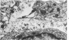
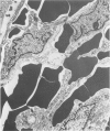
A comparison of the fine structure of subdural neomembranes with the fine structural organization of the normal human dura-arachnoid interface discloses that neomembranes are not de novo proliferations of tissue from a smooth inner dural surface. Rather, a neomembrane is the result of proliferation and excessive thickening of the normal layer of dural border cells. On proliferation, the dural border cells form multilayered tiers and clusters of cells, transfixed by capillaries, with collagen fibrils and elastic fibers between them. Capillaries and collagen fibrils are absent from the normal interface layer. Pathogenetic concepts of chronic subdural hematoma need to be revised. Any pathologic condition inducing cleavage of tissue within the dural border layer at dura-arachnoid interface will be followed by proliferation of fural border cells with production of a neomembrane. There is no compelling reason to postulate that proliferation of the border cell layer is always secondary to traumatic hemorrhage.
Full text
PDF


Images in this article


Selected References
These references are in PubMed. This may not be the complete list of references from this article.
- NELSON E., BLINZINGER K., HAGER H. An electron-microscopic study of bacterial meningitis. I. Experimental alterations in the leptomeninges and subarachnoid space. Arch Neurol. 1962 May;6:390–403. doi: 10.1001/archneur.1962.00450230052007. [DOI] [PubMed] [Google Scholar]
- Nabeshima S., Reese T. S., Landis D. M., Brightman M. W. Junctions in the meninges and marginal glia. J Comp Neurol. 1975 Nov 15;164(2):127–169. doi: 10.1002/cne.901640202. [DOI] [PubMed] [Google Scholar]
- RICHTER G. W. Electron microscopy of hemosiderin; presence of ferritin and occurrence of crystalline lattices in hemosiderin deposits. J Biophys Biochem Cytol. 1958 Jan 25;4(1):55–58. doi: 10.1083/jcb.4.1.55. [DOI] [PMC free article] [PubMed] [Google Scholar]
- SCHOEFL G. I. STUDIES ON INFLAMMATION. III. GROWING CAPILLARIES: THEIR STRUCTURE AND PERMEABILITY. Virchows Arch Pathol Anat Physiol Klin Med. 1963 Nov 8;337:97–141. [PubMed] [Google Scholar]
- Sato S., Suzuki J. Ultrastructural observations of the capsule of chronic subdural hematoma in various clinical stages. J Neurosurg. 1975 Nov;43(5):569–578. doi: 10.3171/jns.1975.43.5.0569. [DOI] [PubMed] [Google Scholar]
- Schachenmayr W., Friede R. L. The origin of subdural neomembranes. I. Fine structure of the dura-arachnoid interface in man. Am J Pathol. 1978 Jul;92(1):53–68. [PMC free article] [PubMed] [Google Scholar]
- Shabo A. L., Maxwell D. S. The subarachnoid space following the introduction of a foreign protein: an electron microscopic study with peroxidase. J Neuropathol Exp Neurol. 1971 Jul;30(3):506–524. doi: 10.1097/00005072-197107000-00013. [DOI] [PubMed] [Google Scholar]