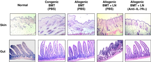
Figure 3.
Histologic analysis of anti–IL-7Rα antibody-treated allogeneic recipients. Skin and small intestine tissues from the recipients were analyzed at day 30 following BMT. Representative tissue samples from each group of mice were stained with H&E. The skin and small intestine from the PBS-treated allogeneic BMT-plus-LN recipients showed evidence of GVHD with lymphocytic infiltration and inflammation. The tissue sections from anti–IL-7Rα antibody-treated allogeneic BMT-plus-LN recipients had normal histology. See “Materials and methods, Histologic analyses” for image acquisition information.

