Introduction
The true incidence of malignant degeneration of hepatic adenoma is not known. A recent review documented only five confirmed cases of a pre-existing adenoma with malignant transformation to hepatocellular carcinoma 1. We report an unusual case of a male patient with no history of steroid use who developed a well-differentiated hepatocellular cancer arising within a pre-existing adenoma, which was diagnosed more than 10 years earlier. Despite complete resection of the malignant component, the tumour recurred nearly six years after resection with multifocal hepatoma at the resection margin. This case not only reinforces previous recommendations supporting routine resection of hepatic adenoma, but also suggests that complete pathological removal of the adenoma and continued long-term follow-up is essential. A review of the recent literature follows the case summary.
Case report
A 52-year-old man presented with sudden, severe midline and left upper quadrant abdominal pain that decreased in intensity over a two-week period. Of note, the patient had had intermittent, mild mid-abdominal pain in the past and had been told 11 years previously that he had an haemangioma with the liver. The most recent radiological imaging of this lesion was a CT scan one year before clinical presentation, which revealed a 5 cm left lateral segment lesion. He had no history of hepatitis, jaundice, or a previous blood transfusion.
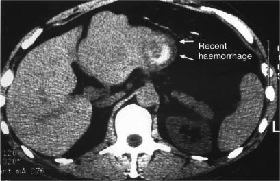
The severity of this episode prompted an investigation that included laboratory tests and abdominal imaging. Bilirubin was normal at 1.2 mg/dL (15.6 micromol/L), while ALT and alkaline phosphatase were mildly elevated at 59 U/L (normal 0–40) and 113 U/L (normal, 35–110), respectively. His haematocrit was normal. An ultrasound scan revealed a 10×11 cm heterogeneous mass in the left lateral segment of the liver. CT scan demonstrated a 12×10 cm mixed cystic and solid mass with a hyperdensity within the cystic portion, which was interpreted as relatively acute haemorrhage (Figure 1). The differential diagnosis based on radiological investigation included haemangioma, focal nodular hyperplasia, and adenoma with a probable recent acute haemorrhage. Given the radiological findings and the patient's symptoms, resection was recommended.
Figure 1. .
CTscan demonstrating left lateral segment cystic and solid mass (a) with recent haemorrhage (b).
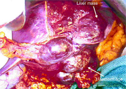
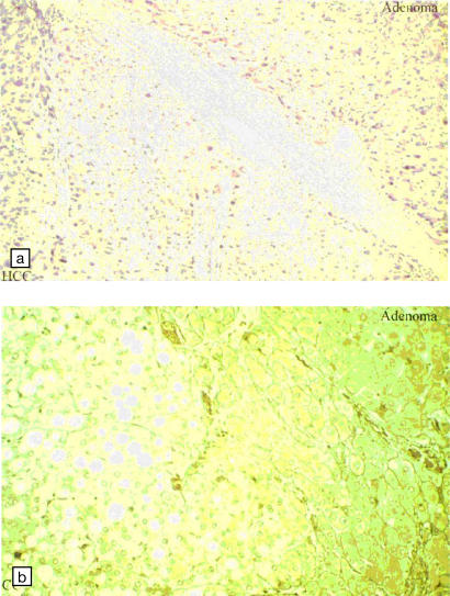
At the time of operation, there was a large non-compressible liver mass in segment III, with recent haemorrhage within the inferior aspect of this lesion (Figure 2). The remainder of the liver was grossly normal, and enucleation was performed. Histological examination of the resected specimen revealed a 5.5 cm well-differentiated hepatocellular carcinoma arising within a 12 cm hepatic adenoma (Figure 3a). Reticulin-stained sections (Figure 3b) differentiated the adenoma, in which the basement membrane was demonstrated, from the hepatocellular carcinoma, which has no basement membrane. The carcinoma was 0.5 cm from the resection margin and was contained entirely within the adenoma. Adenoma was present at the resection margin. The patient recovered from the procedure without incident.
Figure 2. .
Intraoperative evaluation revealed a large liver mass with haemorrhage inferiorly.
Figure 3. .
Hepatocellular carcinoma arising within an adenoma was seen on (a) H & E section and (b) reticulin stain.
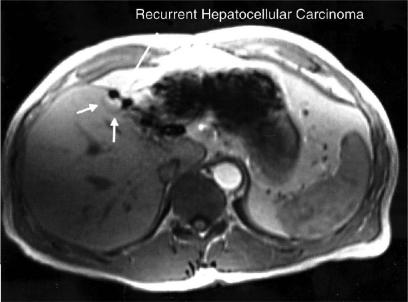
Serum alpha fetoprotein (AFP) and contrast CT scans of the abdomen were obtained every six months for follow-up evaluation. After three negative examinations, the screening interval was expanded to one year and serial abdominal ultrasounds were substituted for the CT scans. Five years after the initial resection, ultrasonography revealed two 2 cm hypoechoic masses within segment IVa of the liver adjacent to the resection margin that were suspicious for metastases. A contrast CT failed to demonstrate these lesions, but both of them were clear on gadolinium-enhanced MRI and were consistent with recurrent hepatoma (Figure 4). At that time, his serum AFP level was normal (1.2 ng/mL), liver function tests were normal, and a hepatitis panel was notable for the presence of hepatitis B core antibody and hepatitis A antibody.
Figure 4. .
Gadolinium-enhanced MRI obtained five years after resection revealed two foci of recurrent hepatocellular carcinoma arising at the resection margin.
Options for treatment of the lesions arising at the resection margin included repeat resection with a formal left lobectomy and ablation with embolisation or alcohol injection. Although resection was recommended, the patient was hesitant to proceed with an operation. Therefore, because of the multifocal nature of his recurrence, an angiogram was performed to assess for other lesions, and embolisation was planned at the same time. Both lesions were seen but there were no other tumours. The segment IV feeding vessels were therefore embolised. On the following day, both lesions were injected with alcohol. The patient tolerated both procedures without difficulty.
Discussion
The management of hepatic adenoma is challenging because of the low but real risk of haemorrhage or malignant transformation. Although malignant transformation of pre-existing adenomata has been reported, the natural history of these lesions is not well understood. Foster and Berman's review of the literature found only five well-documented reports of biopsy-proven hepatic adenoma with malignant transformation to hepatocellular cancer 1. In each case, a liver biopsy confirmed adenoma and the patients were monitored. In four cases, the diagnosis of adenoma was made by percutaneous biopsy 1,2,3,4, while the fifth case involved pathological examination of a left lobectomy specimen, which contained multiple adenomata. This patient developed hepatocellular cancer five years after resection, which presumably arose from residual adenoma in the liver remnant 5. In addition to cases of hepatocellular carcinoma arising within a known adenoma, hepatocellular carcinoma may also arise within a previously undiagnosed adenoma (Table 1) 4,6,7,8,9. Both the presence of hepatocellular cancer arising within an adenoma and dysplasia occurring within an adenoma 10,11 suggest the possibility of a step-wise transformation process from adenoma to carcinoma, although this sequence is difficult to prove.
Table 1. Summary of all known cases of hepatocellular carcinoma arising in an adenoma.
| Reference | Age at Presentation/Sex | Number of Adenomas | Interval prior to malignant transformation (years) | AFP level | Resected | Outcome |
|---|---|---|---|---|---|---|
| Hepatocellular carcinoma arising in a pre-existing adenoma | ||||||
| Gordon 3 | 36/F | solitary | 3 | normal | yes | NED 6 years after resection |
| Gyorffy 4 | 53/F | multiple | 2 | high | no | DOD 7 months after HCC diagnosis |
| Foster1 | 56/F | multiple | 5 | high | no | DOD 5 months after HCC diagnosis |
| Leese 5 | 13/M | multiple | 5 | high | transplant | NED 1 year after operation |
| Tesluk 2 | 34/F | solitary | 3 | – | yes | Postoperative death |
| Present case | 52/M | solitary | II | normal | yes | Recurrent HCC 6 years after operation |
| Hepatocellular carcinoma arising in previously undiagnosed adenoma | ||||||
| Ferrell 9 | 29/F | solitary | n/a | normal | yes | NED 4 years after operation |
| Weimann 7 | not specified | solitary | n/a | normal | yes | NED 1 year after operation |
| Weimann 7 | not specified | solitary | n/a | normal | yes | NED 1 year after operation |
| Belghiti 6 | <50/F | solitary | n/a | normal | yes | NED 6 months after operation |
| Perret [l5] | 24/F | solitary | n/a | normal | yes | NED 1 year after operation |
| Reddy 8 | not specified/F | solitary | n/a | normal | yes | NED 3 years after operation |
Patients with previously undiagnosed adenoma can present with abdominal pain due to intra-abdominal or intratumoural haemorrhage 5,7,12. The risk of haemorrhage is not known because the denominator is not well defined. Although one large review reported that 6 of 44 patients with adenoma presented with haemorrhage 7, Foster and Burman 1 reported no bleeding among 34 patients, 24 of whom had multiple adenoma. Thus, the true incidence of this complication is unknown. An increased risk of rupture is associated with the oestrogenic stimulation of pregnancy or oral contraceptives 13. There is no definite association between size of tumour and the risk of rupture, and although some authors suggest that larger tumours have a greater propensity to bleed 14, massive haemorrhage has been reported even in small lesions 13.
Adenomas are notoriously difficult to diagnose preoperatively by clinical, radiographical, or histological means. The diagnostic challenge involves differentiating adenoma from focal nodular hyperplasia or hepatocellular carcinoma. Clinically, abdominal pain is a frequently presented symptom 6,12, but it is not specific for adenoma versus FNH or HCC 7,8. Adenomas are most commonly diagnosed in young women taking oral contraceptives 15; they occur less commonly in men and are associated with the use of anabolic steroids. Adenomas can decrease in size when oral contraceptives are discontinued 3,16.
Although radiological investigations are the most reliable way of differentiating adenoma from FNH or HCC, definitive diagnosis is still not possible in many cases, even with the use of contemporary imaging modalities. Because these lesions can contain areas of haemorrhage, fibrosis, or necrosis, their heterogeneous appearance can be highly variable and may resemble hepatocellular carcinoma. Multiphasic helical CT scanning appears to be more accurate than conventional scanning in differentiating adenoma from other lesions 17. However, MRI may be the single best radiological test to differentiate adenomas, particularly with gadolinium enhancement, which shows an early and brief blush with contrast, followed by an isointense phase 18,19. Selected use of Tc-99 sulphur colloid nuclear scintigraphy may facilitate differentiation from FNH, which often show colloid uptake because they contain Kupffer cells 20.
Despite extensive radiological evaluation with high quality MRI or helical CT scan, it is impossible to make a definitive diagnosis preoperatively in up to one-third of patients with benign hepatic tumours. Because the ability to achieve a reliable diagnosis by percutaneous biopsy is limited due to sampling error 7,9, and because this technique may cause bleeding or needle-tract seeding 21, it is not routinely recommended. Even when the biopsy is adequate, it is difficult to distinguish reliably adenoma from well-differentiated carcinoma or focal nodular hyperplasia 7. Modern cytopathological techniques such as cytophotometric analysis of DNA content 22 and comparative genomic hybridization 11 may reduce the uncertainty of distinguishing benign lesions from well-differentiated carcinoma, but the experience with these methods is limited. Consequently, some patients are submitted to liver resection in order to obtain a diagnosis.
Because of the risk of haemorrhage, malignant transformation, or missed diagnosis of early malignancy, complete surgical resection of hepatic adenoma is the treatment of choice for all patients who can tolerate surgery. This strategy is particularly important for patients symptomatic lesions or with large, peripheral tumours, in which there is an increased risk of haemorrhage. In addition, women who are using fertility drugs or planning pregnancy should undergo resection. Complete resection may require formal lobectomy, but often these tumours are amenable to segmental resection. The goal is complete resection with a grossly negative margin to avoid the risk of malignancy arising in residual adenoma.
Elevated AFP serum levels strongly suggest carcinoma and can be diagnostic. Unfortunately, in cases of hepatocellular carcinoma arising within an adenoma, the AFP was normal in over 70% of patients (Table 1), as it was in our patient even at the time of recurrence. Thus, close follow-up with serial imaging is essential for all patients with hepatocellular carcinoma arising in an adenoma.
References
- 1.Foster JH, Berman MM. The malignant transformation of liver cell adenomas. Arch Surg. 1994;129:712–17. doi: 10.1001/archsurg.1994.01420310044007. [DOI] [PubMed] [Google Scholar]
- 2.Tesluk H, Lawrie J. Hepatocellular adenoma. Its transformation to carcinoma in a user of oral contraceptives. Arch Pathol Lab Ued. 1981;105:296–9. [PubMed] [Google Scholar]
- 3.Gordon SC, Reddy KR, Livingstone AS, Jeffers LJ, Schiff ER. Resolution of a contraceptive-steroid-induced hepatic adenoma with subsequent evolution into hepatocellular carcinoma. Ann Intern Ued. 1986;105:547–9. doi: 10.7326/0003-4819-105-4-547. [DOI] [PubMed] [Google Scholar]
- 4.Gyorffy EJ, Bredfeldt JE, Black WC. Transformation of hepatic cell adenoma to hepatocellular carcinoma due to oral contraceptive use. Ann Intern Med. 1989;110:489–90. doi: 10.7326/0003-4819-110-6-489. [DOI] [PubMed] [Google Scholar]
- 5.Leese T, Farges O, Bismuth H. Liver cell adenomas. A 12-year surgical experience from a specialist hepato-biliary unit. Ann Surg. 1988;208:558–64. doi: 10.1097/00000658-198811000-00003. [DOI] [PMC free article] [PubMed] [Google Scholar]
- 6.Belghiti J, Pateron D, Panis Y, Vilgrain V, et al. Resection of presumed benign liver tumours. Br J Surg. 1993;80:380–3. doi: 10.1002/bjs.1800800340. [DOI] [PubMed] [Google Scholar]
- 7.Weimann A, Ringe B, Klempnauer J, Lamesch P, et al. Benign liver tumors: differential diagnosis and indicators for surgery. World J Surg. 1997;21:983–90. doi: 10.1007/s002689900337. [DOI] [PubMed] [Google Scholar]
- 8.Reddy KR, Kligerman S, Levi J, Livingstone A, et al. Benign and solid tumors of the liver: relationship to sex, age, size of tumors, and outcome. Am Surg. 2001;67:173–8. [PubMed] [Google Scholar]
- 9.Ferrell LD. Hepatocellular carcinoma arising in a focus of multilobular adenoma. A case report. Am J Surg Pathol. 1993;17:525–9. doi: 10.1097/00000478-199305000-00013. [DOI] [PubMed] [Google Scholar]
- 10.Tao LC. Oral contraceptive-associated liver cell adenoma and hepatocellular carcinoma. Cytomorphology and mechanism of malignant transformation. Cancer. 1991;68:341–7. doi: 10.1002/1097-0142(19910715)68:2<341::aid-cncr2820680223>3.0.co;2-q. [DOI] [PubMed] [Google Scholar]
- 11.Wilkens L, Bredt M, Flemming P, Schwarze Y, et al. Diagnostic impact of fluorescence in situ hybridization in the differentiation of hepatocellular adenoma and well-differentiated hepatocellular carcinoma. J Mol Diagn. 2001;3:68–73. doi: 10.1016/S1525-1578(10)60654-X. [DOI] [PMC free article] [PubMed] [Google Scholar]
- 12.Nagorney DM. Benign hepatic tumors: focal nodular hyperplasia and hepatocellular adenoma. World J Surg. 1995;19:13–18. doi: 10.1007/BF00316973. [DOI] [PubMed] [Google Scholar]
- 13.Rooks JB, Ory HW, Ishak KG, Strauss LT, et al. Epidemiology of hepatocellular adenoma. The role of oral contraceptive use. JAMA. 1979;242:644–8. [PubMed] [Google Scholar]
- 14.Ribeiro A, Burgart LJ, Nagorney DM, Gores GJ. Management of liver adenomatosis: results with a conservative surgical approach. Liver Transpl Surg. 1998;4:388–98. doi: 10.1002/lt.500040505. [DOI] [PubMed] [Google Scholar]
- 15.Perret AG, Mosnier JF, Porcheron J, Cuilleron M, et al. Role of oral contraceptives in the growth of a multilobular adenoma associated with a hepatocellular carcinoma in a young woman. J Hepatol. 1996;25:976–9. doi: 10.1016/s0168-8278(96)80305-9. [DOI] [PubMed] [Google Scholar]
- 16.Gindhart TD. Liver tumors and oral contraceptives: pathology and pathogenesis. Ann Clin Lab Sci. 1978;8:443–6. [PubMed] [Google Scholar]
- 17.Ichikawa T, Federle MP, Grazioli L, Nalesnik M. Hepatocellular adenoma: multiphasic CT and histopathologic findings in 25 patients. Radiology. 2000;214:861–8. doi: 10.1148/radiology.214.3.r00mr28861. [DOI] [PubMed] [Google Scholar]
- 18.Paulson EK, McClellan JS, Washington K, Spritzer CE, et al. Hepatic adenoma: MR characteristics and conelation with pathologic findings. AJR Am J Roentgenol. 1994;163:113–16. doi: 10.2214/ajr.163.1.8010195. [DOI] [PubMed] [Google Scholar]
- 19.Chung KY, Mayo-Smith WW, Saini S, Rahmouni A, et al. Hepatocellular adenoma: MR imaging features with pathologic conelation. AJR Am J Roentgenol. 1995;165:303–8. doi: 10.2214/ajr.165.2.7618545. [DOI] [PubMed] [Google Scholar]
- 20.Cherqui D, Rahmouni A, Charlotte F, Boulahdour H, et al. Management of focal nodular hyperplasia and hepatocellular adenoma in young women: a series of 41 patients with clinical, radiological, and pathological correlations. Hepatology. 1995;22:1674–81. [PubMed] [Google Scholar]
- 21.Jourdan JL, Stubbs RS. Percutaneous biopsy of operable liver lesions: is it necessary or advisable? N Z Med J. 1996;109:469–70. [PubMed] [Google Scholar]
- 22.Ruschenburg I, Hofmann M, Diab E, Droese M. Comparison of the DNA content in liver cell adenoma, hepatocellular carcinoma and regenerative nodules. Anticancer Res. 2000;20:1861–4. [PubMed] [Google Scholar]


