Abstract
Background
Smooth muscle tumours are common in the genito-urinary and gastro-intestinal tracts, but primary leiomyoma of the liver is extremely rare. Only a few cases have been reported to date.
Case outline
We report a case of giant leiomyoma of the liver in a 67-year-old woman that was treated by an extended right hepatectomy. There was no evidence of leiomyoma elsewhere in the abdomen (including the uterus).
Discussion
This appears to be the largest hepatic leiomyoma reported in the literature.
Keywords: hepatic leiomyoma, liver tumour, liver resection
Introduction
Smooth muscle tumours are common in the genito-urinary tract and gastro-intestinal tract, yet primary leiomyoma of the liver is extremely rare 1,2,3,4,5,6,7. We report a case of a patient with a liver leiomyoma of enormous dimensions that was successfully excised.
Case report
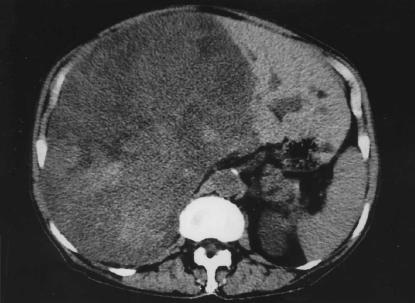
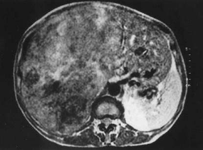
A 67-year-old woman was referred to the surgical department with a large abdominal mass. There was no relevant past medical history. Physical examination revealed a distended abdomen due to a huge, firm mass occupying the entire right upper quadrant. Laboratory findings demonstrated increased serum levels of alkaline phosphatase (850 IU/L) and γGT (215 IU/L), while tumour markers were normal (αFP 1.5 ng/ml; CEA 0.5 ng/ml; TPA 115 ng/ml; CA-125 20 units/ml). Computed tomography (CT) (Figure 1) and magnetic resonance (MR) imaging (Figure 2) showed an enormous mass (approximately 30 cm in diameter) replacing the whole right liver plus segment IV, and extending downwards into the lower abdomen. CT-guided fine needle aspiration biopsy failed to define the nature of the mass. The patient underwent elective operation and an extended right hepatectomy was performed; no extrahepatic lesions were seen, and in particular the uterus was normal. The postoperative course was uneventful and the patient was discharged on day 15. Histological examination showed whirling bundles of well-differentiated regular, spindle-shaped smooth muscle cells with scarce mitotic figures; neither giant cells nor anaplasia were present. The immunoperoxidase staining for vimentin and actin was positive. Four years later she is alive with no sign of local recurrence or distant metastasis.
Figure 1. .
CT scan showing an enormous mass that has completely replaced the right liver and segment IV, extending downward in the lower right abdominal quadrant (max. diameter 31 cm, min. diameter 16 cm).
Figure 2. .
Magnetic resonance scan showing the same case as described in Figure 1.
Discussion
Tumours composed of smooth muscle cells are found most often in the uterus. Indeed, leiomyoma is the most frequent tumour in women. These smooth muscle tumours can sometimes take origin from the muscularis of the gut or the media of blood vessels. They may therefore occur in any organ or tissue of the body.
To consider a leiomyoma of the liver as a primary tumour, it is necessary to exclude the presence of a primitive smooth muscle cell neoplasm at other intra-abdominal sites 1. Thus, a thorough preoperative assessment through imaging modalities is mandatory as well as an accurate intra-operative palpation of the abdominal organs. In particular, the pelvis requires a careful examination, since it is known to be the most probable source of metastases from leiomyomas that otherwise appear benign. Although imaging modalities do not allow a tissue-specific diagnosis, they are very helpful in excluding additional sites of leiomyoma and in planning surgical resection.
As the distinction between benign and malignant smooth muscle tumours of the gastrointestinal tract is not clear-cut, a careful review of the histological sections is imperative. Histological features indicative of malignancy include increased mitotic rate (more than 1/10 high power field), cellular atypia with nuclear pleomorphism, dense cellularity and degenerative changes 1.
In this patient, operation was indicated to define the nature of the lesion as well as to relieve the symptoms deriving from its enormous size. Surgical resection proved to be both diagnostic and curative, as in previously reported cases 1,2,3,4,5,6,7.
The present case is unique in that it represents the only primary leiomyoma of the liver among the few others of such enormous dimensions that are described in the literature.
References
- 1.Hawkins EP, Jordan GL, Mc Gravau MH. Primary leiomyoma of the liver. Successful treatment by lobectomy and presentation of criteria for diagnosis. Am J Surg Pathol. 1980;4:301–4. [PubMed] [Google Scholar]
- 2.Hollands MJ, Jaworski R, Wong KP, Little JM. A leiomyoma of the liver. HPB Surg. 1989;1:337–44. doi: 10.1155/1989/45978. [DOI] [PMC free article] [PubMed] [Google Scholar]
- 3.Bartoli S, Alo P, Leporelli P, Puce E, Di Tondo U, Thau A. Leiomioma epatico primitive. Minerva Chir. 1991;46:777–9. [PubMed] [Google Scholar]
- 4.Herzberg AJ, MacDonald JA, Tucker JA, Humphrey PA, Meyers WC. Primary leiomyoma of the liver. Am J Gastroenterol. 1990;85:1642–5. [PubMed] [Google Scholar]
- 5.Iwatsuki S, Todo S, Starzl TE. Excisional therapy for benign hepatic lesions. Surg Gynecol Obstet. 1990;171:240–6. [PMC free article] [PubMed] [Google Scholar]
- 6.Reinertson TE, Fortune JB, Peters JC, Pagnotta I, Balint J. Primary leiomyoma of the liver. A case report and review of the literature. Dig Dis Sci. 1992;37(4):622–7. doi: 10.1007/BF01307591. [DOI] [PubMed] [Google Scholar]
- 7.Rios-Dalenz JL. Leiomyoma of the liver. Arch Pathol Lab Med. 1965;79:54–6. [PubMed] [Google Scholar]


