Abstract
This report describes an interesting and unusual case of carcinoma gallbladder with skull metastasis.
Keywords: carcinoma gall bladder, skeletal metastasis
Introduction
Disseminated blood-borne metastases from carcinoma of the gall bladder are uncommon and usually occur late. Autopsy studies have reported around 64.8% incidence of distant metastasis. However, most of these metastases are in the liver and only 20% are in other sites. The most common site of extra-abdominal metastasis is the lung followed by the brain. Skeletal metastases in carcinoma gall bladder are very rare. To date there have only been a few case reports of bone metastasis in carcinoma gall bladder at the time of presentation. However, one autopsy series has reported about 10% incidence, which may indicate that the carcinoma gall bladder does metastasize to bone, perhaps in advanced stages. We report here a case of locally advanced but potentially operable gall bladder cancer with skull metastasis as the only site of distant metastasis.
Case report
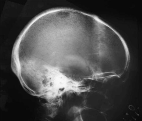
A 60-year-old female presented with pain in the right hypochondrium and jaundice with cholestatic features for 2 months. Abdominal examination revealed hepatomegaly and gall bladder lump. Liver function tests showed serum bilirubin 13.1 mg%, ALT 44 IU/L and alkaline phosphatase 74 IU/L. Ultrasonography showed dilated intrahepatic biliary radicles with a mass in the gall bladder neck. A CT scan of the abdomen showed a mass in the gall bladder neck infiltrating into the liver and common hepatic duct with enlarged lymph nodes along the hepatoduodenal ligament. However, there was no evidence of liver metastasis, ascites, celiac or para-aortic lymph nodes. Therefore the patient was planned for extended cholecystectomy. While the patient was being worked up for surgery, she complained of painful swelling on the scalp. A bony swelling was noted over the right parietal bone. X-ray of the skull revealed a punched out lesion over the parietal bone (Figure 1). A radionuclide whole body bone scan showed increased tracer concentration over the right and left parieto-occipital region, T8–11 vertebrae, left superior iliac spine and left femur near the trochanter. The lesion was confirmed to be a metastatic deposit by fine needle aspiration cytology.
Figure 1. .
Plain X-ray of skull: lateral view showing punched out lesion over the right parietal bone.
Discussion
The incidence of bone metastasis in carcinoma gall bladder is very low and, when present, it is usually associated with advanced disease. Hence, a bone scan is not included in the routine work-up of gall bladder cancer patients. However, in our patient bone metastasis was the only site of distant metastasis, which precluded radical surgery.
In the literature there is another case report in which skeletal metastasis was the only site of distant metastasis 1. The report by Kumar et al. 1 and the present case report raise the question as to whether investigation for bone metastasis should be included in the routine work-up protocol of carcinoma gall bladder. This emphasizes the need to perform a well designed prospective study to look at the incidence of bone metastasis in patients with gall bladder cancer, in particular at a resectable stage, and to decide whether screening for bone metastasis should be included in the routine work-up protocol for gall bladder cancer.
References
- 1.Kumar A, Bhargava SK, Upreti L, Kumar J. Disseminated osteoblastic skeletal metastasis from carcinoma gall bladder – a case report. Ind J Radiol Imaging. 2003;13:37–9. [Google Scholar]


