Abstract
Surgical removal of painful skin was first attempted as a treatment for chronic intractable post-herpetic neuralgia (PHN) more than a century ago, but long-term follow-up has rarely been reported. A patient who underwent surgical excision of 294 cm2 of thoracic skin comprising the entire area of pain and allodynia in October 2000 has been followed for 5.5 years post-surgery. Our initial report presented evidence of benefit in the form of reduced pain, elimination of allodynia, and reduced medication consumption during the first post-operative year. Unfortunately, pain steadily increased and now exceeds pre-surgery levels despite increased medication use. Pain topography and characteristics are different from pre-surgery and may relate to the pathophysiology of PHN. Skin resection cannot be recommended as a treatment for PHN.
Keywords: Excision, allodynia, reinnervation, pathophysiology, PHN, zoster
Introduction
Surgical removal of painful skin was first described by Gowers in 1900 as a treatment for chronic intractable PHN (Gowers 1900). The limited long-term follow-up data that is available regarding this irreversible therapy acknowledges pain recurrence in some patients (Loeser 2001). In one case, pain return was focal and responded to resection of an additional area of skin, but further follow-up was not provided. We previously presented a case of longstanding PHN treated by skin excision of the entire area of pain and allodynia (Petersen et al. 2002); no additional case reports have appeared since then. During the first post-surgical year, the patient reported reduced pain, complete loss of tactile allodynia, and greatly reduced need for analgesic medication. To provide guidance on this type of therapy, we now report the results of post-surgery clinical evaluations over a 5.5 year period.
Case presentation
Pre-operative through 1st post-operative year
The patient is a 70 year-old male with PHN in the right T6 dermatome since 1992. A pre-operative regimen of gabapentin, methadone, nortriptyline, and two lidocaine patches/day did not control pain and allodynia to his satisfaction and he elected to undergo surgical removal of the skin comprising the entire area of pain (MPA) and surrounding allodynic skin in October 2000. Our initial report provides results of pre-operative sensory examination, skin biopsies, and the capsaicin response test, along with pain ratings and medication use through year 1 post-surgery (Petersen et al, 2002).
Placed in context with other patients suffering from severe longstanding PHN that we have studied, this patient’s pattern of modestly reduced cutaneous innervation in the area of pain, severe allodynia, largely preserved thermal sensory function, relief from topical lidocaine, and severe worsening of pain with brief application of topical capsaicin pre-surgery suggested an important contribution from peripheral structures in the maintenance of pain and allodynia (Rowbotham et al. 1996, Rowbotham et al. 1998; Petersen et al. 2000; Petersen et al. 2002).
At surgery, an 11.3 × 26.0 cm2 piece of skin that comprised the entire area of pain and allodynia was excised to the fascia. All surgical margins were in previously non-painful and non-allodynic skin, but to remove such a large region of skin required extending the incision slightly across the posterior midline. The operation reduced pain, eliminated tactile allodynia, and facilitated greatly reduced medication use (summarized in Table 1). At the 32-week and 1-year follow up visits, he described deep and shooting pain located in the anterior chest, a previously asymptomatic area. At 1 year, he considered himself ‘50% improved’ by the surgery.
Table 1.
Visits during the first year with pain ratings and medication.
| Visit Date | VAS Avg (0-100) | Oral Medications |
|---|---|---|
| 1 day (Pre-Surgery) | 30 | gabapentin-1600mg, methadone-20mg, nortriptyline-40mg, lidocaine patch |
| 2 weeks | 9 | gabapentin-1600mg, methadone-20mg, nortriptyline-40mg |
| 4 weeks | 20 | gabapentin-1600mg, methadone-20mg, nortriptyline-25mg |
| 7 weeks | 18 | gabapentin-1200mg, methadone-20mg, nortriptyline-25mg |
| 12 weeks | 2 | gabapentin-800mg, methadone-10mg, nortriptyline-25mg |
| 16 weeks | <1 | gabapentin-600mg, methadone-7.5mg, nortriptyline-25mg (every other day) |
| 20 weeks | <1 | gabapentin-600mg, methadone-7.5mg |
| 32 weeks | 20 | gabapentin-800mg, methadone-10mg |
| 52 weeks | 18 | gabapentin-800mg, methadone-10mg |
2nd-6th post-operative years
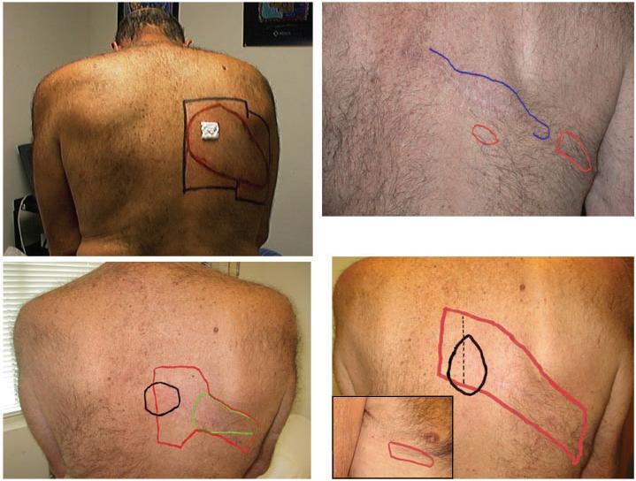
Pain ratings and symptoms, examination findings, and medication use subsequent to our initial report are presented in Table 2 and images of sensory exams are presented in Figure 1. During the second year after surgery, cyclic jabbing pain began to return in the previous MPA location below the right scapula and extended to the posterior midline. Allodynia in the MPA also started to return during the second year, increasing at a slower rate than the jabbing pain. The pain that emerged in the anterior chest below the right nipple during the 1st post-operative year continued unchanged. By the end of the second year, overall pain severity and medication use approached pre-surgical levels.
Table 2.
Clinical assessments from 1 year post-operative to 5 years 6 months post-operative.
| Visit | Pain VAS Avg last 48 hours 0-100 scale [min-max] | Pain Description | Exam | Medications (total daily doses) |
|---|---|---|---|---|
| 1 year 3 months |
12 [2-42] | Cyclic jabbing pain in previous MPA (Most Painful Area) on the back. Each cycle lasts 3-5 days, but pain free between cycles. Reports recurrence of touch sensitivity. | Allodynia to moving tactile stimuli present but mild near scar in previous MPA. | gabapentin-800mg methadone-10mg |
| 1 year 6 months |
40 [20-60] | Cyclic deep and jabbing pain in the previous MPA on the back, occurs less frequent than before surgery, but intensity is as severe as before surgery. Cycles can last 2-3 weeks. Touch sensitivity present, but very mild compared to pre-surgery. |
Pressure is painful in previous MPA. Strip of mild allodynia to moving tactile stimuli along surgical scar. | gabapentin-800-1200mg methadone-10-20 mg |
| 1 year 11 months |
50 [40-80] | Cyclic deep and jabbing pain in the previous MPA are more frequent. No longer pain free between cycles and pain severity approaching pre-surgical levels. Touch sensitivity more severe on back and anterior chest. |
Broader strip of allodynia along the scar. Sensory deficit to pin/cold in anterior chest is mild but detectable. Strip of sensory loss more easily detected along the scar, but is narrower than the area of allodynia. | gabapentin-1200mg methadone-10-15mg lidocaine patch 2 daily nortriptyline-25mg (every other day) |
| 3 years Figure 1 |
15 [4-20] | Cyclic deep and jabbing pain in the previous MPA occur daily with added electrical quality. No pain free moments. Size of area of touch sensitivity on the back remains smaller than pre-surgery. Touch sensitivity in the anterior chest present frequently. | MPA has returned to same location as preoperatively but not as large. Deep pressure in previous MPA produces radiating pain. On the allodynia VAS scale, 3 brush strokes in the MPA rated at 79/100. Infiltration with 5 ml of 1% lidocaine in previous MPA reduces pain intensity by >50% and shrinks area of allodynia around the scar. No change in anterior chest pain. See Figure 1 - bottom left panel. |
gabapentin-1600mg methadone-10-15mg lidocaine patch 2 daily |
| 5 years 6 months Figure 1 |
61 [20-81] | Cyclic deep and jabbing pain in the previous MPA lasting 5-10 minutes, occur multiple times per day and have added electrical quality. Pain radiates cephalad and laterally. No pain free periods. Area of MPA feels numb, tight and very sensitive to touch. Cannot wear clothing unless MPA covered by lidocaine patch. |
MPA remains in same location as preoperatively but not as large. Allodynia ratings same as on 3 year followup visit and area approximately same size as pre-operative. Smaller patch with milder allodynia in the front (Figure 1 - bottom right panel). |
gabapentin 2000 mg methadone 15 mg lidocaine patch 2 daily |
Figure 1.

Panel top left:One-day pre- operative. Area of allodynia (red) and outline of lidocaine patch placement (black). Capsaicin cream 0.075% (9 cm2) shown applied on a small section of the most painful area (MPA).
Panel top right:One-year post-operative: surgical scar visible with area of numbness (blue) and allodynia (red). Only the anterior chest reported as painful.
Panel bottom left:Three years post-operative: The MPA (black) is at posterior midline border of the MPA region prior to surgery. Enlarging area of allodynia (red). Infiltration with 5 ml 1% lidocaine in the MPA reduced the area of allodynia to that shown in green.
Panel lower right:Five years and six months post-operative: Showing MPA (black) and allodynia (red). Posterior midline marked with thin vertical line. Insert shows anterior chest allodynia (red).
On examination near the end of the 3rd post-surgical year the patient rated the unpleasantness of brush stroke in the MPA as 79/100 on the allodynia VAS, essentially identical to pre-operative severity. Infiltration of the MPA with 5 ml of 1% lidocaine reduced pain and allodynia on the back for approximately 6 hours, but did not affect the anterior chest pain.
Examination at 5.5 years post-surgery showed the same findings posteriorly, and mild allodynia anteriorly. He could not tolerate a more detailed sensory and neurological exam and declined skin biopsy and the capsaicin response test in the MPA. Overall, the patient feels that his current pain and pain-related disability exceeds pre-surgery levels. Despite having been nearly pain-free throughout the first post-operative year, he regrets undergoing the operation.
Discussion
In this patient, skin excision produced dramatic improvement during the first year after surgery, but pain and allodynia eventually returned in the prior MPA and new symptoms emerged anteriorly. Skin excision failed to provide long-term pain relief in this patient, and therefore can not be recommended as a treatment for PHN pain and allodynia. This recommendation is consistent with Loeser’s (2001) review of destructive surgical therapies for PHN.
Pathological studies of PHN have demonstrated nervous system damage that varies from mild to severe (for reviews, see Rowbotham et al. 1996; Watson 2001). In many cases of PHN, pain and allodynia may be due to input from abnormal and possibly sensitized primary afferent nociceptors that maintain their central targets in a state of chronic sensitization, a mechanism with much preclinical support (Rowbotham et al, 1998; Devor 2006; Djouhri et al. 2006). This patient’s pre-surgery evaluation suggested an important peripheral contribution to ongoing pain and allodynia. Understandably, but unfortunately, our patient did not wish to undergo skin biopsy assessment of the newly painful and allodynic skin, nor did he wish to undergo the capsaicin response test again. We have no new anatomical and physiological data to support speculations about the mechanisms underlying the return of pain and allodynia in this patient.
At surgery, axons innervating the painful skin were severed below their entry into the dermis by dissecting down to the fascia overlying the thoracic musculature and removing the skin. The surgical margins were in non-painful and non-allodynic skin. Simple undermining of adjacent skin to free skin flaps for rotation to close the surgical defect may have temporarily severed axons to those skin areas as well. Sensory loss in the initial post-operative period was restricted to a narrow strip just above and below the surgical scar.
Return of sensation to the edges of the surgical defect may in theory result from (1) regeneration of nerve fibers from a DRG unaffected by zoster that were denervated during the advancement and rotation of the skin to cover the surgical defect; (2) collateral sprouting of nerve fibers from a DRG unaffected by zoster that were not deafferented during the procedure; (3) collateral sprouting from fibers not involved in either herpes zoster or surgery (intact DRG); (4) regeneration of herpes zoster affected fibers (affected DRG); and (5), a combination of all these fiber types. The time profile of the patient’s return of pain and allodynia followed a time course similar to that of cutaneous reinnervation (Rajan et al. 2003).
The return of pain followed by return of allodynia on the back might be due to excessive activity in either deafferented or intact fibers. Abnormal levels of peripheral input could then either re-sensitize central neurons or sensitize previously unaffected ones. Post-operative clinical evidence that activity in nerve endings in or near the skin surface were contributing to the resurgent pain comes from the temporary relief of pain and allodynia on the back after lidocaine skin infiltration performed 3 years post-surgery. Alternatively, scar neuromas could have formed along the approximately 35 cm long incision suture line or deeper in the region of surgical dissection down to the deep fascia (Devor 2006). In support of this explanation, one area along the surgical scar did show marked sensitivity to pressure.
The emergence of pain in the anterior chest beginning 32 weeks after surgery is difficult to explain and presumably reflects central mechanisms. This portion of the dermatome was distant from the surgical area and asymptomatic pre-operatively. Local infltration with lidocaine on the back in the MPA did not affect pain and allodynia on the anterior chest, suggesting this pain was not dependent on peripheral input from the surgical zone. Although sensory exam 5.5 years after surgery demonstrated allodynia had spread across the posterior midline, this may not be evidence of central mechanims because the surgical incision and dissection required crossing the midline.
In summary, although our patient experienced improvement for one year and some of the patients reported in the literature appear to have benefited from skin resection and skin undermining, the late recurrence of pain and allodynia in our patient and the poor reporting of outcomes in the prior literature compels us to advise against skin resection or undermining for medically intractable PHN pain and allodynia.
ACKNOWLEDGEMENTS
Supported by NINDS grants K24 NS02164, R0-1 NS39521 and an unrestricted grant from the VZV Research Foundation. We wish to thank the patient for his patience and willingness to provide long-term follow-up information.
Footnotes
Publisher's Disclaimer: This is a PDF file of an unedited manuscript that has been accepted for publication. As a service to our customers we are providing this early version of the manuscript. The manuscript will undergo copyediting, typesetting, and review of the resulting proof before it is published in its final citable form. Please note that during the production process errors may be discovered which could affect the content, and all legal disclaimers that apply to the journal pertain.
REFERENCES
- Devor M. Response of nerves to injury in relation to neuropathic pain. In: McMahon SB, Koltzenburg M, editors. Textbook of Pain. 5th Elsevier; Amsterdam: 2006. pp. 905–927. [Google Scholar]
- Djouhri L, Koutsikou S, Fang X, McMullan S, Lawson SN. Spontaneous pain, both neuropathic and inflammatory, is related to frequency of spontaneous firing in intact C-fiber nociceptors. J Neurosci. 2006;26(4):1281–1292. doi: 10.1523/JNEUROSCI.3388-05.2006. [DOI] [PMC free article] [PubMed] [Google Scholar]
- Gowers WR. A manual of diseases of the nervous system. Blakiston Co; Philadelphia, PA: 1900. [Google Scholar]
- Loeser J, Watson CPN, Gershon AA.Surgery for postherpetic neuralgia Herpes Zoster and Postherpetic Neuralgia 2ndRevised and Enlarged Edition200111255–264.Elsevier; Amsterdam [Google Scholar]
- Petersen KL, Fields HL, Brennum J, Sandroni P, Rowbotham MC. Capsaicin activation of ‘irritable’ nociceptors in post-herpetic neuralgia. Pain. 2000;88:125–133. doi: 10.1016/S0304-3959(00)00311-0. [DOI] [PubMed] [Google Scholar]
- Petersen KL, Rice FL, Suess F, Berro M, Rowbotham MC. Relief of post-herpetic neuralgia by surgical removal of painful skin. Pain. 2002;98(12):119–126. doi: 10.1016/s0304-3959(02)00029-5. [DOI] [PubMed] [Google Scholar]
- Rajan B, Polydefkis M, Hauer P, Griffin JW, McArthur JC. Epidermal reinnervation after intracutaneous axotomy in man. J Comp Neurol. 2003;457(1):24–36. doi: 10.1002/cne.10460. [DOI] [PubMed] [Google Scholar]
- Rowbotham MC, Yosipovitch G, Connolly MK, Finlay D, Forde G, Fields HL. Cutaneous innervation density in the allodynic form of post-herpetic neuralgia. Neurobiology of Disease. 1996;3:205–214. doi: 10.1006/nbdi.1996.0021. [DOI] [PubMed] [Google Scholar]
- Rowbotham MC, Petersen KL, Fields HL. Is postherpetic Neuralgia more than one disorder? Pain Forum. 1998;7:231–237. [Google Scholar]
- Watson CP, Oaklander AL, Deck JH.Watson CPN, Gershon AA.The neuropathology of herpes zoster with particular reference to postherpetic neuralgia and its pathogenesis Herpes Zoster and Postherpetic Neuralgia 2ndRevised and Enlarged Edition200111167–182.Elsevier; Amsterdam [Google Scholar]


