

Abstract
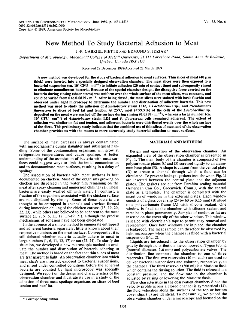
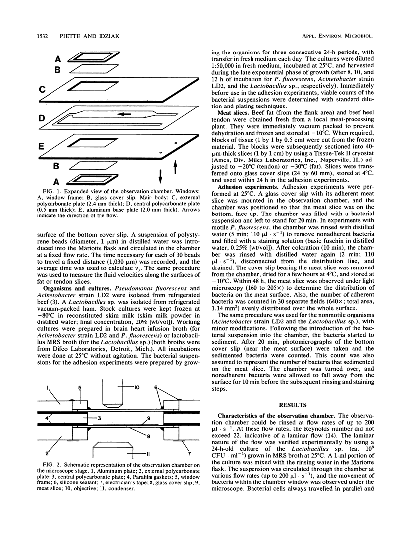
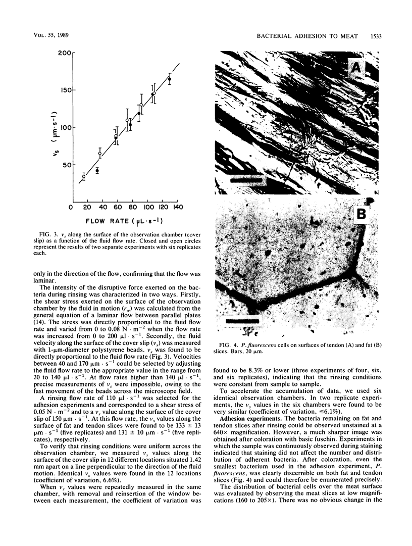
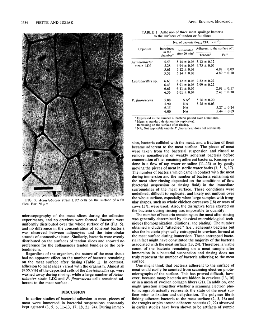
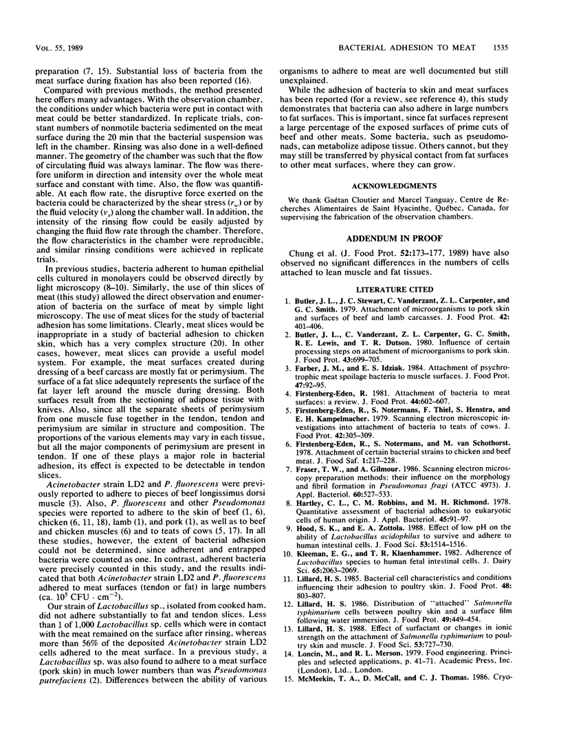
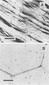
A new method was developed for the study of bacterial adhesion to meat surfaces. Thin slices of meat (40 microns thick) were inserted into a specially designed observation chamber. The meat slices were then exposed to a bacterial suspension (ca. 10(6) CFU.ml-1) to initiate adhesion (20 min of contact time) and subsequently rinsed to eliminate nonadherent bacteria. Because of the special chamber design, the disruptive force exerted on the bacteria during rinsing (shear stress) was uniform over the whole surface of the meat slices, was constant, and could be varied from 0 to 0.08 N.m-2. After being rinsed, the meat slices were stained with basic fuschin and observed under light microscopy to determine the number and distribution of adherent bacteria. This new method was used to study the adhesion of Acinetobacter strain LD2, a Lactobacillus sp., and Pseudomonas fluorescens to slices of beef fat and tendon. At 25 degrees C, most (greater than or equal to 99.9%) of the cells of the Lactobacillus sp. deposited on the meat were washed off the surface during rinsing (0.05 N.m-2), whereas a large number (ca. 10(5) CFU.cm-2) of Acinetobacter strain LD2 and P. fluorescens cells remained adherent. The extent of adhesion was similar on fat and tendon, and adherent bacteria were distributed evenly over the whole surface of the slices. This preliminary study indicates that the combined use of thin slices of meat and of the observation chamber provides us with the means to more accurately study bacterial adhesion to meat surfaces.
Full text
PDF

Images in this article
Selected References
These references are in PubMed. This may not be the complete list of references from this article.
- Hartley C. L., Robbins C. M., Richmond M. H. Quantitative assessment of bacterial adhesion to eukaryotic cells of human origin. J Appl Bacteriol. 1978 Aug;45(1):91–97. doi: 10.1111/j.1365-2672.1978.tb04202.x. [DOI] [PubMed] [Google Scholar]
- Kleeman E. G., Klaenhammer T. R. Adherence of Lactobacillus species to human fetal intestinal cells. J Dairy Sci. 1982 Nov;65(11):2063–2069. doi: 10.3168/jds.S0022-0302(82)82462-4. [DOI] [PubMed] [Google Scholar]
- McMeekin T. A., Thomas C. J., McCall D. Scanning electron microscopy of microorganisms on chicken skin. J Appl Bacteriol. 1979 Feb;46(1):195–200. doi: 10.1111/j.1365-2672.1979.tb02600.x. [DOI] [PubMed] [Google Scholar]
- Notermans S., Kampelmacher E. H. Further studies on the attachment of bacteria to skin. Br Poult Sci. 1975 Sep;16(5):487–496. doi: 10.1080/00071667508416217. [DOI] [PubMed] [Google Scholar]
- Thomas C. J., McMeekin T. A. Attachment of Salmonella spp. to chicken muscle surfaces. Appl Environ Microbiol. 1981 Jul;42(1):130–134. doi: 10.1128/aem.42.1.130-134.1981. [DOI] [PMC free article] [PubMed] [Google Scholar]
- Thomas C. J., McMeekin T. A. Contamination of broiler carcass skin during commercial processing procedures: an electron microscopic study. Appl Environ Microbiol. 1980 Jul;40(1):133–144. doi: 10.1128/aem.40.1.133-144.1980. [DOI] [PMC free article] [PubMed] [Google Scholar]
- Thomas C. J., McMeekin T. A. Spoilage of chicken skin at 2 degrees C: electron microscopic study. Appl Environ Microbiol. 1981 Feb;41(2):492–503. doi: 10.1128/aem.41.2.492-503.1981. [DOI] [PMC free article] [PubMed] [Google Scholar]