

Abstract
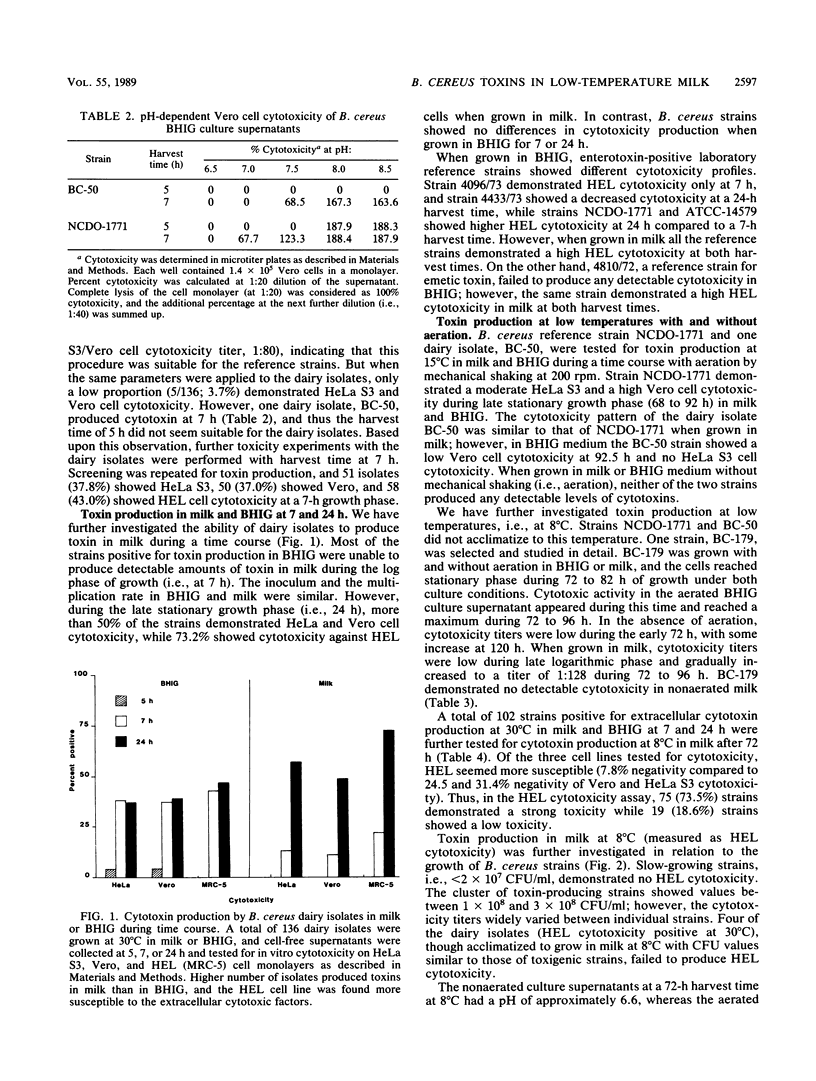
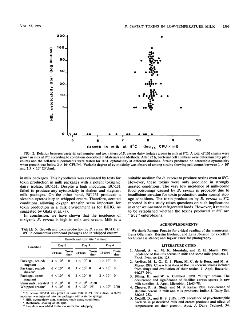
A total of 136 strains of Bacillus cereus isolated from milk and cream were evaluated for toxin production based on HeLa S3, Vero, and human embryonic lung (HEL) cell cytotoxicity in vitro. HEL cell monolayers were more susceptible than the other two cell lines. The percentage of isolates exhibiting HEL cytotoxicity was similar (43.0 and 48.4%) when the strains were grown in brain heart infusion broth containing 0.1% glucose (BHIG) at 7 and 24 h, respectively, at 30 degrees C. In milk, only 21.8% of isolates showed HEL cytotoxicity at 7 h, and the number increased significantly to 73.2% at 24 h at 30 degrees C. Further, 102 toxin-positive isolates were acclimatized to grow at 8 degrees C in milk. Ninety-four (92.2%) of the strains produced HEL cytotoxicity of various degrees with no strict correlation to bacterial cell numbers and also elicited vascular permeability reaction in rabbit skin. Under aerated growth conditions (agitation, 200 rpm) B. cereus elicited cytotoxicity in BHIG and in milk at temperatures of 30, 15, and 8 degrees C. However, in nonaerated (stagnant) cultures toxin production was diminished (BHIG) or completely lost (milk) at all temperatures. Toxin production at 8 degrees C was evaluated in two different types of commercial cardboard milk packages by inoculation with a potent toxigenic dairy isolate. No detectable HEL cytotoxicity was observed in milk in any of the packages either at stagnant conditions or during mechanical shaking. However, the same strain produced cytotoxin in whipped cream at 8 degrees C.
Full text
PDF

Selected References
These references are in PubMed. This may not be the complete list of references from this article.
- García Arribas M. L., Plaza C. J., de la Rosa M. C., Mosso M. A. Characterization of Bacillus cereus strains isolated from drugs and evaluation of their toxins. J Appl Bacteriol. 1988 Mar;64(3):257–264. doi: 10.1111/j.1365-2672.1988.tb03383.x. [DOI] [PubMed] [Google Scholar]
- Glatz B. A., Goepfert J. M. Defined conditions for synthesis of Bacillus cereus enterotoxin by fermenter-grown cultures. Appl Environ Microbiol. 1976 Sep;32(3):400–404. doi: 10.1128/aem.32.3.400-404.1976. [DOI] [PMC free article] [PubMed] [Google Scholar]
- Glatz B. A., Spira W. M., Goepfert J. M. Alteration of vascular permeability in rabbits by culture filtrates of Bacillus cereus and related species. Infect Immun. 1974 Aug;10(2):299–303. doi: 10.1128/iai.10.2.299-303.1974. [DOI] [PMC free article] [PubMed] [Google Scholar]
- Logan N. A., Berkeley R. C. Identification of Bacillus strains using the API system. J Gen Microbiol. 1984 Jul;130(7):1871–1882. doi: 10.1099/00221287-130-7-1871. [DOI] [PubMed] [Google Scholar]
- Marth E. H. Salmonellae and salmonellosis associated with milk and milk products. A review. J Dairy Sci. 1969 Mar;52(3):283–315. doi: 10.3168/jds.S0022-0302(69)86552-5. [DOI] [PubMed] [Google Scholar]
- Melling J., Capel B. J., Turnbull P. C., Gilbert R. J. Identification of a novel enterotoxigenic activity associated with Bacillus cereus. J Clin Pathol. 1976 Oct;29(10):938–940. doi: 10.1136/jcp.29.10.938. [DOI] [PMC free article] [PubMed] [Google Scholar]
- Mortimer P. R., McCann G. Food-poisoning episodes associated with Bacillus cereus in fried rice. Lancet. 1974 May 25;1(7865):1043–1045. doi: 10.1016/s0140-6736(74)90434-6. [DOI] [PubMed] [Google Scholar]
- Mossel D. A., Koopman M. J., Jongerius E. Enumeration of Bacillus cereus in foods. Appl Microbiol. 1967 May;15(3):650–653. doi: 10.1128/am.15.3.650-653.1967. [DOI] [PMC free article] [PubMed] [Google Scholar]
- Raevuori M., Kiutamo T., Niskanen A. Comparative studies of Bacillus cereus strains isolated from various foods and food poisoning outbreaks. Acta Vet Scand. 1977;18(3):397–407. doi: 10.1186/BF03548437. [DOI] [PMC free article] [PubMed] [Google Scholar]
- Shinagawa K., Matsusaka N., Konuma H., Kurata H. The relation between the diarrheal and other biological activities of Bacillus cereus involved in food poisoning outbreaks. Nihon Juigaku Zasshi. 1985 Aug;47(4):557–565. doi: 10.1292/jvms1939.47.557. [DOI] [PubMed] [Google Scholar]
- Spira W. M., Goepfert J. M. Bacillus cereus-induced fluid accumulation in rabbit ileal loops. Appl Microbiol. 1972 Sep;24(3):341–348. doi: 10.1128/am.24.3.341-348.1972. [DOI] [PMC free article] [PubMed] [Google Scholar]
- Spira W. M., Goepfert J. M. Biological characteristics of an enterotoxin produced by Bacillus cereus. Can J Microbiol. 1975 Aug;21(8):1236–1246. doi: 10.1139/m75-185. [DOI] [PubMed] [Google Scholar]
- Spira W. M., Silverman G. J. Effects of glucose, pH, and dissolved-oxygen tension on Bacillus cereus growth and permeability factor production in batch culture. Appl Environ Microbiol. 1979 Jan;37(1):109–116. doi: 10.1128/aem.37.1.109-116.1979. [DOI] [PMC free article] [PubMed] [Google Scholar]
- Thompson N. E., Ketterhagen M. J., Bergdoll M. S., Schantz E. J. Isolation and some properties of an enterotoxin produced by Bacillus cereus. Infect Immun. 1984 Mar;43(3):887–894. doi: 10.1128/iai.43.3.887-894.1984. [DOI] [PMC free article] [PubMed] [Google Scholar]
- Turnbull P. C., Kramer J. M., Jørgensen K., Gilbert R. J., Melling J. Properties and production characteristics of vomiting, diarrheal, and necrotizing toxins of Bacillus cereus. Am J Clin Nutr. 1979 Jan;32(1):219–228. doi: 10.1093/ajcn/32.1.219. [DOI] [PubMed] [Google Scholar]
- Turnbull P. C., Nottingham J. F., Ghosh A. C. A severe necrotic enterotoxin produced by certain food, food poisoning and other clinical isolates of Bacillus cereus. Br J Exp Pathol. 1977 Jun;58(3):273–280. [PMC free article] [PubMed] [Google Scholar]