

Abstract
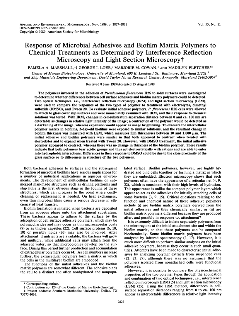
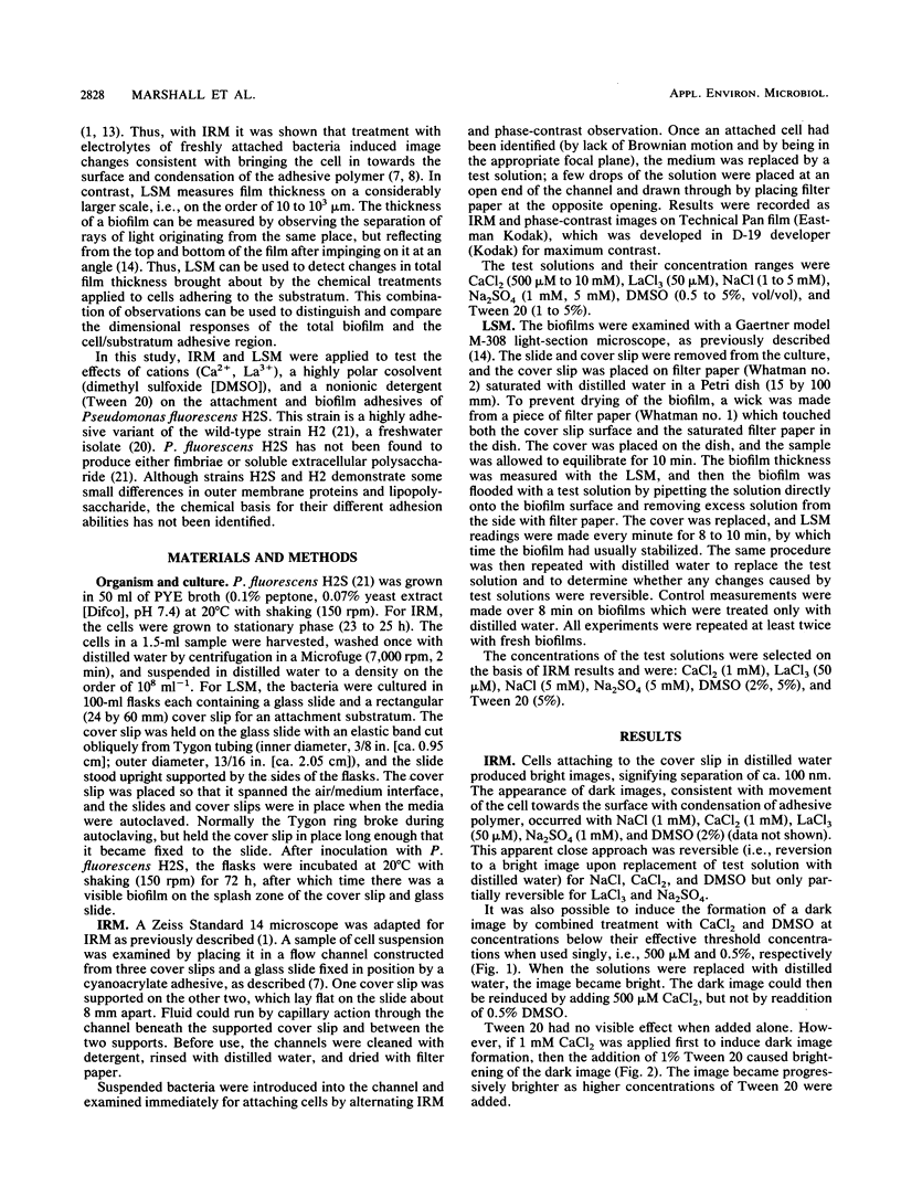
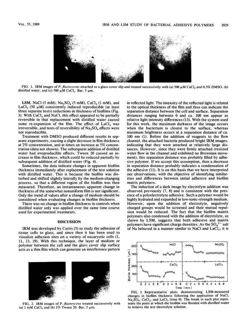
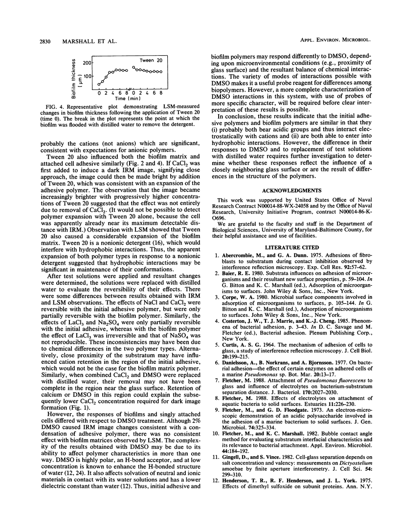
The polymers involved in the adhesion of Pseudomonas fluorescens H2S to solid surfaces were investigated to determine whether differences between cell surface adhesives and biofilm matrix polymers could be detected. Two optical techniques, i.e., interference reflection microscopy (IRM) and light section microscopy (LSM), were used to compare the responses of the two types of polymer to treatment with electrolytes, dimethyl sulfoxide (DMSO), and Tween 20. To evaluate initial adhesive polymers, P. fluorescens H2S cells were allowed to attach to glass cover slip surfaces and were immediately examined with IRM, and their response to chemical solutions was tested. With IRM, changes in cell-substratum separation distance between 0 and ca. 100 nm are detectable as changes in relative light intensity of the image; a contraction of the polymer would be detected as a darkening of the image, whereas expansion would appear as image brightening. To evaluate the intercellular polymer matrix in biofilms, 3-day-old biofilms were exposed to similar solutions, and the resultant change in biofilm thickness was measured with LSM, which measures film thicknesses between 10 and 1,000 microns. The initial adhesive and biofilm polymers were similar in that both appeared to contract when treated with electrolytes and to expand when treated with Tween 20. However, with DMSO treatment, the initial adhesive polymer appeared to contract, whereas there was no change in thickness of the biofilm polymer. These results indicate that both polymers bear acidic groups and thus act electrostatically with cations and are able to enter into hydrophobic interactions.(ABSTRACT TRUNCATED AT 250 WORDS)
Full text
PDF
Images in this article
Selected References
These references are in PubMed. This may not be the complete list of references from this article.
- Abercrombie M., Dunn G. A. Adhesions of fibroblasts to substratum during contact inhibition observed by interference reflection microscopy. Exp Cell Res. 1975 Apr;92(1):57–62. doi: 10.1016/0014-4827(75)90636-9. [DOI] [PubMed] [Google Scholar]
- CURTIS A. S. THE MECHANISM OF ADHESION OF CELLS TO GLASS. A STUDY BY INTERFERENCE REFLECTION MICROSCOPY. J Cell Biol. 1964 Feb;20:199–215. doi: 10.1083/jcb.20.2.199. [DOI] [PMC free article] [PubMed] [Google Scholar]
- Fletcher M. Attachment of Pseudomonas fluorescens to glass and influence of electrolytes on bacterium-substratum separation distance. J Bacteriol. 1988 May;170(5):2027–2030. doi: 10.1128/jb.170.5.2027-2030.1988. [DOI] [PMC free article] [PubMed] [Google Scholar]
- Fletcher M., Marshall K. C. Bubble contact angle method for evaluating substratum interfacial characteristics and its relevance to bacterial attachment. Appl Environ Microbiol. 1982 Jul;44(1):184–192. doi: 10.1128/aem.44.1.184-192.1982. [DOI] [PMC free article] [PubMed] [Google Scholar]
- Henderson T. R., Henderson R. F., York J. L. Effects of dimethyl sulfoxide on subunit proteins. Ann N Y Acad Sci. 1975 Jan 27;243:38–53. doi: 10.1111/j.1749-6632.1975.tb25342.x. [DOI] [PubMed] [Google Scholar]
- Izzard C. S., Lochner L. R. Cell-to-substrate contacts in living fibroblasts: an interference reflexion study with an evaluation of the technique. J Cell Sci. 1976 Jun;21(1):129–159. doi: 10.1242/jcs.21.1.129. [DOI] [PubMed] [Google Scholar]
- Nichols P. D., Henson J. M., Guckert J. B., Nivens D. E., White D. C. Fourier transform-infrared spectroscopic methods for microbial ecology: analysis of bacteria, bacteria-polymer mixtures and biofilms. J Microbiol Methods. 1985;4:79–94. doi: 10.1016/0167-7012(85)90023-5. [DOI] [PubMed] [Google Scholar]
- Paul J. H., Jeffrey W. H. Evidence for Separate Adhesion Mechanisms for Hydrophilic and Hydrophobic Surfaces in Vibrio proteolytica. Appl Environ Microbiol. 1985 Aug;50(2):431–437. doi: 10.1128/aem.50.2.431-437.1985. [DOI] [PMC free article] [PubMed] [Google Scholar]
- Preston T. M., King C. A. Amoeboid locomotion of Acanthamoeba castellanii with special reference to cell-substratum interactions. J Gen Microbiol. 1984 Sep;130(9):2317–2323. doi: 10.1099/00221287-130-9-2317. [DOI] [PubMed] [Google Scholar]
- Pringle J. H., Fletcher M. Influence of substratum wettability on attachment of freshwater bacteria to solid surfaces. Appl Environ Microbiol. 1983 Mar;45(3):811–817. doi: 10.1128/aem.45.3.811-817.1983. [DOI] [PMC free article] [PubMed] [Google Scholar]
- Read R. R., Costerton J. W. Purification and characterization of adhesive exopolysaccharides from Pseudomonas putida and Pseudomonas fluorescens. Can J Microbiol. 1987 Dec;33(12):1080–1090. doi: 10.1139/m87-189. [DOI] [PubMed] [Google Scholar]
- Wrangstadh M., Conway P. L., Kjelleberg S. The production and release of an extracellular polysaccharide during starvation of a marine Pseudomonas sp. and the effect thereof on adhesion. Arch Microbiol. 1986 Aug;145(3):220–227. doi: 10.1007/BF00443649. [DOI] [PubMed] [Google Scholar]