

Abstract
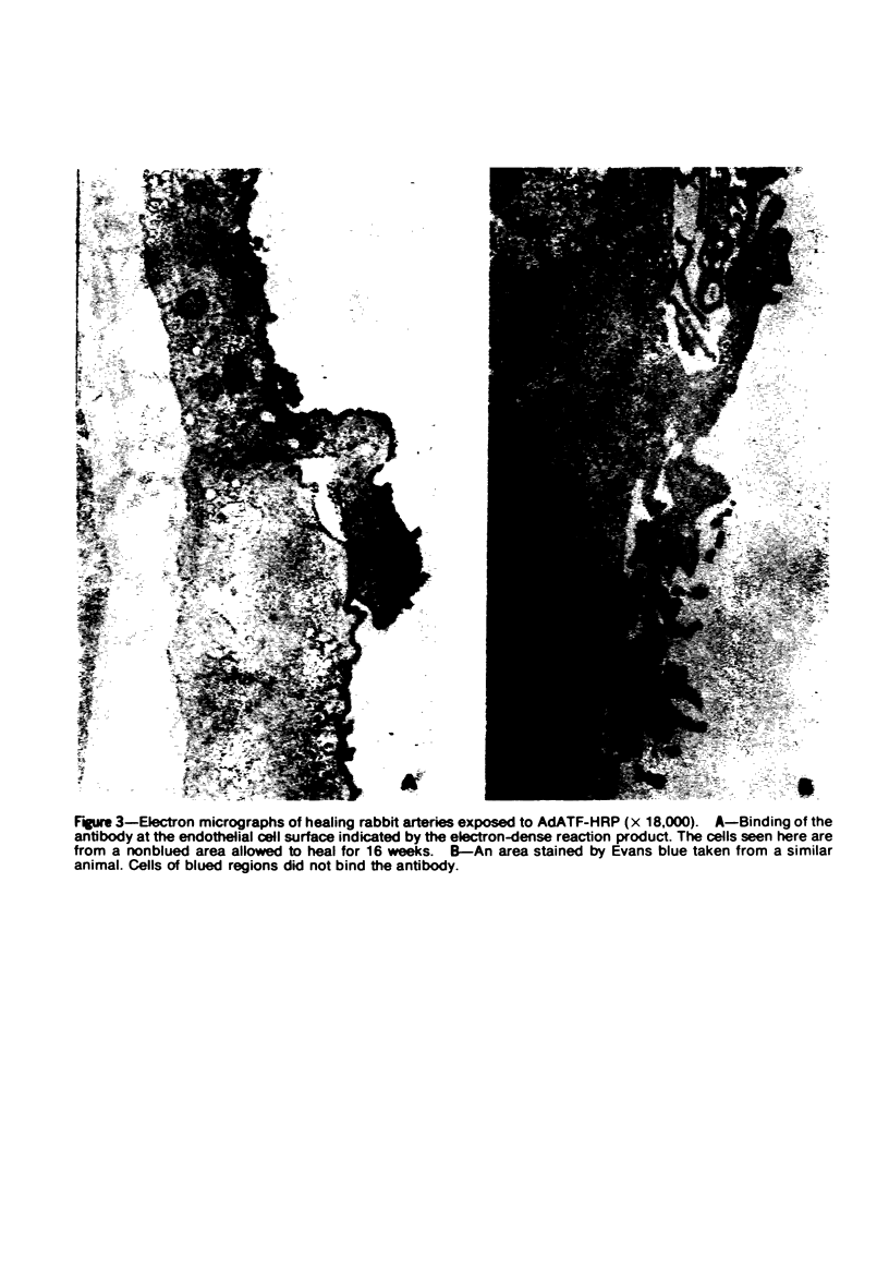
Studies were undertaken to investigate further the basis of intimal proliferation and the identity of the surface lining cell in rabbit aortas subjected to extensive deendothelialization. Endothelial cells were selectively removed by passage of an inflated balloon catheter through the arterial lumen. The healing response was evaluated at intervals up to 36 weeks by several techniques: 1) permeability to Evans blue, 2) reappearance of endothelial cells as indicated by the specific marker, adsorbed goat anti-rabbit tissue factor-horseradish peroxidase, 3) planimetric measurements of intimal thickness, and 4) electron microscopy. The results indicate that endothelial cell recovery progressed slowly and that it extended only from areas spared denudation. The regions not covered by endothelial cells were lined by cells of smooth muscle cell origin. Such areas were permeable to Evans blue-protein complex, and their luminal smooth muscle cells were associated with connective tissue-like material at their luminal surface; this material apparently acted as a base for platelet accumulation. The present findings indicate that the lumen of the extensively denuded vessel is lined by either endothelial or smooth muscle cells and that intimal healing is related to restoration of endothelial cell cover. In addition, intimal thickening reached a maximum well before reendothelialization was complete.
Full text
PDF


Images in this article
Selected References
These references are in PubMed. This may not be the complete list of references from this article.
- BAUMGARTNER H. R. EINE NEUE METHODE ZUR ERZEUGUNG VON THROMBEN DURCH GEZIELTE UBERDEHNUNG DER GEFAESSWAND. Z Gesamte Exp Med. 1963 Sep 12;137:227–247. [PubMed] [Google Scholar]
- BRECHER G., CRONKITE E. P. Morphology and enumeration of human blood platelets. J Appl Physiol. 1950 Dec;3(6):365–377. doi: 10.1152/jappl.1950.3.6.365. [DOI] [PubMed] [Google Scholar]
- Björkerud S., Bondjers G. Arterial repair and atherosclerosis after mechanical injury. I. Permeability and light microscopic characteristics of endothelium in non-atherosclerotic and atherosclerotic lesions. Atherosclerosis. 1971 May-Jun;13(3):355–363. doi: 10.1016/0021-9150(71)90078-5. [DOI] [PubMed] [Google Scholar]
- Hoyer L. W., De los Santos R. P., Hoyer J. R. Antihemophilic factor antigen. Localization in endothelial cells by immunofluorescent microscopy. J Clin Invest. 1973 Nov;52(11):2737–2744. doi: 10.1172/JCI107469. [DOI] [PMC free article] [PubMed] [Google Scholar]
- Huber J. D., Parker F., Odland G. F. A basic fuchsin and alkalinized methylene blue rapid stain for epoxy-embedded tissue. Stain Technol. 1968 Mar;43(2):83–87. doi: 10.3109/10520296809115048. [DOI] [PubMed] [Google Scholar]
- Maynard J. R., Heckman C. A., Pitlick F. A., Nemerson Y. Association of tissue factor activity with the surface of cultured cells. J Clin Invest. 1975 Apr;55(4):814–824. doi: 10.1172/JCI107992. [DOI] [PMC free article] [PubMed] [Google Scholar]
- Muggli R., Baumgartner H. R. Pattern of membrane invaginations at the surface of smooth muscle cells of rabbit arteries. Experientia. 1972 Oct 15;28(10):1212–1214. doi: 10.1007/BF01946177. [DOI] [PubMed] [Google Scholar]
- POOLE J. C., SANDERS A. G., FLOREY H. W. The regeneration of aortic endothelium. J Pathol Bacteriol. 1958 Jan;75(1):133–143. doi: 10.1002/path.1700750116. [DOI] [PubMed] [Google Scholar]
- Ross R., Glomset J. A. Atherosclerosis and the arterial smooth muscle cell: Proliferation of smooth muscle is a key event in the genesis of the lesions of atherosclerosis. Science. 1973 Jun 29;180(4093):1332–1339. doi: 10.1126/science.180.4093.1332. [DOI] [PubMed] [Google Scholar]
- Ross R., Glomset J., Kariya B., Harker L. A platelet-dependent serum factor that stimulates the proliferation of arterial smooth muscle cells in vitro. Proc Natl Acad Sci U S A. 1974 Apr;71(4):1207–1210. doi: 10.1073/pnas.71.4.1207. [DOI] [PMC free article] [PubMed] [Google Scholar]
- Ross R., Klebanoff S. J. The smooth muscle cell. I. In vivo synthesis of connective tissue proteins. J Cell Biol. 1971 Jul;50(1):159–171. doi: 10.1083/jcb.50.1.159. [DOI] [PMC free article] [PubMed] [Google Scholar]
- Schwartz S. M., Stemerman M. B., Benditt E. P. The aortic intima. II. Repair of the aortic lining after mechanical denudation. Am J Pathol. 1975 Oct;81(1):15–42. [PMC free article] [PubMed] [Google Scholar]
- Spaet T. H., Erichson R. B. The vascular wall in the pathogenesis of thrombosis. Thromb Diath Haemorrh Suppl. 1966;21:67–86. [PubMed] [Google Scholar]
- Spaet T. H., Stemerman M. B., Veith F. J., Lejnieks I. Intimal injury and regrowth in the rabbit aorta; medial smooth muscle cells as a source of neointima. Circ Res. 1975 Jan;36(1):58–70. doi: 10.1161/01.res.36.1.58. [DOI] [PubMed] [Google Scholar]
- Stemerman M. B., Baumgartner H. R., Spaet T. H. The subendothelial microfibril and platelet adhesion. Lab Invest. 1971 Mar;24(3):179–186. [PubMed] [Google Scholar]
- Stemerman M. B., Pitlick F. A., Dembitzer H. M. Electron microscopic immunohistochemical identification of endothelial cells in the rabbit. Circ Res. 1976 Mar;38(3):146–156. doi: 10.1161/01.res.38.3.146. [DOI] [PubMed] [Google Scholar]
- Stemerman M. B., Ross R. Experimental arteriosclerosis. I. Fibrous plaque formation in primates, an electron microscope study. J Exp Med. 1972 Oct 1;136(4):769–789. doi: 10.1084/jem.136.4.769. [DOI] [PMC free article] [PubMed] [Google Scholar]
- Stemerman M. B. Thrombogenesis of the rabbit arterial plaque. An electron microscopic study. Am J Pathol. 1973 Oct;73(1):7–26. [PMC free article] [PubMed] [Google Scholar]
- WEIBEL E. R., PALADE G. E. NEW CYTOPLASMIC COMPONENTS IN ARTERIAL ENDOTHELIA. J Cell Biol. 1964 Oct;23:101–112. doi: 10.1083/jcb.23.1.101. [DOI] [PMC free article] [PubMed] [Google Scholar]