Abstract
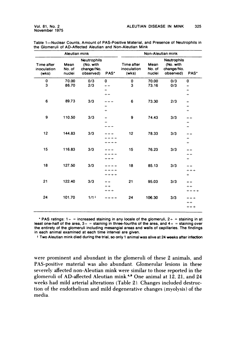
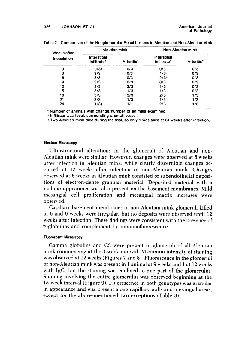
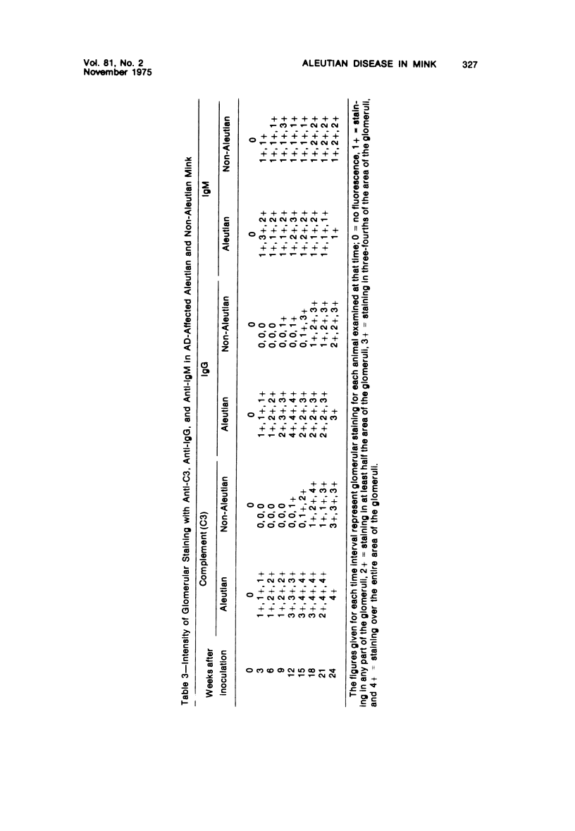
In an attempt to document progression rate differences in the development of glomerular lesions in mink infected with Aleutian disease virus (ADV), the glomeruli of Aleutian and non-Aleutian mink experimentally infected with ADV were evaluated by light, fluorescent, and electron microscopy. The animals were also examined for the presence of interstitial infiltrate, neutrophils, and arterial lesions. One hundred percent of the Aleutian mink had glomerular cell proliferation and interstitial infiltrate, while 95% of the Aleutian and 41% of the non-Aleutian mink had neutrophilic infiltrates and arteritis, respectively. Of the non-Aleutian mink, 91, 83, 42, and 12.5% had glomerular cell proliferations, glomerular neutrophils, interstitial infiltrate, and arterial lesions in, that order. All the Aleutian mink had glomerular depositions of gamma-globulin (IgG) and complement (C3), whereas 75% of non-Aleutian mink had deposits of IgG and C3. One hundred percent of both genotypes had glomerular deposits of immunoglobulin M (IgM). Ultrastructural glomerular changes consisting primarily of depositions of granular electron-dense material on basement membranes were observed in Aleutian mink 6 weeks after infection and 12 weeks after infection in non-Aleutian mink. These findings document progression rate differences in the development of glomerular lesions in Aleutian disease-affected Aleutian and non-Aleutian mink. Further, they emphasize the need for exploration of pathogenetic mechanisms involved in progression rate differences in lesion development.
Full text
PDF
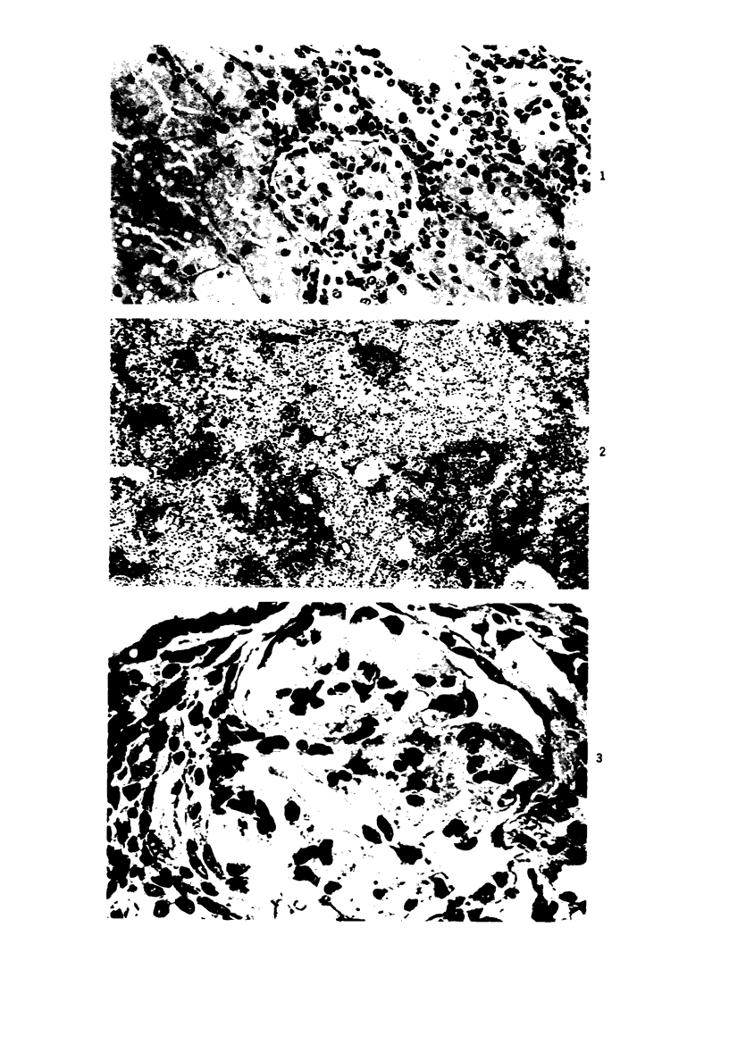
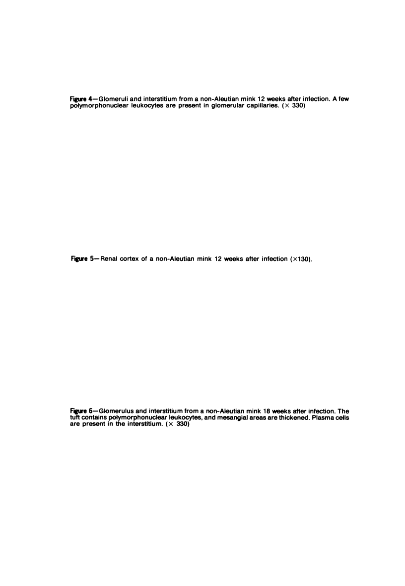
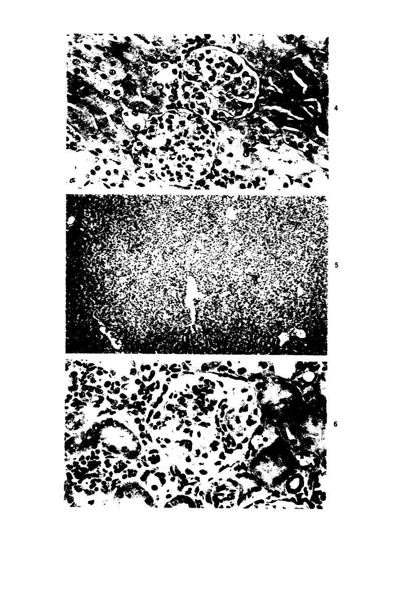
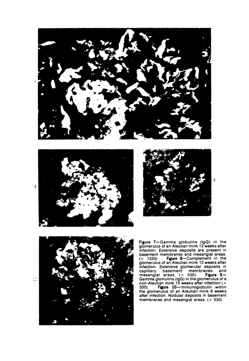
Images in this article
Selected References
These references are in PubMed. This may not be the complete list of references from this article.
- CLARK H. F., SHEPARD C. C. A DIALYSIS TECHNIQUE FOR PREPARING FLUORESCENT ANTIBODY. Virology. 1963 Aug;20:642–644. doi: 10.1016/0042-6822(63)90292-7. [DOI] [PubMed] [Google Scholar]
- Cheema A., Henson J. B., Gorham J. R. Aleutian disease of mink. Prevention of lesions by immunosuppression. Am J Pathol. 1972 Mar;66(3):543–556. [PMC free article] [PubMed] [Google Scholar]
- Cho H. J., Ingram D. G. Antigen and antibody in Aleutian disease in mink. I. Precipitation reaction by agar-gel electrophoresis. J Immunol. 1972 Feb;108(2):555–557. [PubMed] [Google Scholar]
- Cochrane C. G., Hawkins D. Studies on circulating immune complexes. 3. Factors governing the ability of circulating complexes to localize in blood vessels. J Exp Med. 1968 Jan 1;127(1):137–154. doi: 10.1084/jem.127.1.137. [DOI] [PMC free article] [PubMed] [Google Scholar]
- Eklund C. M., Hadlow W. J., Kennedy R. C., Boyle C. C., Jackson T. A. Aleutian disease of mink: properties of the etiologic agent and the host responses. J Infect Dis. 1968 Dec;118(5):510–526. doi: 10.1093/infdis/118.5.510. [DOI] [PubMed] [Google Scholar]
- HELMBOLDT C. F., JUNGHERR E. L. The pathology of Aleutian disease in mink. Am J Vet Res. 1958 Jan;19(70):212–222. [PubMed] [Google Scholar]
- HENSON J. B., LEADER R. W., GORHAM J. R. Hypergammaglobulinemia in mink. Proc Soc Exp Biol Med. 1961 Aug-Sep;107:919–920. doi: 10.3181/00379727-107-26795. [DOI] [PubMed] [Google Scholar]
- Henson J. B., Gorham J. R., Padgett G. A., Davis W. C. Pathogenesis of the glomerular lesions in aleutian disease of mink. Immunofluorescent studies. Arch Pathol. 1969 Jan;87(1):21–28. [PubMed] [Google Scholar]
- Henson J. B., Gorham J. R., Tanaka Y. Renal glomerular ultrastructure in mink affected by Aleutian disease. Lab Invest. 1967 Aug;17(2):123–139. [PubMed] [Google Scholar]
- Henson J. B., Leader R. W., Gorham J. R., Padgett G. A. The sequential development of lesions in spontaneous Aleutian disease of mink. Pathol Vet. 1966;3(4):289–314. doi: 10.1177/030098586600300401. [DOI] [PubMed] [Google Scholar]
- KENYON A. J., TRAUTWEIN G., HELMBOLDT C. F. Characterization of blood serum proteins from mink with Aleutian disease. Am J Vet Res. 1963 Jan;24:168–173. [PubMed] [Google Scholar]
- Lewis E. J., Busch G. J., Schur P. H. Gamma G globulin subgroup composition of the glomerular deposits in human renal diseases. J Clin Invest. 1970 Jun;49(6):1103–1113. doi: 10.1172/JCI106326. [DOI] [PMC free article] [PubMed] [Google Scholar]
- MARDINEY M. R., Jr, MUELLER-EBERHARD H. J. MOUSE BETA-1C-GLOBULIN: PRODUCTION OF ANTISERUM AND CHARACTERIZATION IN THE COMPLEMENT REACTION. J Immunol. 1965 Jun;94:877–882. [PubMed] [Google Scholar]
- PORTER D. D., LARSEN A. E. STATISTICAL SURVEY OF ALEUTIAN DISEASE IN RANCH MINK. Am J Vet Res. 1964 Jul;25:1226–1229. [PubMed] [Google Scholar]
- Padgett G. A., Reiquam C. W., Gorham J. R., Henson J. B., O'Mary C. C. Comparative studies of the Chediak-Higashi syndrome. Am J Pathol. 1967 Oct;51(4):553–571. [PMC free article] [PubMed] [Google Scholar]
- Padgett G. A., Reiquam C. W., Henson J. B., Gorham J. R. Comparative studies of susceptibility to infection in the Chediak-Higashi syndrome. J Pathol Bacteriol. 1968 Apr;95(2):509–522. doi: 10.1002/path.1700950224. [DOI] [PubMed] [Google Scholar]
- Pincus T., Haberkern R., Christian C. L. Experimental chronic glomerulitis. J Exp Med. 1968 Apr 1;127(4):819–832. doi: 10.1084/jem.127.4.819. [DOI] [PMC free article] [PubMed] [Google Scholar]
- Porter D. D., Larsen A. E., Porter H. G. The pathogenesis of Aleutian disease of mink. I. In vivo viral replication and the host antibody response to viral antigen. J Exp Med. 1969 Sep 1;130(3):575–593. doi: 10.1084/jem.130.3.575. [DOI] [PMC free article] [PubMed] [Google Scholar]
- Prieur D. J., Davis W. C., Padgett G. A. Defective function of renal lysosomes in mice with the Chediak-Higashi syndrome. Am J Pathol. 1972 May;67(2):227–236. [PMC free article] [PubMed] [Google Scholar]
- REYNOLDS E. S. The use of lead citrate at high pH as an electron-opaque stain in electron microscopy. J Cell Biol. 1963 Apr;17:208–212. doi: 10.1083/jcb.17.1.208. [DOI] [PMC free article] [PubMed] [Google Scholar]
- Tabel H., Ingram D. G. Immunoglobulins of mink. Evidence for five immunoglobulin classes of 7S type. Immunology. 1972 Jun;22(6):933–942. [PMC free article] [PubMed] [Google Scholar]


