

Abstract
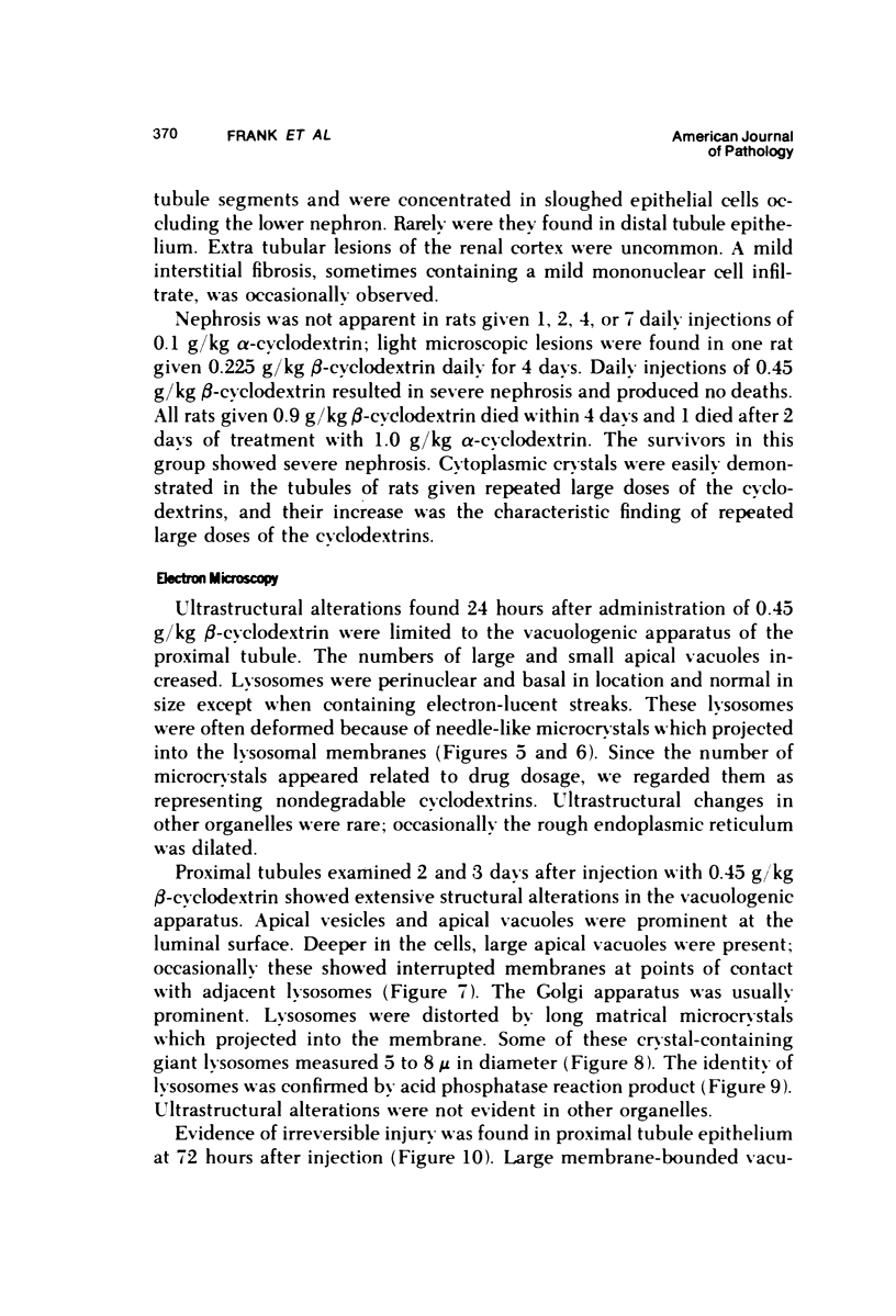
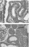
The renal toxicity of the Schardinger dextrins, alpha and beta-cyclodextrin, is manifested as a series of alterations in the vacuolar organelles of the proximal convoluted tubule. These changes begin as an increase of apical vacuoles and the appearance of giant lysosomes. The giant lysosomes characteristic of cyclodextrin nephrosis are notable because of the prominent acicular microcrystals embedded in the lysosomal matrix. Giant vacuoles devoid of acid phosphatase reaction product are found in advanced lesions. The vacuolar apparatus shows advanced changes prior to manifestation of lesions in mitochondria and other organelles. These observations indicate a role of the vacuologenic apparatus in the nephrotic process. Intracellular concentration of toxin via the lysosomal pathway represents a perversion of the physiologic function of the proximal tubule which ultimately leads to cell death.
Full text
PDF


Images in this article


Selected References
These references are in PubMed. This may not be the complete list of references from this article.
- Diomi P., Ericsson J. L., Matheson N. A., Shearer J. R. Studies on renal tubular morphology and toxicity after large doses of dextran 40 in the rabbit. Lab Invest. 1970 Apr;22(4):355–360. [PubMed] [Google Scholar]
- Doolan P. D., Schwartz S. L., Hayes J. R., Mullen J. C., Cummings N. B. An evaluation of the nephrotoxicity of ethylenediaminetetraacetate and diethylenetriaminepentaacetate in the rat. Toxicol Appl Pharmacol. 1967 May;10(3):481–500. doi: 10.1016/0041-008x(67)90088-9. [DOI] [PubMed] [Google Scholar]
- Emanuelli G., Satta G., Perpignano G. Lysosomal damage and its possible relationship with mitochondrial lesion in rat kidney under obstructive jaundice. Clin Chim Acta. 1969 Jul;25(1):167–172. doi: 10.1016/0009-8981(69)90243-5. [DOI] [PubMed] [Google Scholar]
- FRENCH D. The Schardinger dextrins. Adv Carbohydr Chem. 1957;12:189–260. doi: 10.1016/s0096-5332(08)60209-x. [DOI] [PubMed] [Google Scholar]
- Fowler B. A., Brown H. W., Lucier G. W., Krigman M. R. The effects of chronic oral methyl mercury exposure on the lysosome system of rat kidney. Morphometric and biochemical studies. Lab Invest. 1975 Mar;32(3):313–322. [PubMed] [Google Scholar]
- Griffith L. D., Bulger R. E., Trump B. F. The ultrastructure of the functioning kidney. Lab Invest. 1967 Feb;16(2):220–246. [PubMed] [Google Scholar]
- JANIGAN D. T., SANTAMARIA A. A histochemical study of swelling and vacuolation of proximal tubular cells in sucrose nephrosis in the rat. Am J Pathol. 1961 Aug;39:175–193. [PMC free article] [PubMed] [Google Scholar]
- MAUNSBACH A. B., MADDEN S. C., LATTA H. Light and electron microscopic changes in proximal tubules of rats after administration of glucose, mannitol, sucrose, or dextran. Lab Invest. 1962 Jun;11:421–432. [PubMed] [Google Scholar]
- Prieur D. J., Davis W. C., Padgett G. A. Defective function of renal lysosomes in mice with the Chediak-Higashi syndrome. Am J Pathol. 1972 May;67(2):227–236. [PMC free article] [PubMed] [Google Scholar]
- Schwartz S. L., Johnson C. B., Doolan P. D. Study of the mechanism of renal vacuologenesis induced in the rat by ethylenediaminetetraacetate. Comparison of the cellular activities of calcium and chromium chelates. Mol Pharmacol. 1970 Jan;6(1):54–60. [PubMed] [Google Scholar]
- TRUMP B. F., JANIGAN D. T. The pathogenesis of cytologic vacuolization in sucrose nephrosis. An electron microscopic and histochemical study. Lab Invest. 1962 May;11:395–411. [PubMed] [Google Scholar]