

Abstract
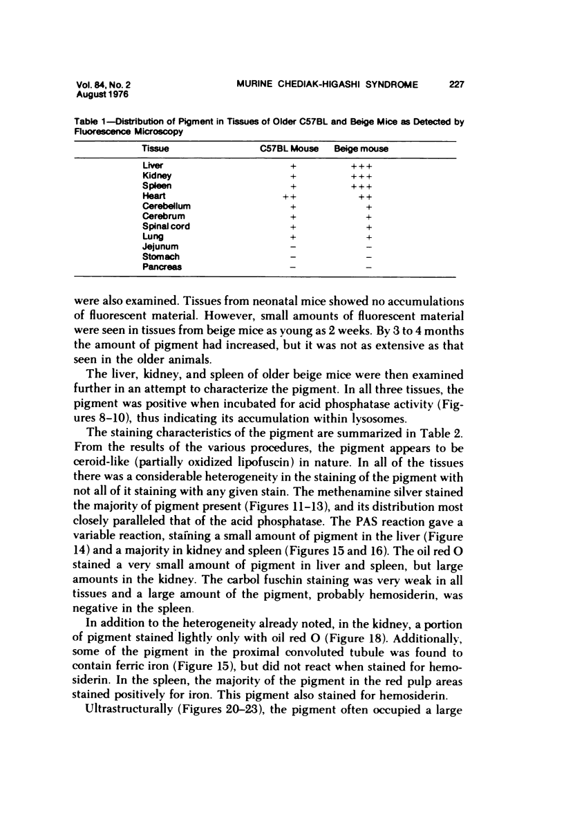
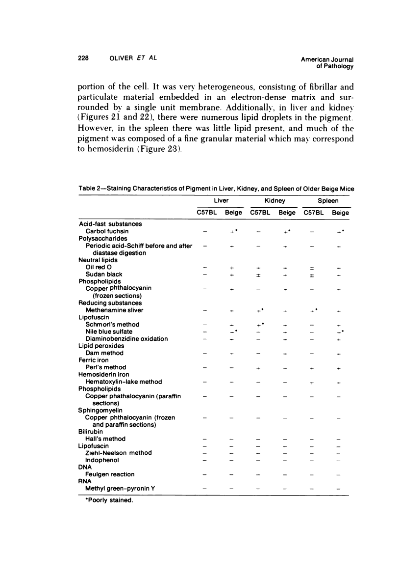
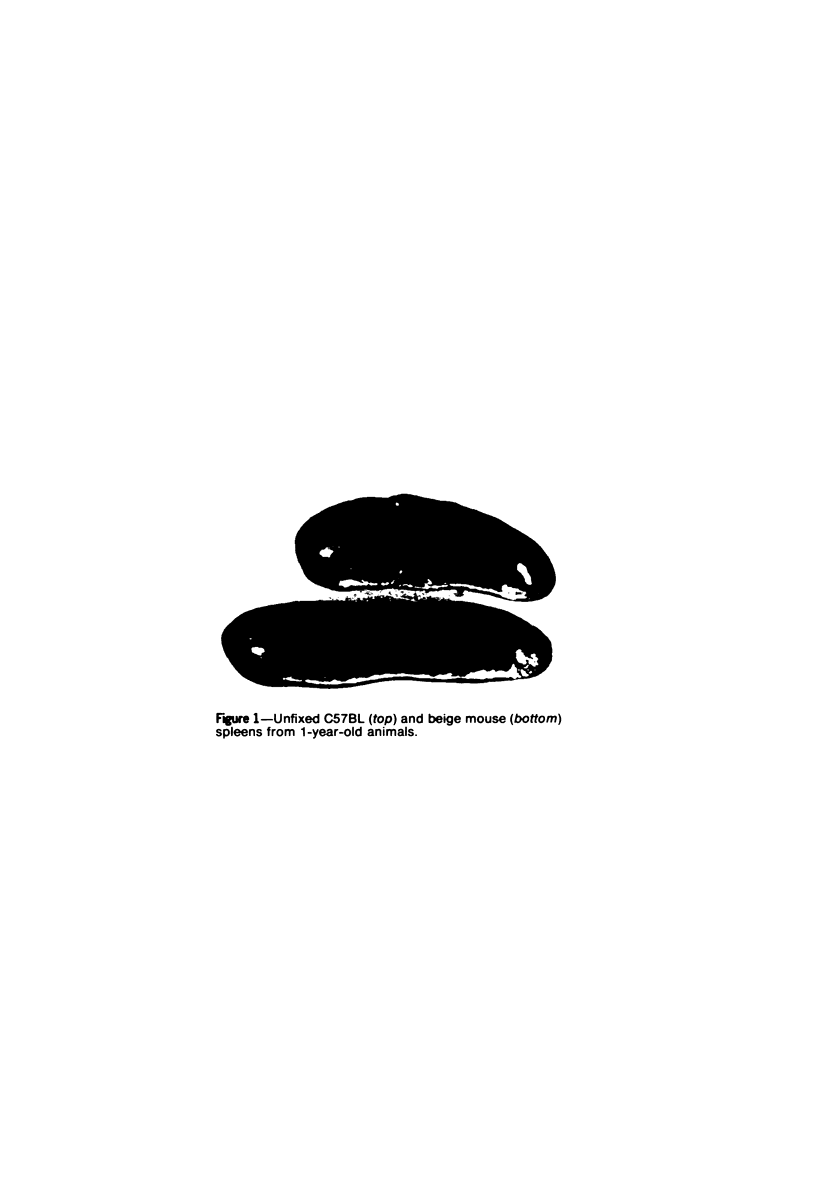
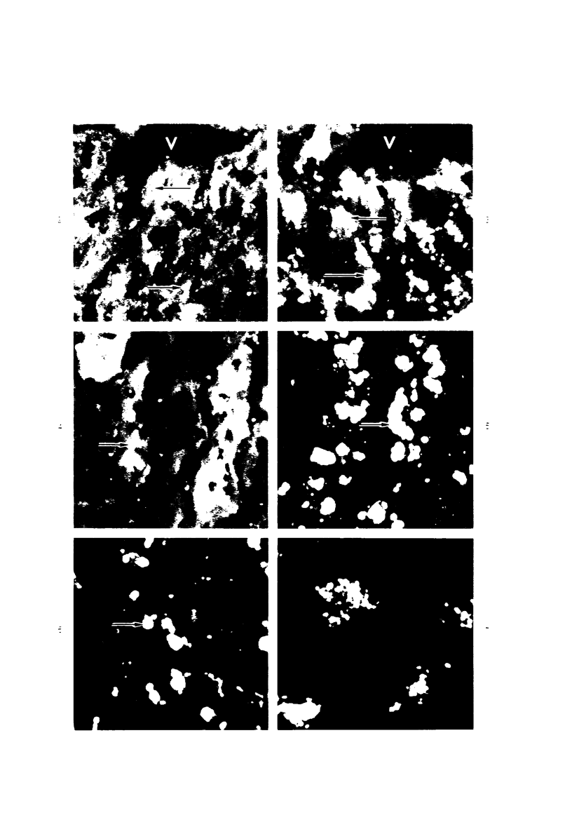
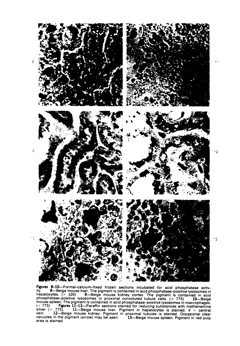
The Chediak-Higashi Syndrome (CHS) is a rare inherited disorder occurring in man and several animal homologs including the beige mouse; it is characterized by pigmentary dilution, susceptibility to pyogenic infections, and the presence of enlarged lysosomes in many cell types. Beige mice 6 months of age and older were found to have darkened livers, kidneys, and spleens, accompanied by splenomegaly. A fluorescence microscopic survey of tissues from beige mice revealed marked accumulations of a microscopic survey of tissues from beige mice revealed marked accumulations of a yellow autofluorescent pigment inhepatocytes, renal proximal tubule cells, and splenic macrophages. Additionally, large amounts of hemosiderin were present in the spleen. In beige mice, the pigment was noted in animals as young as 1 to 2 weeks of age and gradually increased in amount as the animals aged. A histochemical investigation of the pigment showed that it was ceroid-like in nature and contained in lysosomes. Ultrastructurally, the pigment was composed of lipid-like droplets embedded in a dense matrix and surrounded by a limiting membrane. The accumulation of ceroid-like material in beige mice may be a reflection of the metabolic disturbance which underlies CHS.
Full text
PDF
Images in this article
Selected References
These references are in PubMed. This may not be the complete list of references from this article.
- ALPERT M. Hormonal induction of deposition of ceroid pigment in the mouse. Anat Rec. 1953 Aug;116(4):469–493. doi: 10.1002/ar.1091160407. [DOI] [PubMed] [Google Scholar]
- Blume R. S., Wolff S. M. The Chediak-Higashi syndrome: studies in four patients and a review of the literature. Medicine (Baltimore) 1972 Jul;51(4):247–280. [PubMed] [Google Scholar]
- Brandt E. J., Elliott R. W., Swank R. T. Defective lysosomal enzyme secretion in kidneys of Chediak-Higashi (beige) mice. J Cell Biol. 1975 Dec;67(3):774–788. doi: 10.1083/jcb.67.3.774. [DOI] [PMC free article] [PubMed] [Google Scholar]
- CASSELMAN W. G. B. The in vitro preparation and histochemical properties of substances resembling ceroid. J Exp Med. 1951 Dec 1;94(6):549–562. doi: 10.1084/jem.94.6.549. [DOI] [PMC free article] [PubMed] [Google Scholar]
- CAT I., MARINONI L. P., GIRALDI D. J., DE ALMEIDA M. B., SACHELLINETO A., BRAGA H., DA SILVA A. Q. CH'EDIAK-HIGASHI SYNDROME WITH FAMILIAL IDIOPATHIC HYPERLIPAEMIA. Lancet. 1965 Jun 26;1(7400):1398–1398. doi: 10.1016/s0140-6736(65)92203-8. [DOI] [PubMed] [Google Scholar]
- Davis W. C., Douglas S. D. Defective granule formation and function in the Chediak-Higashi syndrome in man and animals. Semin Hematol. 1972 Oct;9(4):431–450. [PubMed] [Google Scholar]
- Davis W. C., Spicer S. S., Greene W. B., Padgett G. A. Ultrastructure of bone marrow granulocytes in normal mink and mink with the homolog of the Chediak-Higashi trait of humans. I. Origin of the abnormal granules present in the neutrophils of mink with the C-HS trait. Lab Invest. 1971 Apr;24(4):303–317. [PubMed] [Google Scholar]
- Davis W. C., Spicer S. S., Greene W. B., Padgett G. A. Ultrastructure of cells in bone marrow and peripheral blood of normal mink and mink with the homologue of the Chediak-Higashi trait of humans. II. Cytoplasmic granules in eosinophils, basophils, mononuclear cells and platelets. Am J Pathol. 1971 Jun;63(3):411–432. [PMC free article] [PubMed] [Google Scholar]
- ESSNER E., NOVIKOFF A. B. Human hepatocellular pigments and lysosomes. J Ultrastruct Res. 1960 Jun;3:374–391. doi: 10.1016/s0022-5320(60)90016-2. [DOI] [PubMed] [Google Scholar]
- ESSNER E., NOVIKOFF A. B. Localization of acid phosphatase activity in hepatic lysosomes by means of electron microscopy. J Biophys Biochem Cytol. 1961 Apr;9:773–784. doi: 10.1083/jcb.9.4.773. [DOI] [PMC free article] [PubMed] [Google Scholar]
- Endicott K. M., Lillie R. D. Ceroid, the Pigment of Dietary Cirrhosis of Rats: Its Characteristics and Its Differentiation from Hemofuscin. Am J Pathol. 1944 Jan;20(1):149–153. [PMC free article] [PubMed] [Google Scholar]
- Essner E., Oliver C. Lysosome formation in hepatocytes of mice with Chèdiak-Higashi syndrome. Lab Invest. 1974 May;30(5):597–607. [PubMed] [Google Scholar]
- Gallin J. I., Bujak J. S., Patten E., Wolff S. M. Granulocyte function in the Chediak-Higashi syndrome of mice. Blood. 1974 Feb;43(2):201–206. [PubMed] [Google Scholar]
- Goldfischer S., Villaverde H., Forschirm R. The demonstration of acid hydrolase, thermostable reduced diphosphopyridine nucleotide tetrazolium reductase and peroxidase activities in human lipofuscin pigment granules. J Histochem Cytochem. 1966 Sep;14(9):641–652. doi: 10.1177/14.9.641. [DOI] [PubMed] [Google Scholar]
- HALL M. J. A staining reaction for bilirubin in sections of tissue. Am J Clin Pathol. 1960 Oct;34:313–316. doi: 10.1093/ajcp/34.4.313. [DOI] [PubMed] [Google Scholar]
- KRITZLER R. A., TERNER J. Y., LINDENBAUM J., MAGIDSON J., WILLIAMS R., PRESIG R., PHILLIPS G. B. CHEDIAK-HIGASHI SYNDROME. CYTOLOGIC AND SERUM LIPID OBSERVATIONS IN A CASE AND FAMILY. Am J Med. 1964 Apr;36:583–594. doi: 10.1016/0002-9343(64)90106-8. [DOI] [PubMed] [Google Scholar]
- Kanfer J. N., Blume R. S., Yankee R. A., Wolff S. M. Alteration of sphingolipid metabolism in leukocytes from patients with the Chediak-Higashi syndrome. N Engl J Med. 1968 Aug 22;279(8):410–413. doi: 10.1056/NEJM196808222790806. [DOI] [PubMed] [Google Scholar]
- LEADER R. W., PADGETT G. A., GORHAM J. R. STUDIES OF ABNORMAL LEUKOCYTE BODIES IN THE MINK. Blood. 1963 Oct;22:477–484. [PubMed] [Google Scholar]
- LEE C. S. Histochemical studies of the ceroid pigment of rats and mice and its relation to necrosis. J Natl Cancer Inst. 1950 Oct;11(2):339–349. [PubMed] [Google Scholar]
- Lutzner M. A., Tierney J. H., Benditt E. P. Giant granules and widespread cytoplasmic inclusions in a genetic syndrome of Aleutian mink. An electron microscopic study. Lab Invest. 1965 Dec;14(12):2063–2079. [PubMed] [Google Scholar]
- MIYAWAKI H. HISTOCHEMISTRY AND ELECTRON MICROSCOPY OF IRON-CONTAINING GRANULES, LYSOSOMES, AND LIPOFUSCIN IN MOUSE MAMMARY GLANDS. J Natl Cancer Inst. 1965 May;34:601–623. [PubMed] [Google Scholar]
- Novikoff A. B., Novikoff P. M., Quintana N., Davis C. Studies on microperoxisomes. IV. Interrelations of microperoxisomes, endoplasmic reticulum and lipofuscin granules. J Histochem Cytochem. 1973 Nov;21(11):1010–1020. doi: 10.1177/21.11.1010. [DOI] [PubMed] [Google Scholar]
- Oliver C., Essner E. Distribution of anomalous lysosomes in the beige mouse: a homologue of Chediak-Higashi syndrome. J Histochem Cytochem. 1973 Mar;21(3):218–228. doi: 10.1177/21.3.218. [DOI] [PubMed] [Google Scholar]
- Oliver C., Essner E. Formation of anomalous lysosomes in monocytes, neutrophils, and eosinophils from bone marrow of mice with Chédiak-Higashi syndrome. Lab Invest. 1975 Jan;32(1):17–27. [PubMed] [Google Scholar]
- PADGETT G. A., LEADER R. W., GORHAM J. R., O'MARY C. C. THE FAMILIAL OCCURRENCE OF THE CHEDIAK-HIGASHI SYNDROME IN MINK AND CATTLE. Genetics. 1964 Mar;49:505–512. doi: 10.1093/genetics/49.3.505. [DOI] [PMC free article] [PubMed] [Google Scholar]
- PIERRO L. J. PIGMENT GRANULE FORMATION IN SLATE, A COAT COLOR MUTANT IN THE MOUSE. Anat Rec. 1963 Aug;146:365–372. [PubMed] [Google Scholar]
- Pappenheimer A. M., Victor J. "Ceroid" Pigment in Human Tissues. Am J Pathol. 1946 Mar;22(2):395–413. [PMC free article] [PubMed] [Google Scholar]
- Prieur D. J., Davis W. C., Padgett G. A. Defective function of renal lysosomes in mice with the Chediak-Higashi syndrome. Am J Pathol. 1972 May;67(2):227–236. [PMC free article] [PubMed] [Google Scholar]
- REYNOLDS E. S. The use of lead citrate at high pH as an electron-opaque stain in electron microscopy. J Cell Biol. 1963 Apr;17:208–212. doi: 10.1083/jcb.17.1.208. [DOI] [PMC free article] [PubMed] [Google Scholar]
- Root R. K., Rosenthal A. S., Balestra D. J. Abnormal bactericidal, metabolic, and lysosomal functions of Chediak-Higashi Syndrome leukocytes. J Clin Invest. 1972 Mar;51(3):649–665. doi: 10.1172/JCI106854. [DOI] [PMC free article] [PubMed] [Google Scholar]
- SARAIVA L. G., AZEVEDO M., CORREA J. M., CARVALHO G., PROSPERO J. D. Anomalous panleukocytic granulation. Blood. 1959 Oct;14:1112–1127. [PubMed] [Google Scholar]
- Spicer S. S. Siderosis Associated with Increased Lipofuscins and Mast Cells in Aging Mice. Am J Pathol. 1960 Oct;37(4):457–475. [PMC free article] [PubMed] [Google Scholar]
- Spurr A. R. A low-viscosity epoxy resin embedding medium for electron microscopy. J Ultrastruct Res. 1969 Jan;26(1):31–43. doi: 10.1016/s0022-5320(69)90033-1. [DOI] [PubMed] [Google Scholar]
- Sung J. H., Okada K. Neuropathological changes in mink with Chediak-Higashi disease. A light and electron microscopic study. J Neuropathol Exp Neurol. 1971 Jan;30(1):33–62. doi: 10.1097/00005072-197101000-00005. [DOI] [PubMed] [Google Scholar]
- TAPPEL A. L. Studies of the mechanism of vitamin E action. III. In vitro copolymerization of oxidized fats with protein. Arch Biochem Biophys. 1955 Feb;54(2):266–280. doi: 10.1016/0003-9861(55)90039-4. [DOI] [PubMed] [Google Scholar]
- White J. G. The Chediak-Higashi syndrome: a possible lysosomal disease. Blood. 1966 Aug;28(2):143–156. [PubMed] [Google Scholar]