

Abstract
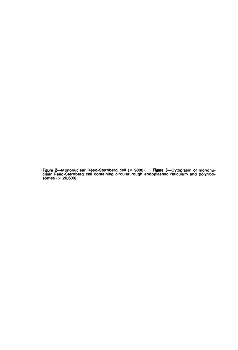
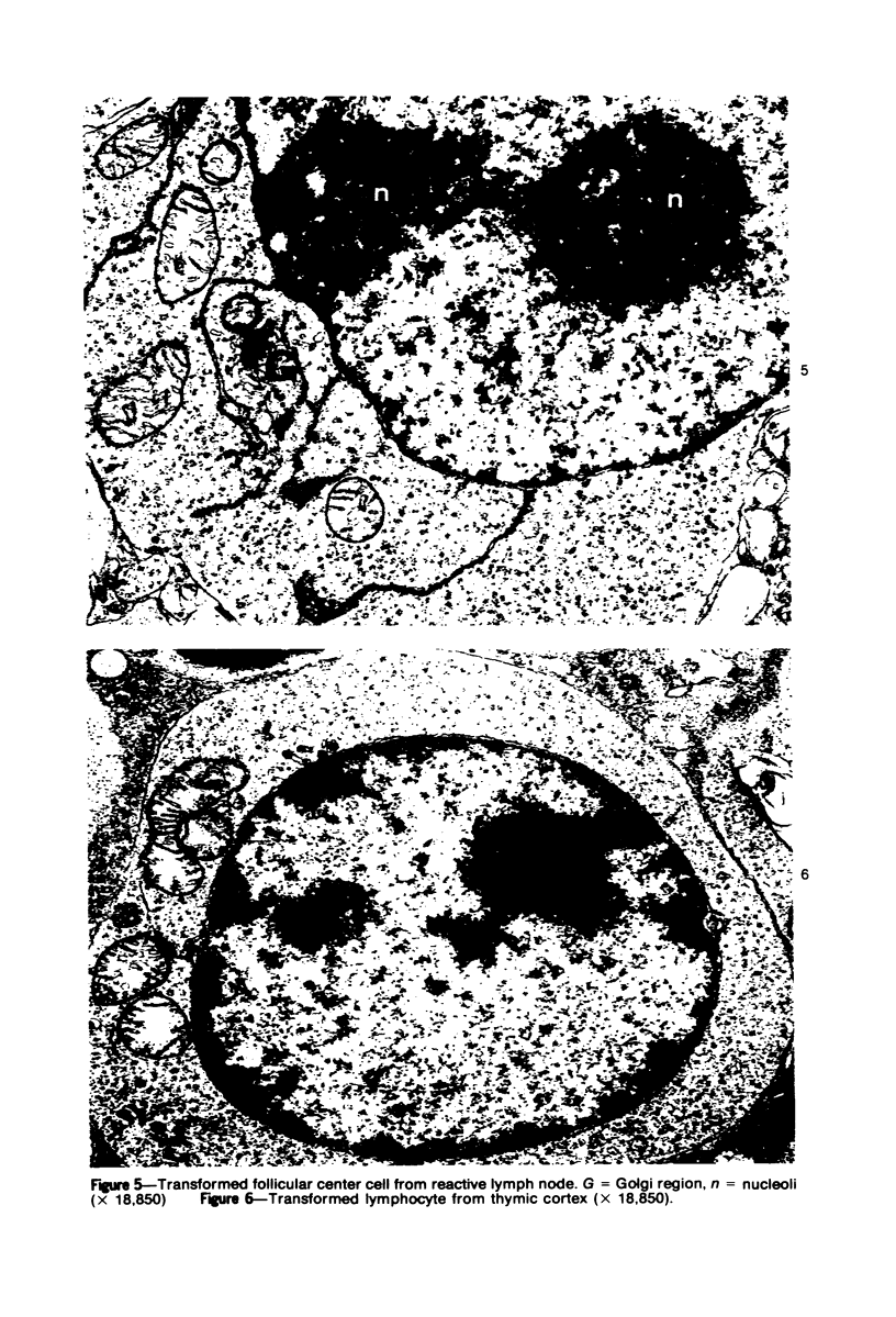
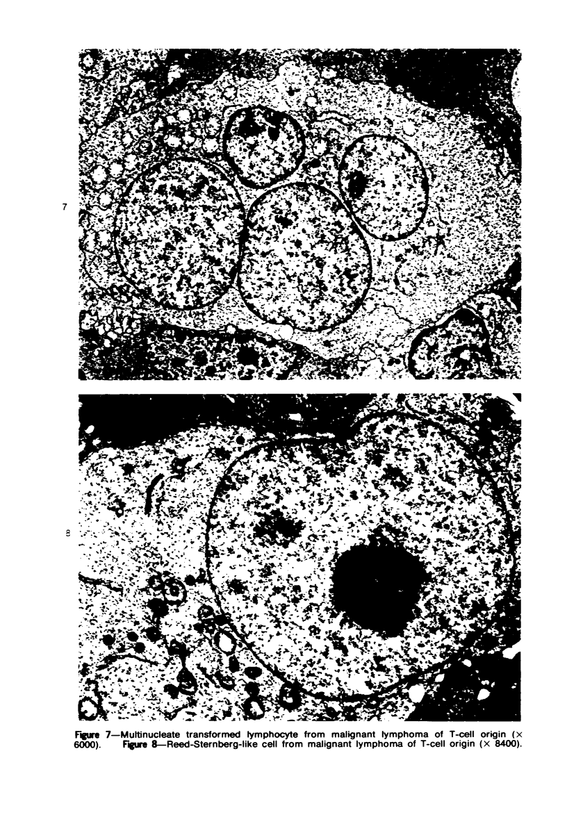
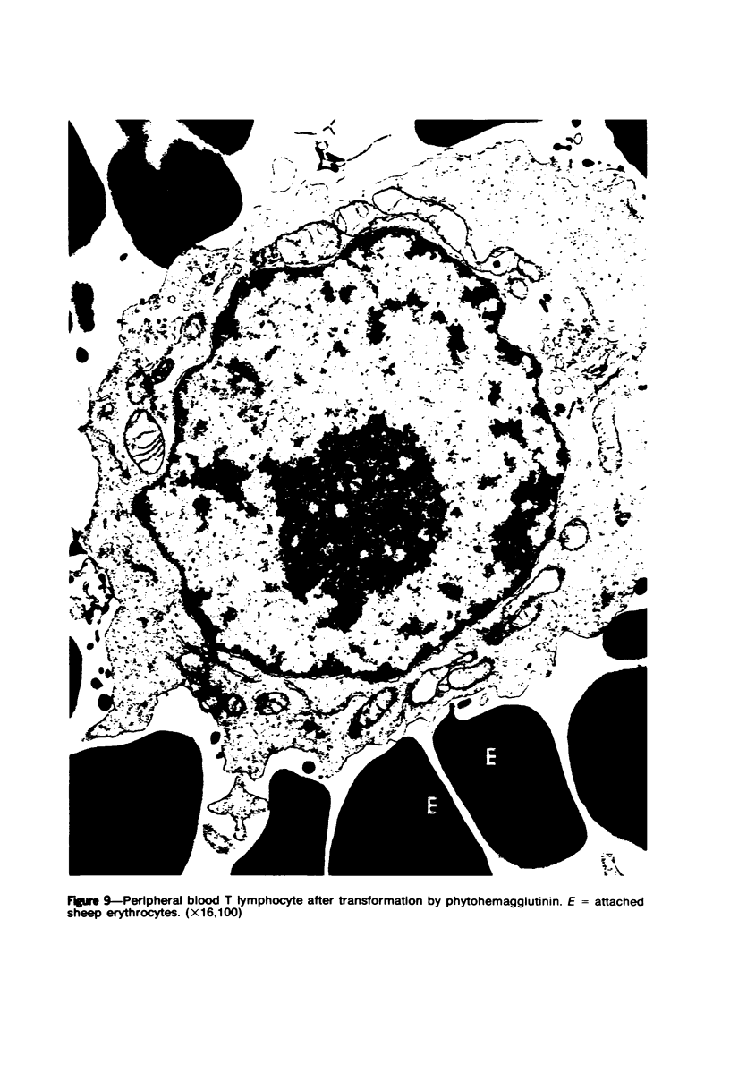
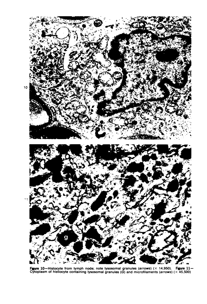
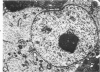
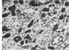
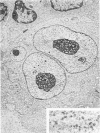
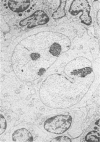
The ultrastructural features of Reed-Sternberg cells from 17 patients with Hodgkin's disease were compared with those of histiocytes and transformed lymphocytes in both benign and malignant conditions. Transformed lymphocytes and Reed-Sternberg cells appeared to have similar features, including large nuclei with dispersed chromatin, large nucleoli, and great numbers of cytoplasmic polyribosomes. Histiocytes contained abundant cytoplasmic lysosomal granules and microfilaments. These results are indicative of the origin of Reed-Sternberg cells from lymphocytes.
Full text
PDF


Images in this article


Selected References
These references are in PubMed. This may not be the complete list of references from this article.
- BRAUNSTEINER H., FELLINGER K., PAKESCH F. Electron Microscopic investigations on sections from lymph nodes and bone marrow in malignant blood diseases. Blood. 1957 Mar;12(3):278–294. [PubMed] [Google Scholar]
- Dorfman R. F., Rice D. F., Mitchell A. D., Kempson R. L., Levine G. Ultrastructural studies of Hodgkin's disease. Natl Cancer Inst Monogr. 1973 May;36:221–238. [PubMed] [Google Scholar]
- Douglas S. D. Human lymphocyte growth in vitro: morphologic, biochemical, and immunologic significance. Int Rev Exp Pathol. 1971;10:41–114. [PubMed] [Google Scholar]
- FRAJOLA W. J., GREIDER M. H., BOURONCLE B. A. Cytology of the Sternberg-Reed cell as revealed by the electron microscope. Ann N Y Acad Sci. 1958 Sep 5;73(1):221–236. doi: 10.1111/j.1749-6632.1959.tb40802.x. [DOI] [PubMed] [Google Scholar]
- Garvin A. J., Spicer S. S., Parmley R. T., Munster A. M. Immunohistochemical demonstration of IgG in Reed-Sternberg and other cells in Hodgkin's disease. J Exp Med. 1974 May 1;139(5):1077–1083. doi: 10.1084/jem.139.5.1077. [DOI] [PMC free article] [PubMed] [Google Scholar]
- Glick A. D., Horn R. G. Identification of promonocytes and monocytoid precursors in acute leukaemia of adults: ultrastructural and cytochemical observations. Br J Haematol. 1974 Mar;26(3):395–403. doi: 10.1111/j.1365-2141.1974.tb00481.x. [DOI] [PubMed] [Google Scholar]
- Glick A. D., Leech J. H., Waldron J. A., Flexner J. M., Horn R. G., Collins R. D. Malignant lymphomas of follicular center cell origin in man. II. Ultrastructural and cytochemical studies. J Natl Cancer Inst. 1975 Jan;54(1):23–36. doi: 10.1093/jnci/54.1.23. [DOI] [PubMed] [Google Scholar]
- Leech J. Immunoglobulin-positive Reed Sternberg cells in Hodgkin's disease. Lancet. 1973 Aug 4;2(7823):265–266. doi: 10.1016/s0140-6736(73)93173-5. [DOI] [PubMed] [Google Scholar]
- Maunsbach A. B. The influence of different fixatives and fixation methods on the ultrastructure of rat kidney proximal tubule cells. I. Comparison of different perfusion fixation methods and of glutaraldehyde, formaldehyde and osmium tetroxide fixatives. J Ultrastruct Res. 1966 Jun;15(3):242–282. doi: 10.1016/s0022-5320(66)80109-0. [DOI] [PubMed] [Google Scholar]
- Order S. E., Hellman S. Pathogenesis of Hodgkin's disease. Lancet. 1972 Mar 11;1(7750):571–573. doi: 10.1016/s0140-6736(72)90360-1. [DOI] [PubMed] [Google Scholar]
- Peckham M. J., Cooper E. H. Proliferation characteristics of the various classes of cells in Hodgkin's disease. Cancer. 1969 Jul;24(1):135–146. doi: 10.1002/1097-0142(196907)24:1<135::aid-cncr2820240118>3.0.co;2-h. [DOI] [PubMed] [Google Scholar]
- van Furth R., Cohn Z. A., Hirsch J. G., Humphrey J. H., Spector W. G., Langevoort H. L. The mononuclear phagocyte system: a new classification of macrophages, monocytes, and their precursor cells. Bull World Health Organ. 1972;46(6):845–852. [PMC free article] [PubMed] [Google Scholar]